Quick answer
Basal cell carcinoma is the most common type of skin cancer, usually caused by long-term sun exposure and typically growing slowly in the skin’s basal cells. At Acibadem in Turkey, evaluation focuses on skin examination and biopsy, and treatment is planned according to the lesion’s size, depth, and location, using options such as surgical removal and other targeted dermatologic therapies.
What is basal cell carcinoma?
Basal cell carcinoma is the most common type of skin cancer. It develops in basal cells, which are small, round cells found in the lowest layer of the epidermis: the outer layer of the skin. These cells normally produce new skin cells as older ones wear away. When the genetic material inside basal cells becomes damaged, most often by ultraviolet (UV) light from the sun or tanning beds, the cells can begin to grow in an abnormal, uncontrolled way. This abnormal growth is what doctors call basal cell carcinoma, sometimes abbreviated as BCC.
To answer the question many patients ask first, what is basal cell carcinoma in practical terms, it is usually a slow-growing skin cancer that rarely spreads to distant parts of the body. However, “rarely spreads” does not mean “harmless.” If left untreated, a basal cell carcinoma can grow deeper into the skin and damage nearby tissue, including muscle, cartilage, and bone. This is why timely diagnosis and treatment matter, even for a cancer that is generally considered less dangerous than other forms.
Basal cell carcinoma most often affects adults, and the risk increases with age because sun damage builds up over a lifetime. It is more common in people with fair skin, light-colored eyes, and blond or red hair, although it can occur in people of any skin tone. Men have historically been diagnosed more often than women, likely reflecting patterns of sun exposure, though this gap has narrowed in many countries. The condition typically appears on parts of the body that receive the most sunlight, the face, ears, scalp, neck, shoulders, and arms, but it can develop anywhere on the skin.
Symptoms of basal cell carcinoma
Basal cell carcinoma symptoms usually appear on the skin itself, which means the condition is often visible before it causes any pain or general illness. Many people first notice a spot, bump, or sore that looks slightly different from the surrounding skin or that fails to heal the way an ordinary scrape or pimple would. Common signs include:
- A pearly or waxy bump, often with a translucent (see-through) quality, sometimes showing tiny visible blood vessels on its surface.
- A sore that does not heal, or one that heals and then reopens, sometimes bleeding, oozing, or crusting repeatedly.
- A flat, scaly, reddish patch, which may slowly grow larger over months or years.
- A shiny pink, red, or skin-colored growth with a slightly raised, rolled edge and a lowered center.
- A white, yellow, or waxy scar-like area without a clearly defined border, which can be easy to overlook.
- A brown, black, or blue lesion with a slightly raised, translucent border, more common in people with darker skin tones.
Symptoms can differ depending on the type of basal cell carcinoma. The nodular type, the most common form, tends to appear as the classic pearly bump on the face. The superficial type often shows up as a flat, scaly, red patch, frequently on the trunk, and can be mistaken for eczema or psoriasis. The morpheaform (also called sclerosing or infiltrative) type looks like a pale scar and tends to grow more deeply and widely under the skin than it appears on the surface, which can make it harder to detect and treat. Pigmented basal cell carcinoma contains darker coloring and can sometimes be confused with melanoma, a different and more aggressive skin cancer.
In its early stage, a basal cell carcinoma is usually small, painless, and easy to dismiss. As it grows, it may itch, become tender, bleed with minor contact, or develop a raw, ulcerated (open-sore) center. In advanced, neglected cases, the tumor can invade deeper structures and cause visible tissue destruction, which is far more difficult to treat. Because early lesions can be subtle, any new or changing skin spot that persists for several weeks deserves medical attention.
Causes and risk factors
The main cause of basal cell carcinoma is damage to the DNA, the genetic instruction code, inside basal skin cells. In most cases, this damage comes from ultraviolet (UV) radiation. Understanding basal cell carcinoma causes helps explain who is most at risk and how the condition can often be prevented.
- Chronic sun exposure: Long-term, cumulative exposure to sunlight is the leading cause. UV rays damage the DNA in skin cells, and over years this damage can lead to cancerous change.
- Sunburns: Intense, intermittent exposure, especially blistering sunburns during childhood or adolescence, increases the risk later in life.
- Tanning beds: Artificial UV light from indoor tanning devices raises the risk of basal cell carcinoma and other skin cancers.
- Fair skin: People with light skin, freckles, blond or red hair, and blue, green, or gray eyes have less melanin, the natural pigment that offers some protection against UV damage.
- Older age: Because sun damage accumulates over decades, most cases occur in middle-aged and older adults, although younger people can also develop the condition.
- Previous skin cancer: Having had one basal cell carcinoma significantly increases the chance of developing another.
- Radiation therapy: Skin that received radiation treatment in the past, for example for acne or another cancer, is at higher risk.
- Weakened immune system: People taking immunosuppressant medication (drugs that lower immune activity), such as organ transplant recipients, are more vulnerable.
- Exposure to arsenic: Long-term contact with arsenic, a toxic element found in some contaminated water sources and industrial settings, increases risk.
- Inherited conditions: Rare genetic disorders, such as basal cell nevus syndrome (Gorlin syndrome) and xeroderma pigmentosum, greatly increase susceptibility, often at a young age.
It is important to understand that having one or more risk factors does not mean a person will definitely develop skin cancer, and some people develop basal cell carcinoma without obvious risk factors. Consistent sun protection, including sunscreen, protective clothing, and avoiding peak-hour sun and tanning beds, remains the most effective known way to lower the risk.
Diagnosis
Basal cell carcinoma diagnosis begins with a careful physical examination. A doctor, often a dermatologist (a physician specializing in skin conditions), will inspect the suspicious spot as well as the rest of the skin, since people with one lesion may have others. The doctor will ask how long the spot has been present, whether it has changed, and whether it bleeds, itches, or fails to heal.
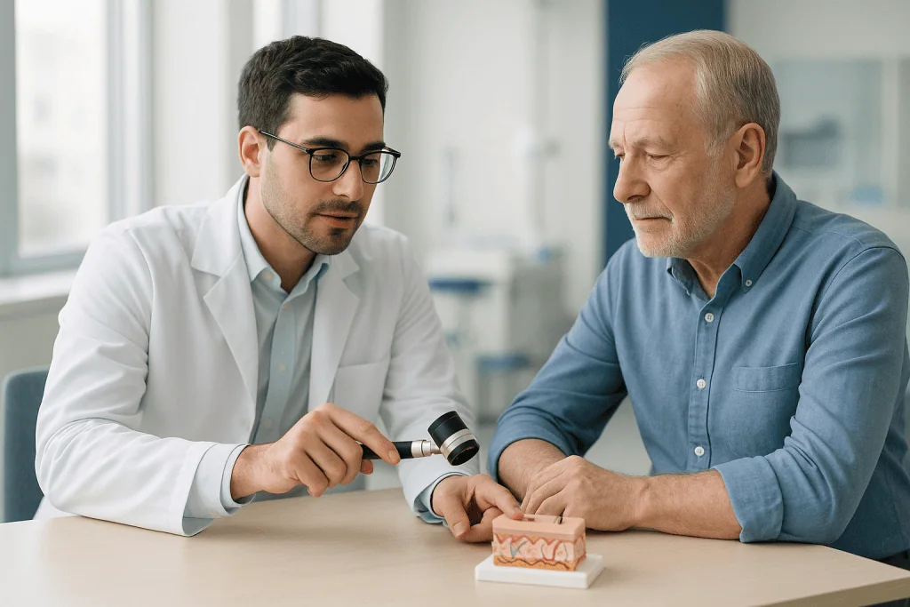
Many dermatologists use a dermatoscope, a handheld magnifying instrument with a light, to examine the lesion’s surface structures and blood vessel patterns in detail. This technique, called dermoscopy, helps distinguish basal cell carcinoma from harmless growths and from other skin cancers, although it cannot confirm the diagnosis on its own.
The definitive test is a skin biopsy. In this procedure, the doctor numbs the area with a local anesthetic (a medication that blocks pain in a small area) and removes all or part of the suspicious tissue. Common biopsy techniques include a shave biopsy, where a thin layer is removed with a blade, and a punch biopsy, where a small cylinder of skin is taken with a round instrument. The sample is then examined under a microscope by a pathologist: a doctor who specializes in identifying disease in tissue samples. The biopsy confirms whether cancer is present and, if so, identifies the specific subtype, which influences treatment planning.
For typical basal cell carcinoma, imaging tests are usually not needed, because the cancer rarely spreads beyond the skin. However, in advanced or aggressive cases, for example, when a tumor is very large, deeply invasive, recurrent, or suspected of involving bone, nerves, or lymph nodes, doctors may order imaging such as CT (computed tomography, detailed X-ray cross-sections), MRI (magnetic resonance imaging, which uses magnetic fields to picture soft tissue), or, in selected situations, PET-CT imaging, which combines metabolic and anatomical scanning to assess whether cancer has spread. These situations are uncommon, and your doctor will explain whether any imaging is appropriate in your case.
Treatment options for basal cell carcinoma
Basal cell carcinoma treatment aims to remove the cancer completely while preserving as much healthy tissue and appearance as possible. The best approach depends on the tumor’s size, location, subtype, and depth, as well as the patient’s age, overall health, and preferences. A general overview of the condition and its management is available on the basal cell carcinoma treatment page. The main options include:
Surgical removal
Surgical excision is one of the most common treatments. Under local anesthesia, the surgeon cuts out the tumor along with a margin of surrounding healthy skin, and the tissue is checked under a microscope to confirm the cancer has been fully removed. For tumors on the face, near the eyes, nose, ears, or lips, or for recurrent or aggressive tumors, doctors may recommend Mohs micrographic surgery. In this specialized technique, the surgeon removes the tumor in thin layers, examining each layer under a microscope during the procedure, until no cancer cells remain. Mohs surgery allows precise removal while sparing as much normal tissue as possible, which is especially valuable in cosmetically sensitive areas.
Destructive procedures
For certain small, low-risk, superficial tumors, doctors may use curettage and electrodesiccation, in which the tumor is scraped away with a spoon-shaped instrument (curette) and the base is treated with an electric needle to destroy remaining cancer cells. Cryotherapy, which freezes the tumor with liquid nitrogen, is another option in selected low-risk cases. These methods do not allow the removed tissue to be fully checked at the margins, so they are generally reserved for lesions where the risk of recurrence is low.
Topical medications and light-based therapy
Some superficial basal cell carcinomas can be treated with prescription creams applied directly to the skin, such as imiquimod (which stimulates the local immune response) or 5-fluorouracil (a topical chemotherapy that destroys abnormal cells). Photodynamic therapy, which combines a light-sensitizing medication with a special light source to destroy cancer cells, may also be considered for certain superficial lesions. These options avoid surgery but may carry a somewhat higher chance of recurrence, so careful follow-up is important.
Radiation therapy
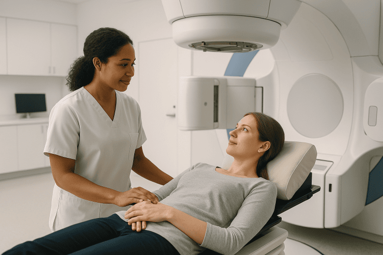
Radiation therapy uses focused high-energy beams to destroy cancer cells. It may be recommended for patients who cannot undergo surgery for health reasons, for tumors in locations where surgery would be difficult, or as an additional treatment after surgery when there is concern that cancer cells may remain.
Systemic therapy for advanced disease
In the uncommon situation where basal cell carcinoma has grown too extensively to be removed surgically, or in the rare cases where it has spread to other parts of the body, doctors may use targeted therapyoral medications known as hedgehog pathway inhibitors, which block the abnormal cell-signaling pathway that drives many basal cell carcinomas. Immunotherapy, which helps the immune system attack cancer cells, may also be considered in certain advanced cases. These treatments are managed by cancer specialists; at some centers, including Acibadem, advanced cases are coordinated through a medical oncology department working together with dermatologists and surgeons.
Watchful waiting
In limited circumstances, for example, in very elderly or frail patients with a slow-growing, low-risk tumor: a doctor may discuss careful monitoring rather than immediate treatment, weighing the burdens of a procedure against the tumor’s expected behavior. Because untreated basal cell carcinoma continues to grow, this approach is chosen cautiously and only after a thorough discussion of the risks.
Living with basal cell carcinoma and outlook
For most people, the outlook after treatment for basal cell carcinoma is favorable. When the cancer is found early and removed completely, treatment is often curative, and the tumor rarely returns at the same site. Spread to distant organs is very rare. That said, no treatment can guarantee that cancer will never come back, and outcomes depend on the tumor’s size, subtype, location, and how early it was treated.
The most important long-term consideration is that people who have had one basal cell carcinoma face a meaningfully higher chance of developing new skin cancers, both additional basal cell carcinomas and other types, in the years that follow. For this reason, doctors typically recommend regular skin examinations after treatment, often every six to twelve months at first, along with monthly self-checks at home. Learning what your own skin normally looks like makes it easier to notice new or changing spots early.
Ongoing sun protection is essential. This includes using broad-spectrum sunscreen daily, wearing wide-brimmed hats and protective clothing, seeking shade during midday hours, and avoiding tanning beds entirely. These habits do not remove risk completely, but they substantially reduce further UV damage to the skin.
Some patients also cope with the cosmetic effects of treatment, particularly after surgery on the face. Scars often fade considerably over time, and reconstructive techniques can improve appearance when needed. If a scar or the diagnosis itself causes distress, discussing these concerns with your care team is a reasonable and common part of follow-up.
Frequently asked questions
What is basal cell carcinoma in simple terms?
Basal cell carcinoma is the most common form of skin cancer. It starts in the basal cells of the skin’s outer layer and usually grows slowly. It most often appears as a pearly bump, a scaly patch, or a sore that will not heal, typically on sun-exposed skin such as the face or neck. It rarely spreads to other organs, but it can damage nearby tissue if left untreated.
How serious is basal cell carcinoma?
Basal cell carcinoma is generally considered the least dangerous of the common skin cancers because it very rarely spreads to distant parts of the body. However, it is still a cancer and should not be ignored. Untreated tumors can grow deeply and destroy skin, cartilage, and even bone, making later treatment more complex. Early treatment is usually straightforward and effective, which is why prompt evaluation matters.
Can basal cell carcinoma heal or go away on its own?
No. Basal cell carcinoma does not go away on its own. A lesion may temporarily crust over or appear to shrink, which can create the false impression of healing, but the cancer cells remain and continue to grow beneath the surface. The only reliable way to eliminate the tumor is through medical treatment, most often removal or destruction of the cancerous tissue.
What causes basal cell carcinoma?
The main cause is DNA damage in skin cells from ultraviolet (UV) radiation, whether from sunlight or tanning beds. Fair skin, a history of sunburns, older age, a weakened immune system, previous radiation exposure, and certain rare inherited conditions all increase the risk. Because UV exposure is the dominant cause, consistent sun protection is the most effective known prevention strategy.
How do doctors confirm the diagnosis?
Diagnosis starts with a skin examination, often using a dermatoscope, a magnifying instrument that reveals surface details. The diagnosis is confirmed with a skin biopsy, in which a small sample of the lesion is removed under local anesthesia and examined under a microscope by a pathologist. Imaging scans are usually unnecessary and are reserved for uncommon advanced cases.
What is recovery like after basal cell carcinoma surgery?
Most surgical removals are performed under local anesthesia as outpatient procedures, meaning patients go home the same day. The wound typically heals over one to several weeks, depending on its size and location, and some scarring is expected. Discomfort is usually mild and manageable. Your doctor will give specific wound-care instructions and arrange follow-up visits to check healing and monitor for any recurrence.
Will basal cell carcinoma come back after treatment?
When a tumor is completely removed, recurrence at the same site is uncommon, though it can happen, particularly with aggressive subtypes or tumors in difficult locations. More importantly, people who have had one basal cell carcinoma are at higher risk of developing new, separate skin cancers over time. Regular skin checks with your doctor and careful sun protection are recommended for the long term.
When to see a doctor
Any skin change that persists or worsens deserves a professional evaluation. You should arrange to see a doctor, ideally a dermatologist, if you notice any of the following:
- A sore that does not heal within about four weeks, or one that heals and then reopens.
- A new bump or spot that is pearly, waxy, shiny, or translucent, especially on the face, ears, scalp, or neck.
- A skin lesion that bleeds, oozes, or crusts repeatedly with little or no injury.
- A flat, scaly, red patch that keeps growing or does not respond to eczema or psoriasis creams.
- A scar-like area appearing without any injury, particularly if it is pale, waxy, or tight-feeling.
- Any existing spot that changes in size, shape, color, or texture, or that starts to itch or become painful.
- A previously treated skin cancer site that changes, develops a new lump, or begins to bleed.
Seek medical attention promptly rather than waiting to see whether a suspicious spot improves. Basal cell carcinoma found early is usually simpler to treat, with smaller scars and better outcomes, than a tumor that has been allowed to grow. If you have a history of skin cancer, significant sun exposure, or a weakened immune system, regular full-body skin examinations with a doctor are a sensible precaution even when no obvious changes are present.
Medically reviewed by the Acıbadem International Medical Board September 2, 2026
See our medical review board →
Update history
- PublishedJune 14, 2026
- Medical review approvedSeptember 2, 2026
- Last content updateSeptember 2, 2026