Basal Cell Carcinoma Treatment
Basal cell carcinoma is the most common skin cancer, usually treated effectively when diagnosed early. Care may include surgical removal, dermatologic procedures, or oncology support depending on tumor size and location.
Quick answer
Basal cell carcinoma (BCC) is the most common form of skin cancer. It starts in the basal cells of the epidermis, grows slowly and very rarely spreads to distant organs. Treatment usually means removing or destroying the tumour, most often by surgical excision under local anaesthetic, with pathology confirming complete removal. Superficial lesions may suit curettage, cryotherapy, photodynamic therapy or prescribed topical treatment.
Basal Cell Carcinoma: What It Is and Why Treatment Matters
Basal cell carcinoma is a cancer that begins in the basal cells: the small, round cells at the bottom of the epidermis that produce new skin. It grows slowly in most cases, very rarely spreads to distant organs, and is usually treated with a short surgical procedure performed under local anaesthetic. It is also the most common cancer in humans, which means a diagnosis, while unwelcome, places you in a very large group of patients for whom well-established treatments already exist.
Of all the cancers doctors diagnose, the one they see most often is basal cell carcinoma. Basal cell carcinoma is strongly linked to ultraviolet light, from the sun and from tanning devices, and it appears most often on skin that has taken decades of exposure: the nose, cheeks, forehead, eyelids, ears, scalp and neck. It can also develop on the chest, back, shoulders, arms and legs. People with fair skin, a history of childhood sunburn, prior radiotherapy, a weakened immune system or certain inherited skin conditions carry a higher risk than the general population.
A slow-growing cancer is not a harmless one. Left alone, a basal cell carcinoma enlarges, bleeds, ulcerates and works its way into the surrounding skin. Given enough time it can invade cartilage, bone and delicate facial structures. The purpose of treatment is straightforward: remove or destroy the tumour completely, confirm that with pathology, and do it in a way that protects how you look and how your face works. Those three goals shape every decision that follows.
What is basal cell carcinoma?
Basal cell carcinoma, usually shortened to BCC, is a malignant tumour of the basal layer of the epidermis. You may also hear it called basal cell cancer or, in older medical texts, rodent ulcer. The cells divide abnormally because their DNA has been damaged, most often by ultraviolet radiation accumulated over many years, and the result is a lesion that keeps growing where healthy skin would stop. Unlike many cancers, BCC almost always stays where it started. That is why local treatment, cutting the tumour out or destroying it in place, is usually all that is required, and why the pathway from diagnosis to treatment is often short.
Is basal cell carcinoma cancer?
Yes. Basal cell carcinoma is a true cancer: its cells grow without normal controls and invade the tissue around them. It is sometimes described casually as “not really cancer” because it so rarely travels to other organs, but that framing is misleading. The correct way to understand BCC is as a locally invasive cancer, one that will keep enlarging and damaging skin, cartilage or bone if it is ignored, but one that responds well to treatment when it is dealt with properly.
Is basal cell carcinoma a serious cancer?
It is serious in a specific way: rarely as a threat to life, but genuinely as a threat to the tissue around it. A BCC on the trunk that is removed early is usually a minor problem, managed in a single outpatient visit. The same tumour left growing on an eyelid, the tip of the nose, a lip or an ear becomes a different matter, because removing it then means removing more tissue in an area where every millimetre affects function and appearance. The seriousness of a basal cell carcinoma, in practice, is largely a question of where it sits and how long it has been allowed to grow.
Is basal cell carcinoma deadly?
Very rarely. Deaths from basal cell carcinoma are uncommon and typically involve tumours that were neglected for many years, tumours in patients who could not receive treatment, or the small group of BCCs with unusually aggressive behaviour. For the overwhelming majority of patients, the danger of BCC lies in local destruction, a growing wound on the face, damage to an eyelid or nostril, rather than in any threat to survival. This is a cancer to take seriously and treat without unnecessary delay, not one to fear as a life-limiting disease.
What is the life expectancy for someone with basal cell carcinoma?
For almost all patients, life expectancy is unchanged by the diagnosis. Because BCC so rarely spreads beyond the skin, a person treated for basal cell carcinoma can generally expect the same lifespan they would have had without it. What does change is the need for ongoing skin surveillance: a person who has developed one BCC has a higher chance of developing another skin cancer later, so regular skin checks become a permanent part of looking after your health.
What Does Basal Cell Carcinoma Look Like?
Basal cell carcinoma often does not look like what patients expect cancer to look like. There is usually no dramatic dark mole and frequently no pain. Instead, most BCCs announce themselves as a small, persistent change in the skin that refuses to behave like an ordinary spot or graze. Common appearances include:
- A small shiny bump with a pearly or translucent quality, sometimes with tiny visible blood vessels running across it
- A pink or red patch that persists for months, often flat and slightly scaly
- A sore that does not heal, or heals and then reopens in the same place
- A lesion that crusts, bleeds with minimal trauma, scabs over and returns
- A pale, waxy, scar-like area with no obvious injury to explain it
- A pigmented brown, blue or black lesion that can be mistaken for a mole
On darker skin tones, basal cell carcinoma may appear brown, black, pink or scar-like, and because the classic “pearly” look is less obvious, diagnosis is sometimes delayed. Any persistent, changing lesion deserves the same attention regardless of skin colour. Pigmented BCCs in particular can resemble moles or other pigmented lesions, which is one of the reasons a biopsy, not appearance alone, settles the diagnosis.
Which skin changes do dermatologists advise having examined?
The pattern that matters most is persistence. A lesion that grows steadily over weeks or months, bleeds when lightly knocked or towelled, forms a scab that keeps coming back, remains open despite creams and wound care, or shows an irregular, rolled, shiny edge is the kind of lesion dermatologists advise having examined. Location adds weight: a suspicious spot near the eye, nose, lip, ear or in the genital area matters more, not because it is more likely to be cancer, but because growth in these places affects function and makes eventual treatment more complex. A lesion that seems small or painless can still be a basal cell carcinoma; the absence of discomfort says nothing about the diagnosis.
Causes and Risk Factors of Basal Cell Carcinoma
The dominant cause of basal cell carcinoma is ultraviolet radiation. Sunlight and tanning devices damage the DNA of basal cells, and over years that damage accumulates until a group of cells escapes normal growth control. This is why BCC concentrates on the head, neck and other chronically exposed skin, and why it becomes more common with age: the tumour of today usually reflects the sun exposure of decades ago, including sunburns in childhood.
UV exposure is not the whole story. Recognised risk factors include:
- Fair skin, light eyes and hair that burns easily and tans poorly
- A history of blistering sunburns, especially in childhood and adolescence
- Regular use of sunbeds or tanning lamps
- Previous radiotherapy to the skin, even many years earlier
- A weakened immune system, for example after organ transplantation or with certain long-term illnesses
- A previous basal cell carcinoma or other skin cancer
- Certain inherited conditions that predispose to multiple BCCs
- Long-term exposure to arsenic or other specific chemical exposures
BCC can occur at any age, but it is far more common in adults with cumulative sun exposure. People who tick several of the boxes above, and especially those who have already had one skin cancer, benefit from a lower threshold for having new lesions checked and from a structured plan of skin surveillance rather than one-off reassurance.
Types of Skin Cancer and Where BCC Fits
There are several types of skin cancer, and understanding where basal cell carcinoma sits among them helps make sense of the treatment decisions. The three most discussed are basal cell carcinoma, squamous cell carcinoma and melanoma. BCC is the most common and the least likely to spread. Squamous cell carcinoma also arises from the epidermis and is usually treatable, though it has a somewhat greater tendency to spread than BCC. Melanoma is less common but more dangerous, because it can metastasise earlier. All three fall under the broader umbrella of skin cancer, and the same habits, sun protection and regular skin checks, help guard against each of them.
Within basal cell carcinoma itself, pathologists recognise distinct subtypes, and the subtype influences treatment. Nodular BCC is the classic pearly bump and is usually well defined. Superficial BCC tends to be a flat, red, scaly patch, often on the trunk, and is the subtype most likely to suit non-surgical treatment. Infiltrative, micronodular and morpheaform (sclerosing) patterns behave more aggressively at a microscopic level: their edges extend beyond what the eye can see, so they typically need wider excision or margin-controlled surgery. A single lesion can also contain a mixture of patterns, which is why the biopsy report matters so much for planning.
Naming varies between countries. In parts of Europe the tumour is called a basalioma; skin specialists use the term interchangeably with basal cell carcinoma, and the treatment principles are identical whichever name appears on your report.
How Basal Cell Carcinoma Is Diagnosed
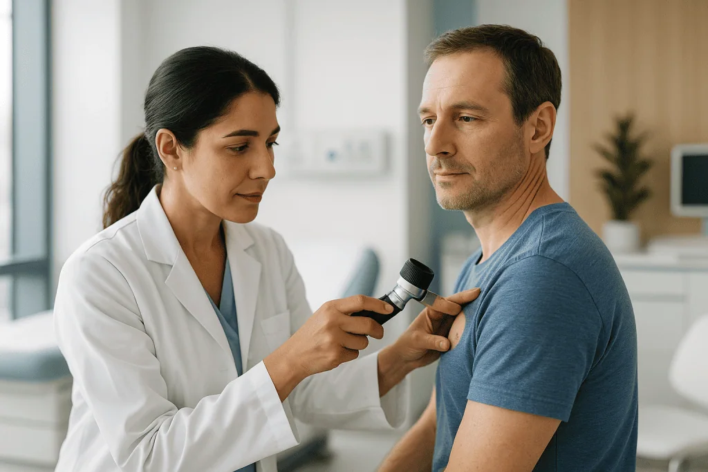
Diagnosis begins with a careful skin examination and a focused history. The physician asks about sun exposure, previous skin cancers, family history, immune status, medications and, importantly, how the lesion has changed over time. The whole skin is usually examined, not just the spot you noticed, because a person with one suspicious lesion often has others, and because sun-damaged skin tends to produce more than one problem over the years.
Dermoscopy, a non-invasive examination with a magnifying instrument and polarised light, lets the clinician see structures beneath the skin surface that are invisible to the naked eye. Certain dermoscopic features, branching vessels, blue-grey globules, shiny white areas, point towards basal cell carcinoma and help decide whether a biopsy is needed and where it should be taken.
A biopsy confirms the diagnosis; appearance alone never does. Depending on the lesion, the physician may perform a shave biopsy (removing a thin surface sample), a punch biopsy (a small cylindrical core) or an excisional biopsy (removing the whole visible lesion). A pathologist examines the tissue, confirms whether BCC is present and, where possible, reports the subtype, nodular, superficial, infiltrative, micronodular or morpheaform. That subtype report is not a formality: it can change the recommended treatment from a simple procedure to margin-controlled surgery.
Imaging is not needed for small, typical basal cell carcinomas. It becomes relevant when a tumour is large, deeply invasive, recurrent after previous treatment, or sitting near critical structures such as the orbit, the ear canal or bone. In those situations ultrasound, CT or MRI can map how far the tumour extends, and the findings may be reviewed in a multidisciplinary setting so that surgery, reconstruction, radiotherapy or systemic therapy can be planned coherently rather than piecemeal.
Who Needs Treatment for Basal Cell Carcinoma?
Anyone with a confirmed basal cell carcinoma should discuss treatment with a qualified skin cancer specialist, because the tumour will not resolve on its own and continues to grow locally. The indication for treatment is the presence of the cancer itself; what varies is how urgent, how extensive and how complex that treatment needs to be.
Common situations in which treatment is planned include:
- A newly diagnosed BCC after biopsy, needing definitive removal
- A clinically suspicious lesion planned for complete excision, with the diagnosis confirmed on the removed tissue
- A superficial BCC suitable for a non-surgical option such as topical therapy or photodynamic therapy
- A tumour in a visible or functionally sensitive area, eyelid, nose, lip, ear, requiring careful cosmetic and reconstructive planning
- A recurrent BCC, meaning a cancer that has returned after previous treatment, where borders are less clear and the risk of further recurrence is higher
Some basal cell carcinomas are classified as high risk and demand particular attention: tumours on the central face, eyelids, nose, lips, ears, hands, feet or genital area; tumours with aggressive histology; large lesions; lesions with poorly defined borders; cancers arising in previously irradiated skin; and BCCs in patients with weakened immune systems. A patient with many BCCs, repeated skin cancers or a genetic predisposition needs more than treatment of the current lesion: they need a longer-term prevention and surveillance strategy built around it.
Although BCC rarely spreads to distant organs, locally advanced basal cell carcinoma does occur, usually when a tumour has grown extensively into deeper tissues or when earlier treatments have not controlled it. These cases may need more than one treatment modality and are best approached through coordinated specialist review rather than a single-department decision.
How Basal Cell Carcinoma Treatment Is Performed
Basal cell carcinoma treatment covers the medical and surgical methods used to remove, destroy or control the cancerous cells. There is no single best method; there is a best method for a specific tumour in a specific patient. The choice depends on the tumour’s location, size, borders, depth and histologic subtype, on whether it is a first-time or recurrent cancer, and on your overall health, medications and preferences. The pathway begins with confirming the diagnosis, grading the tumour’s risk, and then matching the technique to the problem.
Preparation before treatment
Before treatment, the physician reviews the pathology report, examines the lesion and the surrounding skin, and asks about previous skin cancers and previous treatments in the same area. Expect questions about blood thinners, immune-suppressing medications, diabetes, heart conditions, allergies, implanted devices and smoking. None of these usually prevents treatment, but each can influence anaesthesia, bleeding risk and wound healing, and the plan is adjusted accordingly. Any decision about medication belongs to your treating doctors, who weigh the procedure against your wider health.
Photographs are often taken for documentation and surgical planning. If the lesion sits on the face, eyelid, nose, lip or ear, the surgeon may discuss reconstruction options before the procedure rather than after, so you understand what the closure will involve. For larger or complex tumours, imaging may be arranged at this stage. If sedation or general anaesthesia is expected, preoperative blood tests, cardiology review or a formal anaesthesia assessment may be recommended based on your age and history.
Most basal cell carcinoma procedures are outpatient procedures. Many are done under local anaesthesia: the area is numbed and you remain awake throughout. More extensive surgery or reconstruction may call for sedation or general anaesthesia, in which case you will be told whether to fast beforehand and whether to arrange help for the journey home and the first day of recovery.
Surgical excision
Standard surgical excision is the most common treatment for BCC, and for many patients it is the whole treatment. The sequence is simple:
- The area is cleaned and numbed with local anaesthetic.
- The surgeon removes the visible tumour together with a planned margin of surrounding healthy-appearing skin.
- The wound is closed, with stitches, a local skin flap or a graft, or, in selected sites, left to heal naturally.
- The removed tissue goes to pathology for margin assessment.
The pathology report states whether the margins are clear or whether cancer cells reach or approach an edge. Clear margins mean the excision has done its job and the focus shifts to healing and follow-up. Involved margins mean additional treatment is discussed, usually further excision, sometimes a margin-controlled technique. For low-risk BCCs in suitable locations, excision is typically a short, well-tolerated procedure. In cosmetically important areas, incision placement along natural skin lines and the choice of closure technique are treated as part of the cancer operation, not an afterthought.
Micrographic surgery and margin-controlled techniques
For selected tumours, especially on the face and in other high-risk locations, micrographic or margin-controlled surgery may be recommended. In this approach, often known as Mohs surgery, the tumour is removed in stages. After each stage, the margins are examined microscopically with a mapping technique while you wait. If cancer cells remain at one specific point, only additional tissue from that point is removed, and the process repeats until the margins are clear. The logic is to achieve thorough clearance while sparing every millimetre of healthy skin that can be spared.
This approach earns its extra time for recurrent tumours, aggressive subtypes such as infiltrative or morpheaform BCC, poorly defined lesions, and tumours near the eyes, nose, lips or ears, where the visible lesion may represent only part of the cancer’s true extent. Reconstruction is carried out after clearance is confirmed. Depending on the defect, closure may be simple or may require a flap or graft planned by dermatologic, plastic or oculoplastic surgeons.
Curettage, cryotherapy and photodynamic therapy
Some superficial or low-risk basal cell carcinomas can be treated without conventional excision. Curettage and cautery involves scraping the tumour away with a surgical instrument and applying controlled heat to destroy remaining cancer cells and stop bleeding. It can be appropriate for selected low-risk lesions on the trunk or limbs, but it is generally not preferred for high-risk facial sites or aggressive subtypes, and it provides less margin information than excision.
Cryotherapy uses controlled freezing to destroy abnormal cells and may suit carefully selected superficial lesions, though it also yields no tissue specimen for margin assessment. Photodynamic therapy is an option for certain superficial BCCs, particularly when there are multiple lesions or when cosmetic outcome is a priority: a light-activated medication is applied to the skin and then triggered with a specific light source that damages the abnormal cells. Each of these methods trades some certainty of clearance for less tissue disruption, which is why they are reserved for tumours where the biopsy has confirmed a genuinely low-risk subtype.
Topical treatments, radiotherapy and systemic therapy
Topical therapies, creams applied to the skin over several weeks, may be suitable for some superficial basal cell carcinomas. They work by stimulating a local immune response or by directly damaging abnormal cells. They are prescribed only when biopsy has confirmed a superficial subtype, when the site is suitable, and when follow-up is feasible, because the treated area must be monitored to confirm the tumour has actually gone.
Radiotherapy is an option for patients who cannot undergo surgery, who decline it, whose tumours sit in locations where surgery would be particularly difficult, or who need additional treatment after surgery in selected high-risk circumstances. Modern radiation planning shapes the dose around the tumour area while limiting exposure of surrounding healthy tissue, and treatment is typically delivered over a series of short sessions.
For the rare cases of locally advanced or metastatic basal cell carcinoma, medical oncology can offer systemic treatments: targeted drugs that interfere with the growth pathway most BCCs depend on and, in certain situations, immunotherapy. These treatments require careful evaluation, a frank discussion of side effects, and ongoing monitoring within a structured oncology programme. They are the exception in BCC care, not the rule, but for the patients who need them, they exist.
What technology supports diagnosis and treatment?
Technology contributes at several points along the pathway. Dermoscopy sharpens the initial assessment of a suspicious lesion. Digital photography documents the tumour before treatment and provides a baseline for monitoring healing and future skin changes. Pathology techniques, from routine sections to margin mapping, turn removed tissue into the information that drives every subsequent decision. In complex cases, ultrasound, CT or MRI define involvement near bone, cartilage, nerves or the orbit before anyone operates.
In theatre, magnification, precise instruments, electrosurgical tools and meticulous reconstructive technique allow the surgeon to remove the cancer and close the wound with attention to contour and function. In radiotherapy, planning software and image-guided delivery shape treatment around the target. None of this replaces judgement; the value lies in how the tools are matched to the individual tumour.
How long does treatment take, and what happens immediately afterwards?
Many BCC treatments are completed in a single outpatient visit. A small excision may take less than an hour, though your total clinic time is longer once preparation, anaesthesia, dressing and discharge instructions are included. Margin-controlled procedures can take several hours, because tissue is examined during the visit and further stages may be needed. Complex reconstruction may require an operating room and a longer, staged recovery plan.
Afterwards, expect a pressure dressing, stitches in most cases, and some swelling, bruising or tenderness. Discomfort is usually manageable with the medications your care team recommends. You will receive specific instructions on wound care, bathing, activity restrictions and the signs of infection or bleeding to watch for. Stitches, where used, are removed within days to a couple of weeks depending on the site and closure type. The scar itself matures far more slowly, often over several months, and consistent sun protection of the healing area is essential throughout that period.
Why Acting Early Matters
Because basal cell carcinoma usually grows slowly, it can create a false sense of safety. The lesion has been there for a year; surely another few months will not matter. In fact, timing changes almost everything about the treatment except the diagnosis: the size of the excision, the complexity of the reconstruction, the number of options available and the cosmetic result all favour the patient who acts earlier.
How fast does basal cell carcinoma spread?
Most basal cell carcinomas grow slowly, over months to years rather than weeks, and “spread” for BCC almost always means local extension into neighbouring tissue rather than travel through the body. Growth rate varies by subtype: nodular and superficial tumours tend to enlarge gradually and visibly, while infiltrative and morpheaform patterns can extend along tissue planes well beyond their visible edge, so the tumour is larger than it looks. Slow growth is a reason treatment can be planned calmly; it is not a reason to postpone it indefinitely.
What happens if you don’t cut out a basal cell carcinoma?
An untreated basal cell carcinoma keeps growing. Over time it typically ulcerates, bleeds repeatedly, scabs and reopens, and burrows into the surrounding skin. On the face, continued growth can involve the eyelid, the cartilage of the nose or ear, nerves and eventually bone, at which point treatment shifts from a short outpatient excision to major surgery with reconstruction, sometimes combined with radiotherapy. Recurrent cycles of scabbing and bleeding also blur the tumour’s true borders, making eventual removal harder to plan. Nothing about waiting improves the situation; the tumour only becomes a bigger version of the same problem.
Can basal cell carcinoma spread to other parts of the body?
Distant spread is very rare. Basal cell carcinoma is overwhelmingly a locally invasive cancer: it damages what it touches but almost never seeds tumours in lymph nodes, lungs or other organs. The rare metastatic cases usually involve very large, long-neglected or repeatedly recurrent tumours. This is precisely why early, complete local treatment is so effective, dealing thoroughly with the tumour where it sits resolves the problem for the vast majority of patients.
Early action matters most when the BCC is near the eye, nose, lip, ear or scalp. In these areas, even a modest increase in size changes how much tissue must be removed and how complicated the reconstruction becomes. For patients with immune suppression or aggressive tumour features, prompt evaluation carries extra weight, because these tumours are less forgiving of delay.
Benefits of Basal Cell Carcinoma Treatment
The benefits depend on the tumour and the method chosen, but the central goals are constant: control the cancer, preserve healthy tissue, and set up sensible long-term surveillance.
| Benefit | What It Means for You |
|---|---|
| Removal or control of the cancer | Treatment aims to eliminate visible and microscopic basal cell carcinoma cells, reducing the risk that the tumour continues to grow locally. |
| Protection of nearby structures | Early, well-planned care helps preserve the eyelid, nose, lip, ear and other areas where function and appearance are closely connected. |
| Improved wound and cosmetic planning | Choosing the right technique allows closure or reconstruction to be planned around scar position, contour and natural skin movement. |
| Pathology confirmation | Biopsy and excision specimens confirm the subtype, depth and margins: the information that guides follow-up and any additional care. |
| Reduced risk of recurrence | Appropriate treatment and follow-up lower the chance of the cancer returning in the same area, though ongoing surveillance remains important. |
| A plan for future skin health | Because one BCC raises the chance of another skin cancer later, treatment is also the moment to build a prevention and monitoring strategy. |
Recovery After Basal Cell Carcinoma Treatment
Recovery varies with the treatment type, tumour location, wound size and whether reconstruction was needed, but most patients return to light daily activities soon after outpatient care. The timeline below describes a typical course after surgical treatment.
| Time Period | What Patients Can Expect |
|---|---|
| Day 1 | The treated area is covered with a dressing. Mild discomfort, tightness, swelling or bruising may occur. Wound-care instructions and activity guidance are provided. |
| First week | Light activities are usually fine. The wound begins to seal and swelling gradually improves. Strenuous exercise, swimming and heavy lifting may be restricted depending on the procedure. |
| First month | Stitches, if present, are removed earlier in this period according to the site. The incision continues to strengthen. Redness or firmness of the scar can be normal. |
| Several months | Scar maturation continues. The colour, texture and thickness of the scar typically improve gradually. Sun protection reduces discolouration and supports healing. |
| Longer term | Follow-up skin examinations help detect recurrence or new skin cancers early. Continued self-checks and consistent UV protection are advised. |
Two points deserve emphasis. First, the scar you see at stitch removal is not the scar you will have in a year; redness and firmness soften considerably as the tissue remodels. Second, a healing wound and a mature scar both discolour more readily in the sun, so a high-factor sunscreen or physical cover over the site is one of the simplest things you can do to improve the final cosmetic result.
What Influences the Outcome
Most early basal cell carcinomas respond well to treatment, but several factors shape the result. The first is tumour risk. A small, well-defined, low-risk BCC on the trunk is generally straightforward; an infiltrative or recurrent tumour on the nose or eyelid is not. Subtype, depth, borders and location together determine both the choice of technique and the likelihood of recurrence, which is why two patients with “the same diagnosis” can have very different treatment pathways.
The second factor is margin control. For most BCCs, complete removal with appropriate margins is the heart of a good result. In high-risk areas, techniques that assess margins meticulously matter because the visible lesion may represent only part of the cancer’s true extent. Pathology expertise matters equally: the interpretation of biopsy and excision specimens is what tells the team whether the job is finished or not.
The third factor is reconstruction and wound healing. A good outcome is not only a clear pathology report; it is a wound that heals well and lets you blink, breathe, speak, smile and move naturally. Closure technique, tissue quality, blood supply, smoking status, diabetes, medications and aftercare all influence healing. Patients who follow wound-care instructions and attend their follow-up visits give themselves the best chance of a smooth course.
Patient-related factors also count. Immunosuppression, prior radiotherapy, previous surgery in the same area, certain genetic conditions and a history of multiple skin cancers make treatment more complex and long-term surveillance more important. And timing runs through everything: early evaluation preserves options, while delay tends to convert simple procedures into larger ones. The same tumour, six or twelve months apart, can be two quite different surgical problems.
Follow-Up and Preventing the Next Skin Cancer
Treatment of the current lesion is half the task; the other half is watching for the next one. A person who has had one basal cell carcinoma has a higher chance of developing another BCC, a squamous cell carcinoma or a melanoma in the years that follow, not because the treatment failed, but because the same sun-damaged skin remains. Regular full-skin examinations by a clinician, at intervals your dermatologist recommends, are the backbone of follow-up.
Between visits, monthly self-checks help. Learn the sites of your previous lesions and scars, and watch for new persistent bumps, non-healing sores, changing moles or scar-like patches anywhere on the body, not only where the first tumour appeared. Photographs taken at home can make gradual change easier to spot.
Prevention is unglamorous and effective: broad-spectrum sunscreen applied properly and reapplied, hats and clothing on exposed skin, shade during peak hours, and no tanning devices. None of this reverses old damage, but it slows the accumulation of new damage, which is exactly what matters for a cancer driven by lifetime UV dose.
How Basal Cell Carcinoma Care Is Organised at Acibadem
At Acibadem, basal cell carcinoma care is built around matching the treatment to the tumour rather than applying one method to every patient. Depending on the case, that involves dermatologists, plastic and reconstructive surgeons, pathologists, radiation oncologists and medical oncologists. Straightforward lesions are treated efficiently by the appropriate specialist; complex tumours, recurrent, high-risk, near critical facial structures or part of a wider oncology picture, can be discussed in multidisciplinary review so that removal, reconstruction and any further therapy are planned together rather than sequentially.
The same principle applies to the details that shape quality of life. A tumour on the nose, eyelid or lip is a medical problem, but it is also part of how a person sees themselves. Treatment planning therefore weighs tumour control alongside scar placement, facial anatomy, skin type, healing expectations and how follow-up will be organised after treatment, so that surveillance continues seamlessly once the wound has healed.
Making Sense of Your Diagnosis
Basal cell carcinoma is common, but no two cases are interchangeable. A small superficial lesion on the shoulder, a recurrent tumour on the nose and a deeply invasive cancer near the eye call for different levels of expertise and different plans. The essential steps are the same for everyone: an accurate diagnosis with subtype, an honest assessment of the tumour’s risk category, and a treatment choice that balances cancer control against preservation of appearance and function.
When a specialist assesses a basal cell carcinoma, the discussion typically draws on the biopsy report, clinical photographs, any previous treatment records and a current medication list, because each of these changes what is recommended. With that information in hand, the picture usually becomes much clearer than it feels at the moment of diagnosis: for most patients, this is a cancer that can be treated definitively, followed sensibly, and then allowed to recede into the background of an ordinary life.
Preparation
- A dermatologist or oncology specialist evaluates the lesion, reviews medical history, and may perform a skin biopsy to confirm diagnosis. Patients should share medications, allergies, and bleeding risks before treatment. Sun exposure should be avoided before and after the procedure.
Aftercare
- The treated area should be kept clean and protected as instructed, with dressings changed as advised. Follow-up visits check wound healing and pathology results. Long-term skin surveillance and sun protection are important to reduce recurrence and detect new lesions early.
Frequently Asked Questions
What affects the cost of basal cell carcinoma treatment?
The main factors are tumour size, location, subtype, whether it is new or recurrent, the treatment method, pathology requirements, anaesthesia, reconstruction needs, and follow-up care. A personalised quote can only be prepared after specialist review of your medical information.
How can I get a personalised quote from Acibadem?
You can request a free consultation by sharing your diagnosis, biopsy or pathology report if available, photos of the lesion, previous treatment details, and relevant medical history. The team can then guide you on the likely treatment plan and what the package may include.
Does a basal cell carcinoma package usually include pathology?
Many treatment plans include pathology coordination, but the exact scope should be confirmed before travel. Some cases may need biopsy review, margin assessment, or additional testing, which can affect the final cost.
Will I need plastic surgery after basal cell carcinoma removal?
Not always. Small or straightforward lesions may be closed directly, while lesions on the face, eyelids, nose, ears, lips, or other sensitive areas may need flap or graft reconstruction. The specialist will advise based on the expected defect and cosmetic or functional needs.
Is travel to Turkey suitable for every basal cell carcinoma patient?
Travel may be suitable for many patients, but it depends on the urgency of care, general health, tumour characteristics, and follow-up needs. This information is general and not medical or financial advice; a specialist consultation is recommended before making plans.
Medically reviewed by the Acıbadem International Medical Board August 30, 2026
See our medical review board →
Update history
- PublishedJune 8, 2026
- Medical review approvedAugust 30, 2026
- Last content updateSeptember 8, 2026
References1
- Skin Cancer medlineplus.gov
Trusted care for international patients
From the Acibadem Blog
Basal Cell Carcinoma in Older Adults: How Treatment Is Tailored to Health and Skin
Basal cell carcinoma treatment in older adults is tailored to the tumor's size, location, subtype and growth rate, and to the person's overall health,…
How a Basal Cell Carcinoma Excision Heals: Dressings, Stitches and Scar Maturation
After a basal cell carcinoma excision, a stitched wound usually has a sealed surface within about two weeks, with surface sutures removed anywhere from…

When Is Mohs Surgery Chosen Over Standard Excision for Basal Cell Carcinoma?
Mohs surgery is usually chosen over standard excision for basal cell carcinoma when the tumor sits where skin is scarce or cosmetically and functionally…