

When Is Mohs Surgery Chosen Over Standard Excision for Basal Cell Carcinoma?

Key Takeaways
- The core difference between Mohs and standard excision is that Mohs examines the entire edge of each tissue layer during the visit, while excision checks representative slices of the margin days later.
- Mohs is generally favored for basal cell carcinomas on the nose, eyelids, lips, ears, hands, feet and genitals, and for tumors that are large, ill-defined, aggressive in subtype, or recurrent.
- Standard excision is a complete, guideline-endorsed treatment for well-defined low-risk tumors on the trunk and limbs, where a slightly wider margin has no functional cost and the visit takes under an hour.
- Basal cell carcinoma arises in the deepest layer of the epidermis, so excisions typically reach only the subcutaneous fat, a few millimeters down, while Mohs adds depth only where the microscope shows cancer.
- Mayo Clinic advises setting aside the whole day for Mohs because the number of stages, each requiring roughly an hour of laboratory processing, cannot be known in advance.
- The scar from either operation looks its worst around a month after surgery and continues to fade for up to a year, and daily sun protection of the site is part of recovery.
Mohs surgery is usually chosen over standard excision for basal cell carcinoma when the tumor sits where skin is scarce or cosmetically and functionally important, such as the nose, eyelids, lips, ears, fingers or genitals; when it is large, has indistinct edges, an aggressive growth pattern, or has returned after earlier treatment; or when the patient is immunosuppressed. Small, well-defined tumors on the trunk or limbs are often removed by standard excision. The treating team decides.
The spot on the side of Ana’s nose never hurt. It just refused to leave: a shiny pink bump the size of a lentil that bled when she dried her face and then scabbed over, again and again, for the better part of a year. The biopsy came back with two words she had heard of but never expected to own, basal cell carcinoma, and a question she had never heard of at all. Did she want Mohs surgery, or a standard excision?
The Mohs surgery vs excision basal cell carcinoma decision confuses people because it sounds like a ranking, as if one operation must be the premium option and the other the budget one. It isn’t. Both remove the same cancer under local anesthetic. What differs is how, and when, the surgeon confirms the edges are clear, and how much healthy skin is traded away to get there.
That trade matters enormously on a nose. It matters far less on a shoulder blade. Most of what follows comes down to that single idea.
Mohs surgery vs excision for basal cell carcinoma: what is actually different?
Strip away the vocabulary and the two procedures share more than they differ. In each, a surgeon numbs the skin, removes the tumor together with a rim of normal-looking tissue, and closes the wound. The rim is called the margin, meaning the border of apparently healthy skin taken alongside the cancer so that microscopic roots come out with it.
The difference is timing and thoroughness of the margin check. In a standard excision, the surgeon estimates the margin by eye, removes everything in one piece, closes the wound and sends the specimen to a pathology laboratory. Results return days later. If the report shows cancer reaching an edge, a second operation is arranged. Mayo Clinic describes this as the usual approach for basal cell carcinomas judged less likely to return, particularly on the chest, back, arms and legs.
In Mohs micrographic surgery, named for the surgeon who developed it, the tissue is processed on site while the patient waits. The surgeon examines the entire undersurface and edge of each layer under a microscope and removes more tissue only from the precise spot where cancer remains. Mayo Clinic and Cleveland Clinic both describe the procedure as a series of stages completed in a single visit.
Think of excision as measuring carefully and cutting once with a good ruler. Mohs is checking each cut under a magnifying glass before deciding whether to cut again. One method is not intrinsically superior; each answers a different problem. When the surrounding skin is plentiful and the tumor is predictable, the ruler is enough. When every millimeter changes how an eyelid closes or a nostril breathes, the magnifying glass earns its extra hours.
How does Mohs surgery actually work, step by step?
Patients often picture Mohs as a single long operation. It is closer to a loop that repeats until the microscope says stop.
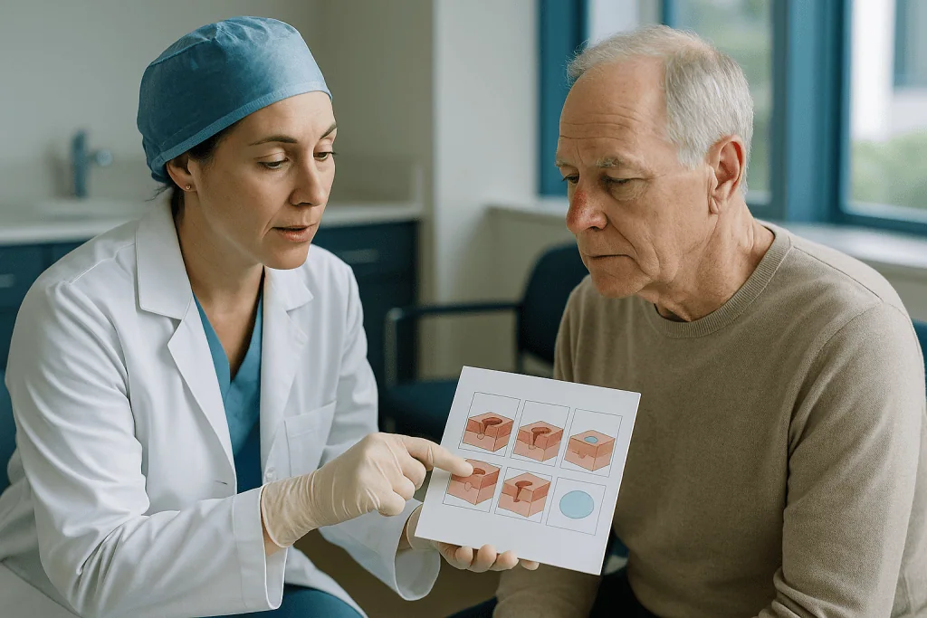
The visit begins with local anesthetic, the same numbing injection used for the biopsy, so the patient is awake throughout. The surgeon removes the visible tumor with a thin rim of surrounding skin, often only a millimeter or two beyond what can be seen, and marks the wound and the specimen with matching orientation cuts so the two can be lined up later like puzzle pieces.
That specimen goes to a laboratory in the same suite. Technicians ink its edges in colors, freeze it and slice it horizontally, a technique called frozen sectioning, which lets the surgeon see the complete outer edge and deep surface rather than the sample cross-sections used in routine pathology. Mayo Clinic notes that this laboratory work takes roughly an hour per stage, which is why the waiting room, not the operating chair, is where most of the day is spent.
The surgeon then reads the slides personally. If every edge is clear, the removal is finished. If cancer cells appear at, say, the two o’clock position on the map, the surgeon numbs the area again and removes another thin layer from that position only, leaving the rest of the wound untouched. The loop repeats until the map is clean.
Only then is repair discussed. Depending on size and site, the wound may be stitched in a straight line, closed with a flap of nearby skin, covered with a graft from elsewhere, or occasionally left to heal on its own under dressings. Mayo Clinic advises setting aside the whole day, since the number of stages cannot be known in advance.
What happens during a standard excision?
A standard excision, sometimes called wide local excision, is the operation most people imagine when they hear the word surgery: one removal, one closure, one lab report.
After local anesthetic, the surgeon draws an outline around the tumor that includes a margin of visibly normal skin. The outline is usually an elongated oval rather than a circle, typically about three times as long as it is wide, because a straight-line closure of an oval lies flat, whereas closing a round hole leaves puckered corners that surgeons call dog-ears. The tissue is removed down to the layer of fat beneath the skin, the wound is closed in layers with dissolving stitches below and fine stitches or adhesive on the surface, and the visit ends within an hour or so for most small tumors.
The specimen travels to a pathology laboratory where it is fixed in preservative, embedded in wax and thinly sliced. These permanent sections give a detailed picture of the tumor’s subtype and depth but are examined at intervals across the specimen rather than along its entire edge, and results take days rather than minutes. Mayo Clinic describes this microscopic check of the margins as a standard part of excision.
When the report reads clear, follow-up is a wound check and stitch removal. When it shows tumor at a margin, the team discusses re-excision or, if the site is delicate, a referral for Mohs to clear what remains.
The trade-off is honest and simple. Excision is quicker and widely available, and for a well-behaved tumor on forgiving skin the extra margin costs the patient almost nothing. Its weakness is uncertainty: a small proportion of patients learn afterward that they need a second procedure.
How deep do they cut to remove basal cell carcinoma?
This is the question people are often too shy to ask aloud, so here it is plainly.

Basal cell carcinoma begins in the basal cells, the round cells forming the deepest layer of the epidermis, the skin’s thin outer sheet. Mayo Clinic describes it as the most common type of skin cancer, appearing most often on sun-exposed areas such as the head and neck. Because it starts so superficially, the tumor is usually a few millimeters deep, not centimeters.
In a standard excision, the surgeon removes the full thickness of skin down to the subcutaneous fat, the soft cushioning layer beneath the dermis. That is not because the cancer is expected to be that deep; it is because taking a complete, even block of skin gives the pathologist an unbroken deep margin to inspect and gives the wound a clean base to close.
Mohs works in the opposite direction. Each layer is deliberately thin, and depth is added only where the microscope shows cancer still present. On a nose, where skin sits directly on cartilage, that restraint can mean the difference between a wound that closes in a line and one that needs a graft.
Depth also depends on the tumor’s personality. Nodular basal cell carcinomas, the classic pearly bump, tend to grow as a compact ball with fairly clear edges. Infiltrative and morpheaform subtypes, which look like a pale scar rather than a lump, send thin strands between collagen fibers well beyond what is visible, so surgeons expect to go wider and deeper than the surface suggests. Your biopsy report names the subtype, and it is one of the strongest reasons a team leans toward one operation or the other.
Which basal cell carcinomas usually point toward Mohs?
Guidelines from major dermatology bodies converge on a short list, and Mayo Clinic and Cleveland Clinic summarize it in similar terms. Mohs is generally favored when a basal cell carcinoma has one or more of the following features.
- It sits in a high-risk or tissue-scarce location: the nose, eyelids, lips, ears, temples, fingers, toes, genitals, or the central face generally.
- It is large for its site, or its edges cannot be clearly seen or felt.
- The biopsy shows an aggressive subtype such as infiltrative, morpheaform or micronodular growth.
- It has come back after a previous treatment, whether surgery, scraping or a cream.
- It arose in skin previously treated with radiation or in a long-standing scar.
- The patient’s immune system is weakened, for example after an organ transplant, because tumors in this setting behave less predictably.
Dermatologists sometimes describe the face’s high-risk region as the H-zone, a rough letter H drawn across the temples, ears, eyes, nose and lips. Skin here is thin, mobile and close to structures that must keep working: tear ducts, nostril rims, the muscles that close the eye. It is also where embryonic fusion planes lie, seams in facial development along which tumors can track deeper than expected.
The logic is the same in every bullet. Where the cost of removing too much is high, or the odds of leaving some behind are elevated, checking every edge before closing is worth a long day. Where neither is true, the same long day buys little. None of these features is an automatic ticket to Mohs; the surgeon weighs them against the patient’s health, preferences and the practical realities of the visit.
Why excision instead of Mohs? When the simpler operation fits better
People sometimes hear a recommendation for standard excision as a downgrade, as if their cancer had been judged not worth the fuss. The opposite is closer to the truth: it usually means the tumor is predictable enough that the fuss adds nothing.
Mayo Clinic lists surgical excision as a common treatment for basal cell carcinomas less likely to return, especially those on the chest, back, arms and legs. On these sites the skin is thick and generous, a few extra millimeters of margin leave no functional consequence, and a linear scar hidden under clothing is rarely a concern. Taking a slightly wider margin than strictly necessary is a small price for a single, short visit.
There are practical reasons too. Excision can be done by dermatologists, plastic surgeons and general surgeons, often on the day of consultation, while Mohs requires a surgeon with specific fellowship training and an on-site laboratory. In many regions that means a wait. For a small nodular tumor on a calf, the team may reasonably judge that removing it next week by excision serves the patient better than removing it by Mohs in two months.
Permanent-section pathology also offers an advantage the frozen sections of Mohs do not: a leisurely, detailed look at the tumor’s architecture and depth by a pathologist, which can be reassuring when the biopsy raised any doubt about the diagnosis.
Finally, some patients simply cannot, or prefer not to, spend a full day in a clinic. Standard excision respects that. The right question is not which procedure is more sophisticated but which one removes this tumor, in this place, in this person, with the least burden. Often the answer is the quieter operation.
When is Mohs surgery not recommended, and who is asked to wait?
Mohs is a superb tool with real limits, and a good team names them.
It is not recommended when it is simply unnecessary: a small, well-defined, low-risk tumor on the trunk gains nothing from margin mapping that excision would not deliver more efficiently. Professional appropriate-use guidance from dermatology societies exists precisely to discourage Mohs in these settings, and surgeons generally follow it.
It is also poorly suited to tumors that have grown beyond skin. When imaging or examination suggests invasion of bone, major nerves or the orbit, the case usually moves to a multidisciplinary team involving head and neck surgeons, oncologists and radiation specialists, because the layer-by-layer method cannot safely chase cancer through those structures. MedlinePlus notes that basal cell carcinoma rarely spreads to distant sites, but when it is locally advanced, surgery alone may not be the plan.
Some patients are poor candidates for the day itself. A person with advanced dementia, severe anxiety about procedures, or difficulty sitting for several hours may not tolerate repeated stages. Others have medical conditions that need optimizing first: an infection at the surgical site, unstable heart or lung disease, or a recent event that makes any elective procedure unwise. Blood-thinning medicines are usually continued rather than stopped, since the bleeding risk of skin surgery is generally manageable, but that decision belongs to the prescribing clinician and surgeon together, never to the patient alone.
Being asked to wait is different from being turned away. Common reasons include a biopsy still under review, a repair that needs a second surgeon’s availability, or a frail patient for whom the team wants to weigh a non-surgical option first. The NHS notes that treatment for non-melanoma skin cancer is planned around the individual, and a pause for planning is part of that care, not a sign of neglect.
Mohs surgery vs excision for basal cell carcinoma: side-by-side comparison
The table below gathers the practical differences in one place. It describes typical patterns drawn from Mayo Clinic and Cleveland Clinic patient guidance; individual experiences vary, and your team’s plan takes precedence.
| Feature | Mohs micrographic surgery | Standard excision |
|---|---|---|
| When margins are checked | During the visit, before the wound is closed | Days later, after closure |
| How much of the edge is examined | Entire outer edge and deep surface of each layer | Representative cross-sections through the specimen |
| Amount of healthy skin removed | Designed to be minimal; added only where cancer persists | Predetermined margin set by the surgeon |
| Typical length of visit | Several hours; plan for the day | Usually under an hour for small tumors |
| Usual sites | Face, ears, scalp, hands, feet, genitals; recurrent or aggressive tumors | Trunk, arms, legs; well-defined low-risk tumors |
| Who performs it | Dermatologic surgeon with Mohs training and on-site lab | Dermatologist, plastic surgeon or general surgeon |
| If cancer remains at an edge | Removed the same day, same spot | Second procedure scheduled |
| Pathology detail | Frozen sections read by the surgeon | Permanent sections read by a pathologist |
Notice what the table does not contain: a column marked better. A tumor’s location and behavior fill in the answer more reliably than any general preference. A reader with a pearly bump on the upper back will likely find their situation in the right-hand column and can feel entirely confident in it. A reader with a scar-like patch beside the eye will probably find theirs on the left, and the extra hours are well spent.
Where a case sits between the columns, which is common, the conversation with the surgeon becomes the deciding factor, and the questions later in this article are meant to make that conversation easier.
What are the alternatives when neither surgery fits?
Surgery is the mainstay for basal cell carcinoma, but it is not the only option, and knowing the alternatives helps you understand why a surgeon might steer you toward or away from the operating chair. The NHS and Mayo Clinic describe the following approaches.
Scraping and cautery, often abbreviated C&E, uses a spoon-shaped instrument to scrape away the soft tumor and an electric needle to seal the base. It is quick and suits small, superficial tumors on the trunk, but there is no margin to examine, so it is not used where certainty matters.
Cryotherapy freezes the tumor with liquid nitrogen. It is reserved for very superficial lesions and, like scraping, provides no pathology.
Topical treatments apply a cream for several weeks. One class is an immune response modifier, which prompts local immune cells to attack the tumor; another is a topical chemotherapy agent, which interferes with rapidly dividing cells. Both are limited to superficial basal cell carcinoma and cause expected redness and crusting during treatment. Whether either suits a given lesion is a prescribing decision for the dermatologist.
Photodynamic therapy combines a light-sensitizing cream with a specific wavelength of light that activates it, destroying superficial tumor cells. It is used for superficial disease and for patients with many lesions.
Radiation therapy directs targeted X-rays at the tumor over a series of sessions. It is an option for people who cannot undergo surgery, for tumors in locations where surgery would be disfiguring, or as an addition after surgery when microscopic disease may remain.
For the rare locally advanced or metastatic tumors, oral targeted drugs known as hedgehog pathway inhibitors block a signaling route that basal cell carcinomas depend on to grow. Those decisions rest with an oncology team and are beyond the scope of any surgical comparison.
Mohs surgery recovery time: what do the days and weeks afterward look like?
Recovery after either procedure is more about patience than pain, though the first week asks for both.
The day of surgery ends with a bulky pressure dressing that usually stays in place for a day or two, per the team’s instructions, to limit bleeding and swelling. Numbness wears off over a few hours; after that, most people describe soreness rather than sharp pain. Your team will tell you which over-the-counter options are appropriate for you, and that guidance should come from them, not from a general article.
Bruising and swelling peak in the first two to three days and are dramatic near the eye, where loose tissue lets fluid collect; a black eye after Mohs on the nose or cheek is common and expected. Cleveland Clinic advises avoiding strenuous activity, heavy lifting and bending for a period the surgeon specifies, because raised blood pressure in the head can reopen small vessels.
Surface stitches typically come out within one to two weeks, sooner on the face and later on the trunk and limbs, according to patient guidance from Mayo Clinic and Cleveland Clinic. Dissolving stitches beneath the skin disappear on their own over weeks. Wounds left to heal without closure fill in from the base over roughly four to six weeks, a range these same sources describe for smaller defects, and need daily cleaning and ointment throughout.
The scar is the long game. It looks its worst at around a month, red and slightly raised, then fades and softens over many months; Cleveland Clinic notes that scar maturation can continue for up to a year. Daily sun protection of the healing site is part of care, and the team may suggest silicone gel or massage later. Regular skin checks continue afterward, because a person who has had one basal cell carcinoma is more likely to develop another, as Mayo Clinic notes.
What are the risks of Mohs and excision?
Both operations are considered low-risk outpatient procedures, and both carry the same family of complications. Mayo Clinic and Cleveland Clinic list them plainly, and so should any surgeon.
Bleeding is the most frequent early problem. It usually responds to firm, uninterrupted pressure, and patients taking anticoagulants are coached in advance on what to expect. Infection is uncommon in clean facial skin but more likely on the lower legs, where circulation is slower; it shows itself as spreading redness, warmth and discharge a few days in.
Nerve injury deserves specific mention. Sensory nerves are unavoidably cut in any skin surgery, so numbness around the scar is normal and often improves over months, though it can be permanent. Motor nerves, which move muscles, run close to the surface at the temple, the angle of the jaw and the neck; injury there can weaken an eyebrow or the corner of the mouth. Surgeons plan around these zones and will tell you if your tumor lies near one.
Scarring is universal. Its appearance depends on site, skin type, wound size and repair method more than on which operation was chosen. Flaps and grafts carry their own risks of partial failure, color mismatch and contour irregularity.
The tumor can return with either method. Mohs is designed to reduce that possibility by confirming clear margins before closure, and excision accepts a small chance of positive margins in exchange for speed. No technique eliminates risk entirely, which is why follow-up skin examinations are part of every plan.
One risk unique to Mohs is psychological rather than physical: the wound can end up markedly larger than the visible spot suggested, because hidden roots were found and followed. Knowing this beforehand turns a shock into an expected outcome.
What people often get wrong about Mohs and excision
Myths gather around any procedure people research at midnight. A few deserve gentle correction.
Mohs is always the better choice. It is the better choice for particular tumors in particular places. For a small nodular basal cell carcinoma on the back, it offers no advantage over excision and costs a full day. Appropriate-use criteria exist to keep it where it helps.
Being offered excision means the cancer isn’t being taken seriously. Excision with margin examination is a complete, guideline-endorsed treatment. The recommendation reflects the tumor’s predictability, not a shortcut.
Mohs guarantees the cancer will never return. No treatment carries a guarantee. Mohs lowers the chance of leaving tumor behind by checking every edge, but new tumors can arise in the same sun-damaged skin, and rare regrowth still occurs.
Basal cell carcinoma is harmless, so treatment can wait indefinitely. MedlinePlus notes that it rarely spreads to other organs, which is genuinely reassuring. But it keeps growing locally, and a tumor that could have been closed in a line at six months may need a flap at two years. Untreated, it can destroy cartilage and bone.
A small bump means a small wound. The visible part of a basal cell carcinoma can be the tip of a wider, shallow spread. Surgeons cannot promise a wound size before the microscope has spoken.
You will be asleep. Both procedures are done under local anesthetic. You are awake, can talk, and can ask the surgeon what they are seeing.
The bigger the margin, the safer. Beyond a certain point, extra margin removes only healthy tissue. The aim is a clear margin, not a large one, which is precisely why Mohs exists.
What I wish I knew before Mohs surgery: practical preparation
Ask anyone who has spent a day in a Mohs suite and the advice is remarkably consistent, and remarkably ordinary.
Eat a real breakfast. Local anesthetic does not require fasting, and a long morning on an empty stomach makes everything feel worse. Bring snacks, water, a book or downloaded episodes, a phone charger and reading glasses; the waiting between stages is where the hours go. Mayo Clinic’s own advice is to plan for the whole day even though many people finish sooner.
Dress for the site. A button-front shirt avoids pulling a collar over a fresh dressing on the face or scalp. For surgery near the eye, arrange a driver, since swelling and a bulky dressing can affect vision on the way home. Skip makeup, lotion and jewelry near the area.
Bring an accurate list of every medicine and supplement you take, and do not stop anything on your own beforehand. Blood thinners, in particular, are usually continued for skin surgery; the surgeon and prescriber decide together, and a patient who stops one unprompted may take on a far greater risk than a bruise.
Ask, before the first cut, how the wound will be repaired and whether that will happen the same day or later with another surgeon. Knowing the plan removes the most common source of surprise. Ask, too, whether photographs will be taken; many people find it helpful to see the tumor’s true extent afterward.
Clear the week, not the day. Heavy exercise, gardening and lifting are typically off the table for a period the team defines, and a facial bandage invites questions at work. Some people schedule for a Thursday or Friday to buy quiet weekend healing.
Finally, expect the wound to look larger than the spot did, and expect the scar to look worse before it looks better.
Questions to ask your care team
A ten-minute consultation moves quickly, and the questions that matter tend to arrive in the parking lot. Bring these on paper.
- What subtype of basal cell carcinoma did my biopsy show, and does that change which procedure you recommend?
- Why are you suggesting Mohs, or excision, for this tumor in this location? What would make you choose the other?
- If we choose excision and the margins come back positive, what is the plan?
- How large might the final wound be, and how do you expect to repair it? Same day or a separate visit?
- Are there nerves near this site that could be affected, and how would that show itself?
- Should I continue all my usual medicines, including blood thinners and supplements? Who makes that decision?
- What is your typical timeline for stitch removal, activity restrictions and returning to work for a tumor like mine?
- Are there non-surgical options that would be reasonable for me, and what are their trade-offs?
- How often should I have skin checks afterward, and what should I watch for at the scar?
- Who do I call if something worries me after hours?
The answers you receive should be specific to you. If a surgeon explains why Mohs suits a temple tumor with indistinct edges, or why excision is entirely adequate for a well-defined bump on the forearm, you are hearing a reasoned decision. If the recommendation comes without a reason, it is fair to ask for one.
It is also entirely reasonable to ask whether a second opinion would be useful, particularly for tumors on the central face or those that have returned. Good surgeons welcome the question. Whatever is decided, the plan belongs to you and your treating team together.
When to call your doctor
Most recoveries are uneventful, and most late-night worries turn out to be ordinary healing. A few signs, though, warrant a call the same day rather than a wait for the next appointment.
In the first days after either procedure, contact your team if bleeding soaks through the dressing and does not stop after the period of firm, continuous pressure your team instructed, or if the wound edges separate. Call for signs of infection, which Mayo Clinic and Cleveland Clinic describe as increasing rather than settling pain after the second or third day, redness spreading outward from the wound, warmth, swelling that keeps growing, cloudy or foul-smelling discharge, or a fever. Seek prompt attention for any change in vision after surgery near the eye, new weakness of the face such as a drooping brow or mouth corner, or a rapidly enlarging tense purple swelling under the skin, which can signal a collecting blood clot that needs draining.
Chest pain, sudden shortness of breath, or a painful swollen calf are emergencies unrelated to the skin wound and need urgent care regardless of recent surgery.
Weeks and months later, call if the scar develops a new lump, a pearly bump, an ulcer that will not heal, or bleeding without injury; regrowth at a treated site is uncommon but is exactly what follow-up exists to catch. Any new spot elsewhere that bleeds, crusts and returns over more than a few weeks deserves a look, as does a scar-like patch you do not remember earning.
Before treatment, if you are still deciding, a sore that has not healed within about a month is reason enough to ask for an examination, according to MedlinePlus. Your treating team, not a symptom list, makes the diagnosis and the plan.
Frequently asked questions
Why excision instead of Mohs for my basal cell carcinoma?
Excision is usually recommended when the tumor is small, has clear edges, shows a non-aggressive subtype on biopsy, and sits on skin with room to spare, such as the trunk, arms or legs. In those situations a modest extra margin costs nothing functionally, the visit is short, and the same microscopic margin check happens a few days later. It reflects a predictable tumor, not a less serious approach.
When is Mohs surgery not recommended?
Mohs is generally not recommended for low-risk basal cell carcinomas on the trunk and limbs, where excision achieves the same goal more efficiently, or for tumors that have invaded bone, major nerves or the eye socket, which need a multidisciplinary plan. It may also be unsuitable for people who cannot tolerate several hours of staged surgery, or when an active infection or unstable medical condition should be addressed first.
How long is Mohs surgery recovery time?
The first two to three days bring the most swelling and bruising, especially near the eye. Surface stitches typically come out within one to two weeks depending on the site, and wounds left to heal without stitches fill in over roughly four to six weeks, according to Mayo Clinic and Cleveland Clinic guidance. Scars continue to soften and fade for up to a year. Your surgeon sets your specific activity restrictions.
What are typical basal cell carcinoma excision margins?
A margin is the rim of normal-looking skin removed alongside the tumor to capture microscopic roots. In standard excision the surgeon sets a margin of a few millimeters based on the tumor’s size, subtype and location, then removes the block down to the fat layer. In Mohs the initial margin is deliberately thin, and more is taken only where the microscope shows cancer remaining. Your surgeon can tell you the planned margin for your lesion.
How deep do they cut to remove basal cell carcinoma?
Basal cell carcinoma begins in the bottom layer of the epidermis, so most tumors are only a few millimeters deep. Standard excision removes the full thickness of skin down to the underlying fat to provide a clean deep margin and a wound that closes well. Mohs removes thin layers and goes deeper only at spots where cancer persists. Aggressive subtypes can extend further than the surface suggests.
Is Mohs surgery painful?
Both Mohs and excision are performed under local anesthetic, so you are awake but numb during the procedure, and the injection is usually the most uncomfortable moment. Afterward most people describe soreness and tightness rather than sharp pain, peaking in the first two to three days. Your team will advise which over-the-counter options are appropriate for you; that guidance should come from them rather than a general source.
Will the wound be bigger than the spot I can see?
Often, yes. The visible bump is frequently the tip of a wider, shallow spread beneath the surface, and Mohs follows those hidden roots until the microscope shows clear edges. Surgeons cannot promise a final wound size before the tissue has been examined. Asking in advance how the wound is likely to be repaired, and whether that will happen the same day, prevents most of the surprise.
Can basal cell carcinoma come back after Mohs or excision?
It can, though regrowth at a treated site is uncommon with either method when margins are clear. Mohs is designed to reduce that chance by confirming every edge before closure, while excision accepts a small possibility of positive margins in exchange for speed. Separately, people who have had one basal cell carcinoma are more likely to develop new ones elsewhere, which is why ongoing skin checks are recommended.
Should I stop blood thinners before skin cancer surgery?
Not on your own. For most skin surgery, including Mohs, blood-thinning medicines are continued because the bleeding risk is generally manageable with pressure and careful technique, and stopping them can carry greater risks. Any adjustment is a joint decision between your surgeon and the clinician who prescribes the medicine. Bring a complete list of medicines and supplements to your consultation so the team can plan.
What is the difference between Mohs and a staged excision?
Both remove tissue in rounds guided by microscopy, but Mohs processes frozen sections within about an hour so all rounds happen in one visit, with the surgeon reading the slides. Staged excision uses permanent sections read by a pathologist over days, so each round is a separate appointment and the wound is temporarily dressed in between. Staged excision is sometimes chosen when permanent-section detail is particularly valuable.
References
This article is for general information only and is not a substitute for professional medical advice. Please consult a qualified doctor about your individual situation.

More from the Blog

How Lymphoma Treatment Is Planned: Hodgkin Versus Non-Hodgkin and What Subtype Changes
Hodgkin and non-Hodgkin lymphoma are treated differently because they are different diseases under a microscope. Hodgkin lymphoma is usually planned around a small number…
What Robotic Surgery Changes for Kidney Cancer Patients: Incisions, Precision, Recovery
Robotic kidney cancer surgery is keyhole surgery in which the surgeon removes part or all of a kidney using instruments controlled from a console…
Building Strength Before Esophageal Cancer Treatment: Nutrition and Swallowing Checks
Nutrition before esophageal cancer surgery means keeping weight, muscle and swallowing as stable as possible in the weeks before an esophagectomy. Care teams usually…

When Radiation Therapy Is Used Before Surgery, After Surgery or on Its Own
Radiation therapy is used before surgery to shrink a tumor so it can be removed more safely, after surgery to destroy microscopic cancer cells…

Targeted Therapy vs Chemotherapy: Why One Aims at a Mutation and the Other at Dividing Cells
Targeted therapy vs chemotherapy comes down to what each drug aims at. Chemotherapy damages cells that are dividing quickly, which includes cancer cells but…

Fever During Leukemia Treatment: Why It Is Treated as Urgent and What Your Team Does
A fever during leukemia treatment is treated as urgent because chemotherapy and the disease itself lower neutrophils, the white blood cells that fight bacteria,…