PET-CT Imaging
PET-CT Imaging combines positron emission tomography and computed tomography to show metabolic activity and detailed anatomy. It is commonly used for cancer detection, staging, treatment response assessment, and recurrence evaluation.
Quick answer
PET-CT imaging combines a PET scan, which shows how metabolically active tissues are, with a CT scan, which maps anatomy in detail. A small amount of radioactive tracer is injected; after an uptake period, the scanner produces fused images. Doctors use it mainly in cancer care to help detect disease, stage it, assess treatment response and investigate suspected recurrence.
PET Scan and PET-CT Imaging: The Essentials
PET-CT imaging is a combined diagnostic test that joins a PET scan, positron emission tomography, which shows how active your tissues are at a cellular level, with computed tomography (CT), which maps your anatomy in fine cross-sectional detail. A small amount of radioactive tracer is injected into a vein, and the scanner records where that tracer accumulates, fusing the metabolic picture with the anatomical one. Doctors use PET-CT mainly in cancer care: to help detect disease, work out how far it has spread, check whether treatment is working, and investigate suspected recurrence after treatment has finished.
The reason this combination matters is simple. Ordinary imaging can show you a lump or an enlarged lymph node, but it cannot always tell you what that finding means. Is it active disease, scar tissue, inflammation, or something less serious? For many patients, this uncertainty is one of the hardest parts of the journey, because the next treatment decision often depends on the answer. A PET scan addresses the question a purely structural image cannot: not just where something is, but whether it behaves like metabolically active tissue.
It helps to be clear about what this test is not. PET-CT is not a treatment, and it does not replace ultrasound, MRI, standard CT, biopsy or blood tests. In most cases it works alongside them, and one or more of those tests may still be needed after the scan. Its value lies in giving your doctors a more integrated picture of structure and function, and that picture can influence whether surgery is appropriate, whether radiotherapy should target a specific region, whether chemotherapy appears to be working, or whether a biopsy is needed at all.
Because a PET scan is ordered to answer important clinical questions, the process should be deliberate. The right scan at the right time, interpreted by experienced specialists and read alongside pathology, blood tests and prior imaging, can help avoid unnecessary delays and support better-informed decisions. A scan ordered at the wrong moment, or without a clear question behind it, is far less useful: a point worth returning to later on this page.
What is a PET scan?
A PET scan is a nuclear medicine imaging test that uses a small amount of radioactive tracer to show metabolic activity inside the body. The most widely used tracer is a form of glucose labelled with a radioactive atom. Many cancer cells process energy differently from normal tissue and take up this glucose-based tracer more intensely, so they can appear as areas of increased activity on the images. Importantly, the scanner itself does not send radiation into your body to create the PET picture; it detects the signal that the tracer emits from within. The tracer dose is calculated for diagnostic purposes and kept within established standards, and the material decays quickly on its own.
On its own, a PET image is a functional map with relatively coarse anatomy. Bright areas tell you something is active, but not always exactly which structure is involved. That is why modern scanners acquire PET and CT together in a single session. Almost every clinical PET scan performed today is in fact a PET-CT, and the two terms are often used interchangeably in referral letters, appointment paperwork and reports. If your doctor has requested “a PET scan”, a combined PET-CT is almost certainly what will be performed.
What is the difference between a PET scan and a CT scan?
A PET scan and a CT scan measure different things: PET shows how tissues behave, while CT shows how they are built. CT uses X-rays to produce detailed cross-sections of organs, bones and vessels, and it is excellent at showing size, shape and precise location. PET shows biochemical activity, so it can suggest whether a structure seen on CT is metabolically active or quiet. Neither test replaces the other, which is exactly why they are combined into one examination.
A residual mass after cancer treatment illustrates the difference well. On CT alone, that mass may look essentially unchanged from one scan to the next. PET can add the missing dimension: whether the mass still contains active tissue, or whether it is more likely to represent scar. The same logic applies at diagnosis. A small nodule may look indeterminate on CT, and metabolic information can shift the balance of probability in one direction or the other, although a biopsy often remains the definitive step.
Who May Need a PET Scan
PET-CT is generally recommended when there is a specific clinical question that physical examination, blood tests or standard imaging cannot fully answer. In cancer care, this often includes people with a new diagnosis, people whose doctors need to stage disease more accurately before choosing a treatment, and people being reassessed during or after treatment to see whether therapy has worked as expected.
You may be referred for PET-CT if you have symptoms or findings that raise concern for cancer, unexplained weight loss, a persistent mass, abnormal lymph nodes, unexplained pain, or a suspicious finding on another scan. Sometimes the concern begins after a screening test or a routine imaging study shows an abnormality that needs better characterisation. In other cases, PET-CT is used after a biopsy has already confirmed cancer, and the next step is to understand whether the disease is localised or has spread.
Treatment response assessment is another common reason. After chemotherapy, immunotherapy, radiotherapy or combined treatment, your doctors may want to know whether the tumour remains active. Structural imaging alone may show a residual mass, but that mass may be scar tissue rather than living tumour. PET-CT can help make this distinction in many situations, although timing relative to treatment matters and interpretation must be careful.
PET-CT is also used when recurrence is suspected. Someone who has completed cancer treatment may develop new symptoms, rising tumour markers, or an unclear finding on another scan. In these circumstances, PET-CT can assess much of the body in a single session and help determine whether there is evidence of active disease and, if so, where it is located.
The decision to order the scan usually follows a consultation and a review of your medical history, symptoms, pathology reports, prior imaging and blood work. It depends on the type of cancer or suspected condition, the timing relative to any treatment, and the specific question that needs answering. A well-chosen scan is more useful than simply ordering more imaging, which is why specialist input matters. Common situations include:
- Patients with a newly diagnosed cancer who need accurate staging
- Patients with suspicious findings on CT, MRI, ultrasound or X-ray
- Patients being evaluated for possible recurrence after treatment
- Patients whose doctors need to assess response to therapy
- Patients in whom biopsy planning or radiotherapy planning may depend on metabolic imaging
- Selected patients with inflammatory, infectious, neurological or cardiac indications
Is it serious if a doctor orders a PET scan?
Not necessarily. A PET scan is ordered to answer a question, not because the answer is already known. Doctors request PET-CT in a wide range of situations: to characterise a finding that might turn out to be harmless, to confirm that a completed treatment has done its job, to plan the details of a therapy that has already been decided, or to stage a disease whose extent is genuinely uncertain. In some of these situations the scan brings reassuring news; in others it changes the plan. What the referral does tell you is that your doctors believe the result will meaningfully inform your care, otherwise the scan would not be worth its cost in time, radiation exposure and uncertainty. If you are unsure why the scan has been requested, asking your treating doctor what question it is meant to answer is entirely reasonable.
PET Scan for Cancer: Where It Helps Most
A PET scan for cancer is typically used at four points in the care pathway: initial characterisation of a suspicious finding, staging of a confirmed diagnosis, assessment of response during or after treatment, and investigation of suspected recurrence. Each of these uses answers a different question, and each is interpreted differently, which is one reason the scan sits within a broader oncology and cancer treatment pathway rather than standing alone.
For a patient with a newly diagnosed cancer, PET-CT may help determine whether disease is confined to one area or has spread to lymph nodes or distant organs. This distinction can decide whether treatment should begin with surgery, systemic therapy, radiotherapy or a combination. A disease that appears limited on one test may prove more extensive on PET-CT, and, just as usefully, a finding that looks concerning on standard imaging may show no meaningful metabolic activity, which can spare a patient an unnecessary procedure.
During and after treatment, PET-CT may show whether therapy is reducing metabolic activity in the tumour. This can be clinically meaningful because metabolic changes sometimes appear before a tumour visibly shrinks. In lymphoma, for example, PET-CT is often valuable both at diagnosis and after treatment, because changes in activity help assess how the disease has responded. That said, treatment itself can cause inflammation that appears active on the scan, so the timing of response imaging is chosen carefully and the images are read in context.
What cancers will a PET scan show?
PET-CT with the standard glucose-based tracer shows many, but not all, cancers well. It is commonly used in lymphoma, lung cancer, colorectal cancer, head and neck cancers, melanoma, oesophageal cancer and cervical cancer, among others where metabolic imaging adds clinically useful information. In some of these cancers, PET-CT is central to staging; in others, it is reserved for specific questions or specific stages of the disease.
Honesty requires the other half of the answer too. Some tumour types take up the standard tracer weakly, either because they grow slowly or because of their particular biology. In those situations, PET-CT may be less informative, a different tracer may be considered, or other imaging, MRI, contrast CT, ultrasound, may simply be the better tool. There are also normal tissues, such as the brain and the bladder, that take up or excrete tracer intensely as a matter of course, which affects how findings in those regions are assessed. This is why the choice of scan and tracer belongs to specialists who know both the disease and the limits of each test.
In lung cancer specifically, PET-CT may help characterise a suspicious nodule, evaluate lymph nodes in the chest and look for disease elsewhere in the body, all questions that bear directly on whether surgery is possible. In colorectal cancer, it is often used when recurrence is suspected or when surgery for limited spread is being considered. In melanoma and oesophageal cancer, it contributes to staging in selected situations. The pattern across all of these is the same: the scan is used where its answer changes what happens next.
Outside cancer care, PET-CT has selected roles in evaluating certain infections and inflammatory conditions, in carefully chosen cases of fever of unknown origin, and in some neurological and cardiac assessments. These uses depend on the clinical setting, the tracer chosen, and whether the expected information will genuinely change management.
How a PET Radiology Scan Is Performed
A PET radiology scan is carried out in a nuclear medicine or radiology department, and the appointment has three distinct phases: preparation, a quiet uptake period after the tracer injection, and the scan itself. Understanding these phases in advance removes most of the surprises. At Acibadem, this examination sits within the radiology and nuclear medicine services, where the same teams that acquire the images also handle protocol selection and reporting.
Before the Scan: Preparation and Review
PET-CT begins before you enter the scanner, because preparation directly affects image quality. Most patients are asked not to eat for several hours before the exam, typically four to six, although drinking water is usually encouraged. Depending on the tracer and the reason for the scan, the team may also give you specific guidance about exercise in the days beforehand, about what to wear, and about blood sugar. This last point is particularly relevant for people with diabetes, because elevated blood glucose can change how the standard tracer distributes in the body; the imaging team will discuss the practical arrangements with you and your treating doctor in advance.
Before the scan, the imaging team reviews your medical history, current symptoms, prior scans, pathology results, recent treatments, and pregnancy or breastfeeding status where relevant. This step ensures the study is appropriate and correctly timed. If a contrast-enhanced CT is planned as part of the examination, your kidney function and any allergy history are also reviewed. Bringing prior imaging with you, ideally the actual images, not just the written reports, makes comparison possible and improves the value of the new scan.
On the day, a technologist places a small intravenous line and injects the tracer. You then rest quietly during the uptake period while the tracer circulates and is absorbed by tissues. You will usually be asked to sit or lie still and avoid talking or moving around much; muscle activity during this window takes up tracer and can complicate interpretation. Many departments provide a quiet, dimly lit room for exactly this reason.
During the Scan: PET and CT in One Session
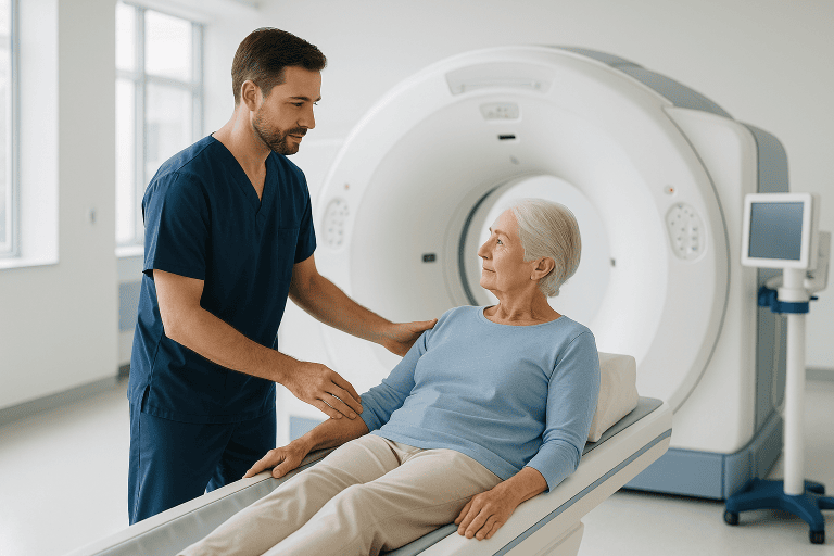
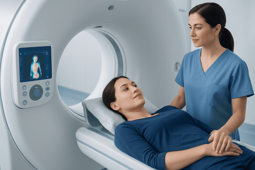
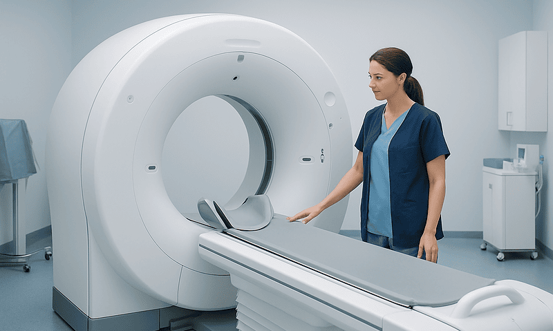
Once the uptake period is complete, you are positioned on the scanner table. You will not feel the imaging itself; your only task is to lie still while the table moves slowly through the scanner. The CT component is acquired first or as part of the combined protocol, using X-rays to create detailed cross-sectional images. In some cases, intravenous contrast is given during the CT to improve visualisation of blood vessels and organs.
The PET component then detects the signals emitted by the tracer within your body. Sophisticated software reconstructs those signals into images showing areas of differing metabolic activity, and the system fuses them with the CT so that each area of activity can be matched to an exact anatomical structure. This fused view is the core advantage of the technique and the main reason PET-CT has become so central in oncology and other complex diagnostic settings.
How long does a PET scan take?
Plan for roughly two to three hours in the department, although the image acquisition itself usually takes only about twenty to forty minutes. The rest of the visit is taken up by preparation, the tracer injection, and the uptake period, which commonly lasts between forty-five and ninety minutes depending on the tracer and the protocol. The scanned body area matters too: a limited study is quicker than a skull-to-thigh survey, and occasionally additional delayed images are needed, which extends the appointment. Your department will give you a realistic time estimate for your specific protocol, but as a rule, the waiting takes longer than the scanning.
The Technology Behind the Images
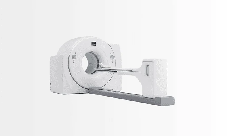
PET-CT relies on integrated scanner hardware, specialised radiotracers produced under strict pharmaceutical standards, advanced image reconstruction, and, most importantly, expert interpretation. Modern systems are designed to improve image quality, reduce motion artefacts and optimise radiation dose where possible. For patients, the practical benefit of the technology is clarity: a combined study can help distinguish active disease from inactive tissue, identify small areas that deserve attention, sharpen staging, guide biopsy targets and help define radiotherapy fields. Technology alone does not deliver that clarity; protocol selection, careful preparation and experienced reading do. If you want more background on radiation dose and imaging safety in general, the guide on radiology and imaging safety for international patients covers the topic in plain terms.
After the Scan: What Happens Next
There is no recovery period in the traditional sense, because PET-CT involves no surgery and, in almost all cases, no sedation. Most people return to their usual activities shortly afterwards. You may be advised to drink fluids to help clear the tracer, and you may receive simple, temporary guidance about close contact with pregnant women or small children for a short period, since the tracer decays within hours. These instructions are precautionary and short-lived.
Interpretation takes longer than the scan itself. A nuclear medicine physician, often working closely with radiologists and the referring team, reviews the images carefully and compares them with earlier scans and your clinical records. The final report addresses the question that prompted the study: whether disease is present, whether it appears localised or has spread, whether a response to treatment is evident, or whether recurrence is likely. The most useful reports do more than list abnormalities: they help your doctors decide what to do next.
Understanding the Images and the Report
Many patients look at their scan images and wonder what they are seeing. It is worth knowing what specialists look for, and also why reading these studies is a trained skill rather than a matter of spotting bright dots.
How do specialists read PET-CT scan images?
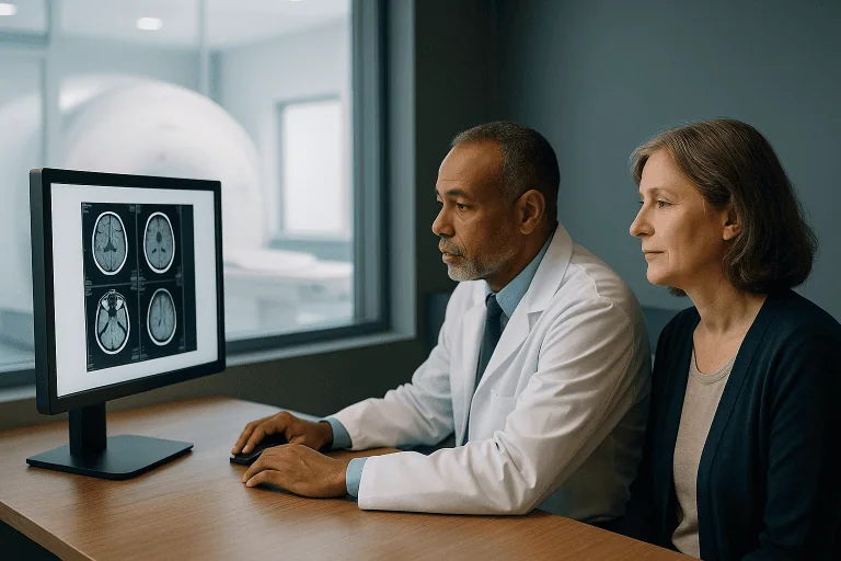
Specialists read PET-CT by reviewing the PET data, the CT data and a fused PET-CT image together, moving through the body systematically rather than jumping to the most obvious finding. A fused PET-CT image overlays the metabolic map, usually in colour, on the grey-scale anatomy of the CT, so that an area of uptake can be tied to a precise structure: a lymph node, a segment of lung, a length of bowel. The reader assesses the intensity of uptake, often described with a standardised uptake value, alongside the size, shape and location of the corresponding structure on CT.
Crucially, not every bright area is cancer. The brain, heart, kidneys and bladder show strong uptake as a matter of normal physiology. Infection, inflammation, healing tissue after surgery or radiotherapy, and even tense muscles can also take up tracer. Conversely, a genuinely important finding can be subtle. This is why reading a PET-CT properly requires comparison with prior imaging, knowledge of your treatment history, and correlation with pathology and laboratory results, and why the written report, produced by a specialist who has all of that context, is a more reliable guide than the raw images. In complex cases, the findings are discussed in multidisciplinary meetings where imaging specialists, oncologists, surgeons and other physicians weigh them together before a recommendation is made.
Why Timing Matters
In diagnostic imaging, timing can matter as much as technology. PET-CT is most valuable when it is used early enough to shape a treatment plan rather than after decisions have already been made. If cancer is suspected or newly diagnosed, accurate staging at the outset helps avoid both under-treatment and over-treatment. Disease that appears limited on one test may prove more extensive on PET-CT, changing the recommended strategy; a finding that looks alarming on standard imaging may show no meaningful activity, changing it in the other direction.
Delays carry their own cost, because disease biology does not pause while decisions are deferred. In some situations, a condition can progress from a localised stage to a more advanced one, narrowing the available options. When PET-CT is used to assess response to therapy, waiting too long can postpone a necessary change of approach; recognising earlier that a regimen is not working as hoped supports a timely adjustment by the treating team.
There is also a timing question in the other direction: scanning too soon. Imaging performed shortly after surgery, radiotherapy or certain systemic treatments can be dominated by inflammation, which takes up tracer and muddies the picture. Specialists deliberately schedule response scans at intervals when the images are most interpretable. If your scan has been set for a date that seems later than you expected, this is often the reason.
Potential Benefits of PET-CT Imaging
The value of PET-CT lies in how it refines diagnosis and supports better-informed decisions. The benefits below are the most common reasons doctors recommend this study.
| Benefit | What It Means for You |
|---|---|
| Combines function and anatomy | Your doctors can see both where an abnormality is and whether it appears metabolically active, which can improve diagnostic clarity. |
| More accurate staging in many cancers | Treatment planning may be more precise because PET-CT can help show whether disease is localised, in lymph nodes, or present elsewhere in the body. |
| Assessment of treatment response | The scan may help determine whether chemotherapy, radiotherapy, immunotherapy or other treatment is reducing active disease. |
| Evaluation of suspected recurrence | If symptoms return or another test is unclear, PET-CT can help identify whether active disease is present and where it may be located. |
| Support for targeted planning | Results can help guide biopsy decisions, radiotherapy planning, and the sequence of further investigations or therapies. |
What Recovery and Follow-Up Typically Look Like
Although PET-CT requires no surgical recovery, patients often want to know what the days after the appointment involve. The timeline below reflects the usual experience.
| Time Period | What Patients Can Expect |
|---|---|
| Day 1 | Arrival, preparation review, tracer injection, uptake period, and imaging. Most patients go home or return to their accommodation the same day. |
| First 24 Hours | Normal daily activity is usually possible. Drinking fluids may be recommended, and brief precautionary guidance about close contact in specific situations may be given while the tracer decays. |
| First Week | Your physician reviews the report in the context of symptoms, pathology and prior scans. This is often when treatment planning or next-step decisions are made. |
| First Month | If the scan was part of cancer staging or response assessment, further consultations, biopsy, surgery, systemic therapy or radiotherapy planning may follow based on the findings. |
| Longer Term | Some patients need a repeat PET-CT or other imaging later to monitor disease status, assess response over time, or investigate new concerns. |
What Influences the Usefulness of a PET-CT Scan
The quality of a PET-CT result depends on more than the machine. The first factor is whether the scan is appropriate for your specific condition. Different cancers behave differently: some are highly visible on PET, others are better assessed with different imaging or different tracers. The clinical question matters too. A PET-CT ordered to stage a newly diagnosed lymphoma is not read the same way as one ordered months after radiotherapy for a head and neck tumour, and a report written without knowledge of that context is worth much less.
Timing is the second factor. Performed too soon after surgery, radiotherapy or certain systemic treatments, the scan is clouded by inflammation; performed too late, it may miss the window in which a treatment could still be adjusted. The third factor is preparation. Fasting as instructed, keeping blood sugar in a workable range where relevant, avoiding strenuous exercise beforehand and following tracer-specific guidance all contribute to clearer images. These sound like small details, but they directly affect what the reader can and cannot say with confidence.
The fourth and arguably most important factor is expert interpretation. PET-CT findings are powerful, but they are also full of potential traps: physiological uptake, post-treatment inflammation, infection and healing tissue can all mimic disease, while some genuine disease shows only modest activity. Correlation with the CT anatomy, prior imaging, pathology, clinical history and laboratory data is essential, and in complex cases a multidisciplinary review adds a further layer of scrutiny. Experienced readers also know when the honest answer is that another test, a biopsy, an MRI, or simply a follow-up scan at an interval, will give a more reliable answer than the current images can.
For you as a patient, a good result does not simply mean a normal scan. It means information that is accurate, clinically meaningful and usable in decision-making. Sometimes that confirms a suspected diagnosis. Sometimes it changes the treatment plan. Sometimes it helps avoid an intervention that would not have helped. The goal is clarity that leads to appropriate care.
PET-CT Within a Wider Care Pathway at Acibadem
Diagnostic imaging is rarely an isolated service. For most patients, a PET-CT is one step within a larger need: confirming a diagnosis, staging a cancer, evaluating a response to treatment, or making sense of unclear or conflicting earlier results. The usefulness of the scan therefore depends not only on the scanner, but on how the imaging is integrated into clinical care around it.
At Acibadem, PET-CT imaging sits within broader diagnostic and treatment pathways. Depending on the disease area, findings can be assessed with input from oncologists, surgeons, radiation oncologists, radiologists, nuclear medicine physicians and other relevant specialists. That kind of joint review matters most exactly when imaging findings could alter the course of treatment, which is, after all, the reason the scan was ordered.
Prior records play a large role in this process. A PET-CT read alongside your earlier scans, pathology reports and treatment history answers more questions than the same images read in isolation, so gathering and sharing that documentation before the appointment is genuinely worthwhile. Technology contributes, but it works best when paired with experience: teams who understand both the strengths of PET-CT and its limits, and who know when a different study or a defined follow-up interval will serve you better than another scan.
Moving Forward with Better Information
PET-CT imaging can be an important step when the next medical decision depends on understanding both the structure and the activity of a disease process. For many patients, it answers questions that standard imaging cannot answer completely: whether disease is present, how far it extends, whether treatment is working, or whether a suspected recurrence is real.
If a PET scan has been suggested to you, the useful questions to think through with your treating doctor are the same ones experienced teams ask themselves: why this scan, why now, and how will the result change the plan? When those three questions have clear answers, the scan is likely to earn its place in your care. When they do not, that conversation itself is valuable, because the point of PET-CT is never the image, but the decision it makes possible.
Preparation
- Patients are usually asked to fast for several hours before the scan and drink water unless told otherwise. Blood sugar may be checked, especially in people with diabetes. Comfortable clothing is recommended, and metal objects may need to be removed. Your doctor may advise avoiding strenuous exercise before the appointment.
Aftercare
- Most patients can return to normal activities the same day after the scan. Drinking extra fluids may help clear the tracer from the body more quickly. Follow any specific instructions about contact with pregnant women or small children for a short period if advised. Results are reviewed by imaging specialists and shared with your referring doctor.
Frequently Asked Questions
What is PET-CT imaging and why might my doctor recommend it?
PET-CT imaging combines two scans in one session: PET shows how tissues and organs are functioning, while CT provides detailed anatomical images. Doctors often recommend PET-CT to help detect cancer, evaluate how far a disease has spread, monitor response to treatment, or investigate certain neurological and cardiac conditions. Because it shows both structure and metabolic activity, it can reveal important information that may not appear on standard imaging alone.
How should I prepare for a PET-CT scan before traveling to Turkey?
Preparation depends on the reason for your scan, but patients are often asked to avoid strenuous exercise for 24 hours and to fast for several hours beforehand. You should usually drink water unless told otherwise. If you have diabetes, special instructions may be needed to manage blood sugar safely. Bring your previous imaging, pathology reports, medication list, and doctor’s notes. Acibadem specialists provide a personalized assessment and clear pre-scan guidance before your appointment.
How long does a PET-CT scan take from start to finish?
A PET-CT visit usually takes a few hours in total, although the actual scanning time is often shorter. After arrival, a small amount of radiotracer is injected, and there is typically a waiting period to allow it to circulate in the body. The scan itself commonly takes around 20 to 45 minutes, depending on the area being examined. Your care team will explain the timeline in advance so you can plan your day comfortably.
Is PET-CT imaging painful or uncomfortable?
PET-CT imaging is generally not painful. You may feel a brief needle prick when the radiotracer is injected, but the scan itself is noninvasive. During imaging, you will need to lie still on a table for a period of time, which some people find mildly uncomfortable if they have pain or difficulty staying in one position. The team will help you feel as comfortable as possible and can explain each step before the scan begins.
Is the radiation from a PET-CT scan safe?
PET-CT uses radiation from both the CT scan and the injected radiotracer, but the amount is carefully controlled and used only when medically appropriate. For many patients, the diagnostic value outweighs the potential risks. Your doctor will consider your age, medical history, and the reason for the scan before recommending it. If you are pregnant, breastfeeding, or think you might be pregnant, tell the team in advance so they can advise you appropriately.
Can I eat, drink, or take my usual medications before PET-CT imaging?
Many patients are asked not to eat for several hours before PET-CT, although plain water is usually encouraged. Some medications can be taken as normal, but this depends on your condition and the type of scan. Diabetic patients often need a tailored plan because blood sugar levels can affect image quality. It is important to follow the exact instructions provided by your imaging team. Acibadem specialists can review your medications and create a personalized preparation plan.
When will I get my PET-CT results and who explains them to me?
PET-CT images are reviewed by experienced radiology and nuclear medicine specialists who analyze both the PET and CT findings together. In many cases, a formal report is prepared within a short time, though timing can vary depending on the complexity of the scan and whether prior studies need comparison. Your referring doctor or Acibadem specialist will explain what the results mean, answer your questions, and discuss whether any further tests or treatment steps are needed.
What conditions can PET-CT help diagnose or monitor?
PET-CT is most commonly used in oncology to detect suspected cancer, stage disease, assess recurrence, and evaluate response to chemotherapy, radiotherapy, or other treatments. It can also support assessment of some neurological conditions, such as certain memory disorders, and selected heart problems by showing tissue viability and function. The exact role of PET-CT depends on your symptoms and medical history. A personalized review helps determine whether this scan is the most suitable imaging choice for you.
Can international patients have PET-CT imaging at Acibadem in Turkey without staying long?
Yes, many international patients travel to Turkey specifically for advanced imaging and do not need a long stay. PET-CT is usually performed as an outpatient procedure, so patients can often attend the appointment and return to their accommodation the same day. The exact schedule depends on your medical needs, preparation requirements, and whether consultations are planned around the scan. Acibadem’s international patient services can help coordinate appointments, language support, and travel-related planning.
What should I do after a PET-CT scan, and are there any restrictions?
After a PET-CT scan, most patients can return to normal activities unless their doctor advises otherwise. You may be asked to drink extra water to help flush the radiotracer from your body. As a precaution, some patients are advised to limit close contact for a short time with pregnant women or very young children, depending on the scan type. If you received a sedative, you may need someone to accompany you. Your team will give clear post-scan instructions.
Medically reviewed by the Acıbadem International Medical Board September 14, 2026
See our medical review board →
Update history
- PublishedJuly 19, 2026
- Medical review approvedSeptember 14, 2026
- Last content updateSeptember 14, 2026
From the Acibadem Blog
What a PET-CT Scan Shows That a CT Alone Cannot: Metabolic Activity in Cancer Staging
A PET-CT scan shows how actively tissues are using energy, mapped onto a detailed anatomical picture. The PET part tracks a mildly radioactive sugar…

How to Prepare for a PET-CT Scan: Fasting, Blood Sugar Checks and What to Wear
To prepare for a PET-CT scan, most people are asked to stop eating for about six hours beforehand, drink only plain water, avoid sugar,…