

Quick answer
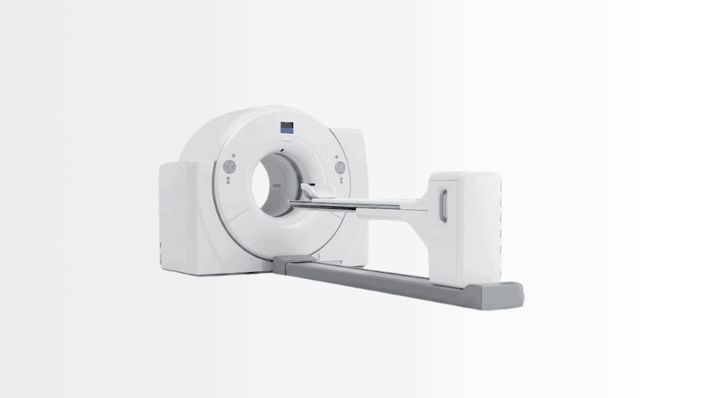
A PET-CT scanner combines metabolic imaging from positron emission tomography with detailed anatomical images from computed tomography, helping specialists identify and assess many conditions, particularly cancer. At Acibadem in Turkey, PET-CT imaging is used to support diagnosis, staging, treatment planning and follow-up after specialist evaluation.
A PET-CT scan answers the question anatomy alone cannot: not just where something sits, but whether it is metabolically alive. By fusing PET’s map of cellular activity with CT’s precise anatomy in a single pass, the scan shows active disease as bright signal on an exact location, which is why PET-CT sits at the centre of modern cancer staging, treatment-response assessment and recurrence hunting. Across the Acibadem network, PET-CT runs within nuclear medicine departments whose reports feed directly into the group’s tumour boards.
What is a PET-CT scan and how does it work?
PET (positron emission tomography) traces a tiny amount of radioactively labelled sugar, FDG, injected into a vein. Because many cancer cells consume glucose faster than the tissue around them, they light up on the PET image. CT (computed tomography), acquired in the same session on the same machine, supplies the anatomical map. The scanner fuses the two: activity from PET, address from CT. Other tracers extend the method beyond sugar, prostate-specific tracers such as PSMA, and receptor tracers such as DOTA compounds for neuroendocrine tumours, each chosen by the nuclear medicine physician for the question being asked.
What a PET-CT scan shows, and is used for
The scan’s four classic jobs: staging a newly diagnosed cancer, one whole-body study showing whether disease has spread, and where; assessing treatment response, comparing activity before and after chemotherapy or radiotherapy, often earlier and more truthfully than size changes alone; hunting recurrence when a tumour marker rises but conventional imaging sees nothing; and characterising ambiguous findings, where activity helps separate the suspicious from the innocent. Beyond oncology, PET-CT contributes to selected cardiac viability and inflammation questions: your physician defines the indication, because the tracer and protocol follow from it.
The scan: preparation and what actually happens
Preparation protects the image: fasting for several hours beforehand, avoiding strenuous exercise the previous day, and, for diabetic patients, glucose management coordinated in advance, because sugar in the blood competes with the tracer. On the day: a small injection, then a quiet resting hour while the tracer distributes (resting genuinely matters, active muscles steal signal), then the scan itself, typically 20–30 minutes on the couch. The whole visit spans two to three hours. Afterwards you may resume normal life; drinking water helps clear the tracer, and close, prolonged contact with pregnant women and small children is limited for the rest of the day as a courtesy of caution.
PET-CT results: how they are read
The report describes where activity concentrates and how intensely, often expressed as SUV values, against the anatomical background. Two honest truths about interpretation: not everything bright is cancer (infection, inflammation and healing tissue also consume sugar), and not every cancer is bright (some low-grade tumours sip rather than gulp). This is precisely why PET-CT reports at Acibadem are read by nuclear medicine physicians and then weighed in tumour boards alongside pathology and the clinical story: the scan informs the decision; it does not make it alone.
Radiation, safety and honest limits
A PET-CT involves a real but managed radiation dose, from both the tracer and the CT, justified when the answer changes treatment, and minimised by modern scanners and protocols. The examination is generally avoided in pregnancy, and breastfeeding requires a planned pause; tell the team beforehand. The tracer itself causes no side effects patients feel: it is a sugar, not a drug.
What determines the cost of a PET-CT scan?
“PET scan cost” is one of the most-searched phrases around this technology, and the honest answer is that price varies enormously by country, tracer and setting. The real variables: which tracer the question needs (FDG versus speciality tracers like PSMA), whether the study is part of a staging work-up read into a tumour board, and insurer or national-coverage arrangements. At Acibadem, planned imaging follows the group’s written-estimate practice, the cost is stated before the appointment, and for international patients the scan is usually scheduled within a records-first plan so one visit covers imaging and the consultation that acts on it.
PET-CT at Acibadem
PET-CT operates within the group’s nuclear medicine departments, with corporate-verified installations at campuses including Altunizade in Istanbul, City Clinic Mladost in Sofia and Sistina in Skopje. Reports flow into the group’s tumour-board oncology, and images travel in your file, readable at every campus, comparable scan to scan.
PET-CT cancer by cancer: where it changes decisions
The scan’s value is easiest to see in specifics. In lymphoma, PET-CT is woven into the standard of care, staging at diagnosis, and the interim scan whose Deauville score can escalate or de-escalate chemotherapy mid-course. In lung cancer it polices the mediastinum and hunts distant spread before surgery is promised, and it flags which nodes EBUS should sample. In melanoma, head-and-neck, oesophageal and cervical cancers, whole-body staging routinely redraws maps that CT alone had settled. In colorectal cancer it earns its place when markers rise but scans stay silent, and before liver-metastasis surgery: the operation nobody wants to perform twice. Prostate cancer belongs increasingly to PSMA tracers, which see recurrence at PSA levels conventional imaging cannot touch; neuroendocrine tumours to DOTA tracers reading their receptors. And everywhere, the response question, is this treatment working?, is answered in metabolic weeks rather than anatomical months. None of this replaces the tumour board; it feeds it the map.
What separates a good PET-CT from a scan
Three quiet factors decide quality. Preparation discipline: glucose control, genuine rest during uptake, warmth (shivering muscles and brown fat both light up and can mimic or mask disease): the protocols exist because physics punishes shortcuts. Reading depth: a nuclear medicine physician who knows the pitfalls, post-treatment inflammation, physiological uptake patterns, the artefacts of motion and blood sugar, separates signal from story. Continuity: response assessment is a comparison, and comparisons are honest when acquisition and platform match scan to scan, which is why serial studies belong in one system. At Acibadem all three are house rules, and every oncological read lands in a file the group’s tumour boards and every campus can open.
Frequently Asked Questions
What is the Deauville score?
A five-point scale grading lymphoma’s metabolic response on interim and end-of-treatment PET, one of the clearest examples of the scan directly steering chemotherapy decisions.
Why must I rest quietly after the injection?
Because working muscles consume the tracer: an hour of genuine rest keeps the sugar signal for the tissues being questioned.
My blood sugar runs high, can I still have the scan?
Yes, with planning, glucose competes with the tracer, so diabetic preparation is coordinated in advance rather than improvised on the day.
How long does a PET-CT scan take?
The scan itself typically takes 20–30 minutes; with the tracer’s quiet distribution hour and preparation, plan two to three hours at the department.
Do I need to fast before a PET-CT?
Yes, several hours without food, and no strenuous exercise the day before; your appointment instructions state the exact protocol, and diabetic patients receive a coordinated plan.
Is the injection or scan painful?
Only the small intravenous injection is felt; the scan is simply lying still on the couch.
Is a PET-CT scan safe?
It carries a managed radiation dose justified when the answer changes treatment; the tracer itself has no felt side effects. Pregnancy is generally a reason not to scan, tell the team.
Can I be around people afterwards?
Yes, ordinary contact is fine; as a courtesy of caution, prolonged close contact with pregnant women and small children is limited for the rest of the day.
What does it mean if something “lights up”?
That the tissue is metabolically active, which includes cancers but also infection, inflammation and healing; the nuclear medicine physician reads brightness in context, never alone.
Can PET-CT miss a cancer?
Some low-activity tumours show faintly or not at all, one honest reason the scan is weighed with pathology and other imaging in a tumour board rather than trusted in isolation.
What is an SUV value?
A standardised measure of how intensely a spot takes up tracer, useful for comparing scans over time, interpreted by the reporting physician rather than as a stand-alone verdict.
What is a PSMA PET-CT?
A prostate-specific tracer study that maps prostate cancer with high sensitivity, chosen by the physician when the clinical question calls for it.
How soon are results ready?
Reports are typically completed within a working day and enter your Acibadem file immediately, and, where a tumour board is waiting on them, they are read into it directly.
How is PET-CT different from CT or MRI alone?
CT and MRI show structure; PET adds behaviour. Fused, they show whether an abnormality is metabolically active and exactly where it is: the combination neither achieves alone.
How often can PET-CT be repeated?
As often as the clinical benefit justifies the dose, response assessment at planned treatment milestones is routine, and the decision is your oncology team’s.
Will my scan be compared with previous ones?
Yes, prior studies are integrated wherever usable, and same-platform follow-up makes SUV comparisons meaningful scan to scan.
Can I get a PET-CT as an international patient?
Yes, through the group’s records-first process: the indication confirmed from your records, a written plan and estimate, and the scan scheduled so the consultation that acts on it follows in the same visit.
Related Medical Units
Available at these Hospitals










Related Treatments
Speak with our medical team
Find out how this technology could support your diagnosis or treatment.