

What a PET-CT Scan Shows That a CT Alone Cannot: Metabolic Activity in Cancer Staging
Key Takeaways
- A PET-CT adds a metabolic layer to anatomy: it maps how much of a glucose-like tracer each tissue absorbs, so a normal-sized lymph node that glows can change the N stage without any change in size on CT.
- FDG-PET misses some cancers by design, including many prostate cancers, some kidney and mucinous tumors and low-grade lymphomas, which use too little sugar to stand out; different tracers exist for several of these.
- Infection, healing surgical sites, arthritis, brown fat and tense muscles all take up tracer, which is why the Mayo Clinic lists inflammation among causes of false-positive results and why bright spots usually need biopsy confirmation before treatment changes.
- Expect about six hours of fasting, roughly an hour of quiet tracer uptake and about 30 minutes in the scanner, with around two hours in the department overall (NHS, Mayo Clinic, Cleveland Clinic).
- The tracer leaves the body within a few hours, per the NHS, so keeping a few hours' distance from pregnant women and young children and drinking plenty of water are the only real aftercare steps.
- SUV, the standardized uptake value in the report, varies with scanner, timing and blood sugar, so no single number separates benign from malignant and comparisons are most meaningful between scans done the same way.
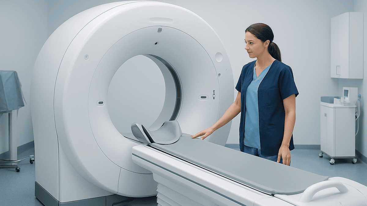
A PET-CT scan shows how actively tissues are using energy, mapped onto a detailed anatomical picture. The PET part tracks a mildly radioactive sugar tracer that many cancers absorb faster than normal cells; the CT part shows where that activity sits. Together they reveal metabolically active disease, including spread to lymph nodes or distant organs that a CT alone may show only as size or shape, or not at all.
The CT report had already been read twice, once by the radiologist and once, slowly, at a kitchen table. It listed a lung nodule, two lymph nodes “of borderline size,” and a liver spot “of uncertain significance.” Three phrases, three unknowns. Then the oncologist said the sentence that so many people hear at this point: “Before we decide anything, I’d like a PET-CT.”
Anyone asking what does a PET-CT scan show is really asking a sharper question: what can this test tell me that the scan I already had could not? The honest answer is that a CT is superb at anatomy, shapes, sizes and positions, while a PET-CT adds a second layer, a map of how hard each tissue is working. Cancer tends to work hard.
This explainer walks through what that metabolic map is, how it changes cancer staging, where it fails, and what the days around the appointment usually look like.
What does a PET-CT scan show that a CT alone cannot?
A CT scan (computed tomography, an X-ray technique that builds cross-sectional pictures of the body) answers one kind of question: what is there, how big is it, and what does it sit next to? It sees a lymph node as an oval of a certain diameter. It cannot say whether the cells inside are quietly filtering lymph or dividing at speed.
PET (positron emission tomography) answers a different question: what is that tissue doing? Before the scan, a small amount of a radioactive tracer, most often a glucose look-alike called fluorodeoxyglucose or FDG, is injected into a vein. Cells that burn a lot of sugar draw the tracer in. Many cancer cells burn far more than their neighbors, so they light up on the image, according to the Mayo Clinic.
On its own, a PET image is blurry and hard to locate, a glowing cloud without landmarks. Fusing it with a CT taken in the same session pins each bright spot to a precise anatomical address. That fusion is what the hyphen in PET-CT means.
So the added value comes in four forms. A normal-sized node that glows becomes suspicious. An enlarged node that stays dark becomes less worrying. A distant spot that a CT called “indeterminate” can be re-read in the light of its metabolic behavior. And activity in places nobody had looked, a bone, an adrenal gland, a node above the collarbone, can appear on a single whole-body sweep.
None of these findings is proof. Inflammation, infection and healing tissue also consume sugar, and the MedlinePlus guidance is clear that PET results are interpreted alongside other tests and, where needed, a biopsy (removal of a tissue sample for laboratory examination). What the PET-CT adds is direction: it tells the team where to look harder and what to weigh more heavily.
How does a PET-CT scan actually work, in plain language?
Think of the tracer as sugar wearing a tiny beacon. FDG is chemically close enough to glucose that cells pull it inside through the same doorways they use for ordinary fuel. Once inside, a chemical difference stops it being fully broken down, so it accumulates. The more sugar a cell was taking up at that moment, the more tracer stays trapped there.
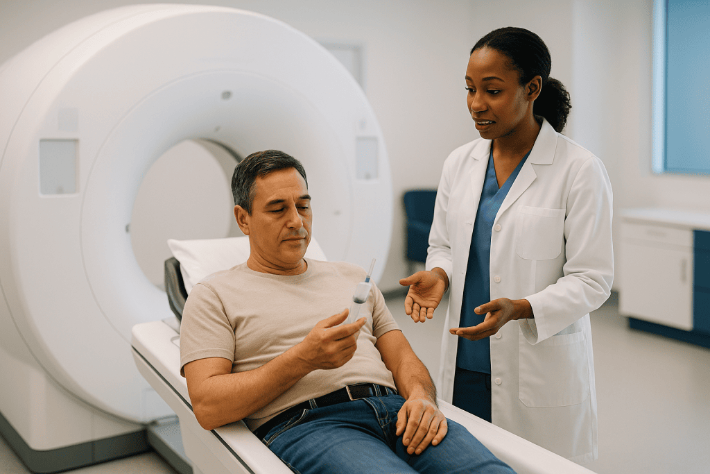
The beacon part is a radioactive fluorine atom attached to the sugar. As it decays, it releases a positron, a particle that behaves like an electron with a positive charge. Within a fraction of a millimeter the positron meets an electron and both vanish, sending out two gamma rays in exactly opposite directions. The PET scanner is a ring of detectors that waits for those paired signals and works out, from their timing, where in the body they originated.
That waiting is why the appointment has a quiet middle. After the injection, you rest for roughly an hour while the tracer distributes, and both the NHS and the Mayo Clinic describe the scan itself as taking around 30 minutes or so. During that hour you are asked to stay still and warm, because working muscles and cold-activated brown fat (a heat-generating tissue in the neck and shoulders) also soak up sugar and can create confusing bright patches.
The CT component runs on the same table, usually just before the PET acquisition. Some centers use a low-dose CT purely for mapping; others add iodinated contrast (a dye that sharpens blood vessels and organs) for a fuller diagnostic CT. A computer then overlays the two datasets so a radiologist or nuclear medicine physician can scroll through the body and see, slice by slice, both the anatomy and the color-coded intensity of metabolic activity.
Why metabolic activity matters so much in cancer staging
Staging is the process of describing how far a cancer has spread, and it drives almost every decision that follows. Most solid tumors are staged with the TNM system: T for the size and local extent of the primary tumor, N for regional lymph node involvement, and M for metastasis, meaning spread to distant organs. The stage that emerges from those three letters shapes whether surgery is sensible, whether treatment is aimed at the whole body or one region, and how a team frames what to expect.
Here is the difficulty. On a CT, a lymph node has to enlarge before it looks abnormal, and nodes enlarge for many reasons, including old infections. A node stuffed with tumor cells can still measure within normal limits. Metabolic imaging steps around size. If a small node is metabolically avid (a radiology term meaning it takes up tracer strongly), that finding can move a patient from one N category to another without a millimeter of change on the CT.
The M category benefits even more. Chasing distant spread by CT means imaging region after region and still guessing about the bones. A PET-CT sweep typically covers the base of the skull to the mid-thigh in one pass, as the Cleveland Clinic describes, so an unexpected focus in a rib or an adrenal gland is caught in the same session.
In practice, this is why oncology guidelines in several major cancers position PET-CT as a staging step before decisions about surgery or radiotherapy. The scan does not replace tissue confirmation. Its role is to make sure a treatment with real side effects is not planned around an incomplete map, and to prompt a biopsy when a single distant spot would change the whole plan.
What problems can a PET scan detect beyond cancer?
Cancer is the headline use, but the underlying idea, imaging how tissue behaves rather than how it looks, has a wider reach.
In the heart, a PET scan can show which areas of muscle are still alive but starved of blood after a heart attack and which are scar. The Mayo Clinic notes that this distinction helps cardiologists judge whether a procedure to restore blood flow is likely to help a given region. Tissue that still takes up sugar is worth rescuing; scar is not.
In the brain, patterns of reduced sugar use can support the evaluation of certain dementias, and localized changes can help pinpoint where seizures begin in people being considered for epilepsy surgery, according to Johns Hopkins Medicine. Specialized tracers that bind to abnormal proteins are also used in research and some clinical settings, though interpretation sits firmly with neurology teams.
Infection and inflammation are the double-edged sword. Because immune cells are metabolically busy, PET can reveal hidden sources of infection, unexplained fevers, or inflammation in blood vessels. The same property is the leading cause of false alarms in cancer imaging: a healing surgical bed, a recent chest infection or an inflamed joint can glow as brightly as a tumor.
Within oncology, the tracer has broadened beyond FDG. Prostate cancer often uses little sugar, so tracers that bind to a protein on prostate cancer cells are now used for staging. Well-differentiated neuroendocrine tumors, which grow slowly and use sugar modestly, are imaged with tracers that attach to hormone receptors on their surface. These are named here only to explain why “a PET scan” is not a single test. Which tracer, if any, suits a particular situation is a decision for the treating team.
Does a PET-CT scan show all cancers?
No, and this is the most useful thing to understand before the appointment.
The first limit is biology. FDG-PET depends on a tumor consuming more sugar than the tissue around it. Some cancers do not cooperate. Many prostate cancers, some kidney cancers, mucinous tumors that produce a lot of gel-like fluid, and slow-growing or low-grade lymphomas can take up tracer weakly or not at all. A normal-looking FDG-PET in these settings does not rule disease out, which is why other tests or different tracers are used.
The second limit is background noise. The brain uses sugar constantly, so it glows on every scan and small brain metastases can hide in that glare; MRI (magnetic resonance imaging, which uses magnets and radio waves rather than radiation) is usually preferred for the brain. The bladder fills with excreted tracer, which can obscure the pelvis. The heart and, in some people, brown fat add their own bright areas.
The third limit is size. A PET scanner has a resolution floor, and very small clusters of tumor cells simply do not produce enough signal to stand out. A scan reported as negative cannot promise that no microscopic disease exists, and clinicians read it as “no detectable metabolically active disease,” a narrower statement.
The fourth limit is timing. Recent chemotherapy can temporarily quiet a tumor’s sugar use, and recent surgery or radiotherapy creates inflammation that mimics disease. The Mayo Clinic flags that infection and inflammation can produce false-positive results. Scheduling, therefore, is part of the test’s accuracy, and teams often wait a set interval after treatment before re-imaging.
Put together, a PET-CT is a powerful staging tool with well-mapped blind spots, not an all-seeing eye.
Which cancers are most commonly evaluated on a PET scan?
People often search for “the cancer most found on PET scans,” which slightly misreads how the test is used. A PET-CT is rarely the first test that discovers a cancer. It is more often ordered after a suspicious finding on another scan, to stage a known cancer, to plan radiotherapy, to check how a treatment is working, or to investigate a possible recurrence.
With that in mind, the cancers in which FDG-PET-CT plays the largest routine role are those that reliably take up sugar and where distant spread changes treatment. Lung cancer is the classic example: a solitary nodule’s metabolic activity helps judge how suspicious it is, and whole-body staging can reveal nodes or distant spread that alter the case for surgery. Lymphomas, cancers of the lymphatic system, are staged and reassessed with PET-CT because their metabolic response is a recognized way to gauge treatment effect. Head and neck cancers, esophageal cancer, colorectal cancer when recurrence is suspected, melanoma at higher stages, and cervical cancer are other frequent settings, as the Cleveland Clinic summarizes.
Another common use is the “unknown primary”: a person has cancer confirmed in a lymph node, but nobody knows where it started. A whole-body metabolic map can suggest a hidden origin that focused imaging missed.
The scan is also used to watch for treatment response. A tumor that shrinks slowly on CT may nonetheless have gone metabolically quiet, an earlier signal that a therapy is working. The reverse, a stable-sized mass that keeps glowing, can prompt a rethink sooner. Whether and when to use PET-CT for response assessment varies by cancer type and is set out in disease-specific guidelines, so the oncology team’s choice may differ from a neighbor’s experience with a different diagnosis.
PET scan vs CT scan vs MRI: how they compare for staging
Each imaging method answers a different question, and staging often uses more than one. The table below sets out the practical differences people most often ask about.
| Feature | CT | PET-CT (FDG) | MRI |
|---|---|---|---|
| What it measures | Density and shape of tissue using X-rays | Metabolic activity (sugar uptake) plus CT anatomy | Water and fat content and tissue structure using magnets and radio waves |
| Best at | Fine anatomical detail, lungs, bones, quick whole-body survey | Distinguishing active disease from scar, finding unsuspected spread, normal-sized nodes | Soft tissue contrast, brain, spinal cord, liver, pelvis |
| Main blind spots | Cannot tell active tumor from scar or benign enlargement | Low-sugar tumors, brain background glow, tiny lesions, inflammation mimics | Lungs, calcified bone detail, long scan times |
| Radiation | Yes | Yes (tracer plus CT) | None |
| Typical time in scanner | Minutes | About 30 minutes, after roughly an hour of tracer uptake (NHS, Mayo Clinic) | Often 30 to 60 minutes or longer |
| Preparation | Sometimes fasting if contrast is used | Fasting several hours, avoid strenuous exercise, blood sugar check | Metal safety screening |
Two points deserve emphasis. First, “pet scan vs ct scan” is not a contest with a winner; the PET-CT literally contains a CT, and radiologists read the two layers together. Second, MRI’s lack of radiation makes it the preferred tool where repeated imaging of the brain or pelvis is needed, and it frequently sits alongside a PET-CT in a full staging work-up rather than replacing it.
Who is usually offered a PET-CT, and who is asked to wait?
In cancer care, a PET-CT is generally offered when its result would genuinely change a decision. That includes staging a newly diagnosed cancer known to take up FDG, judging whether a lung nodule or other indeterminate finding is suspicious enough to biopsy, planning the target for radiotherapy, assessing response in cancers where metabolic change is a recognized marker, and investigating rising tumor markers or symptoms that suggest recurrence.
Equally, there are situations where teams hold back or adjust timing.
Pregnancy is the clearest. The tracer and CT both involve radiation, so the NHS advises telling the department if you are or might be pregnant; the scan may be delayed or an alternative chosen. People who are breastfeeding are asked to discuss it in advance, since expressed milk may need to be set aside for a period.
Recent treatment matters. Because chemotherapy can temporarily suppress tumor metabolism and radiotherapy or surgery stirs up inflammation, scanning too soon can mislead in both directions. Teams typically schedule response scans after a defined interval that depends on the treatment and the cancer type.
Blood sugar is a practical gatekeeper. High circulating glucose competes with the tracer for entry into cells and degrades image quality, so people with diabetes are given individualized instructions about meals and medicines, and centers check a fingerstick glucose on arrival. Any adjustment to diabetes medicines is decided by the prescribing clinician, never improvised.
Kidney function comes into play only when iodinated CT contrast is planned, and known contrast allergies are reviewed beforehand. Severe claustrophobia, inability to lie still, or very recent acute infection are discussed case by case. The MedlinePlus summary notes the radiation exposure is small, but the guiding principle remains that each scan should earn its place in the plan.
How long does a PET scan take, and what happens on the day?
Set aside a good part of the day. The scan itself is short; the preparation around it is not.
Instructions usually begin the day before. The Mayo Clinic advises avoiding strenuous exercise for 24 hours beforehand, because working muscles pull in sugar and can glow on the images. The NHS describes fasting for about six hours before the appointment, with plain water allowed and encouraged. Most departments ask you to wear warm, comfortable clothing without metal, and to arrive having taken regular medicines unless told otherwise.
On arrival, a technologist checks your history, confirms fasting, and tests blood glucose. A small cannula goes into a vein, usually in the arm or hand. The tracer injection takes moments and does not cause a warm flush or taste the way CT contrast sometimes does.
Then comes the uptake period. You rest in a quiet, dimly lit room for around an hour, wrapped in blankets, asked not to read, talk much or use your phone, since even jaw and hand muscles take up tracer. It is an oddly peaceful stretch of a stressful day.
You empty your bladder, then lie on the narrow table with arms usually raised above the head. The table slides through a ring that is more open than an MRI tunnel. The CT sweep takes under a minute; the PET acquisition follows, in a series of positions from the skull base to the thighs, typically around 30 minutes. Breathing normally and staying still are the only jobs.
Including check-in, uptake and scanning, the Cleveland Clinic suggests planning for roughly two hours in the department. Afterward the cannula comes out and, in most cases, you can eat, drink and leave straight away.
What not to do after a PET scan
The tracer’s radioactivity fades quickly, and the NHS states it leaves the body within a few hours, mainly through urine. A few simple measures cover that window.
- Avoid prolonged close contact with pregnant women, babies and young children for the few hours the department specifies. This is a precaution for them, not a sign that you are hazardous; the dose to others is very small, but small children have longer lives ahead and more sensitive tissues.
- Do not skip fluids. Drinking well and emptying the bladder often clears the tracer faster, as the MedlinePlus guidance advises.
- Do not breastfeed until the interval your team gives you has passed; expressed milk from that period is usually discarded and the timing should be confirmed with the department rather than assumed.
- Do not travel through airport or border radiation detectors without the card or letter some centers provide; residual tracer can trigger sensitive alarms for a short period.
- Do not rely on a sedative-free drive home if you were given medicine to manage anxiety or claustrophobia; arrange a companion if that applies.
There is nothing to avoid in terms of food. Most people resume normal meals immediately, and those with diabetes return to their usual routine as instructed by their own clinician.
What you do not need to do is wait for a symptom. Allergic reactions to FDG are extremely rare, and the tracer has no known effect on how you feel. Any lingering soreness is almost always from the cannula site. If CT contrast was used, a mild warmth or metallic taste passes within minutes; any hives, facial swelling, wheeze or breathlessness in the hours afterward is a reason to seek urgent care, covered in the final section.
Reading the report: SUV, "avid," "hypermetabolic" and incidental findings
PET-CT reports have their own vocabulary, and a few terms carry most of the weight.
Hypermetabolic or FDG-avid means a region took up more tracer than the surrounding tissue. It is a description, not a diagnosis. Cancer, infection, inflammation and normal structures such as the heart or vocal cords can all be hypermetabolic.
SUV, the standardized uptake value, is a number that estimates how concentrated the tracer is in a region compared with an even spread through the body. Reports often quote an SUVmax, the brightest single point in a lesion. Higher values raise suspicion, but the number depends on the scanner, the timing after injection, body size and blood sugar, so radiologists interpret it in context. Comparing SUV between two scans is most meaningful when both were done the same way on the same equipment. No single cut-off separates benign from malignant, and clinicians resist treating SUV as a verdict.
Physiologic uptake flags glow that is expected: brain, kidneys and bladder, bowel, heart muscle, and active brown fat. Recognizing these is a core skill, and it is why the reading physician wants to know about recent exercise, cold exposure, or a fresh cup of coffee.
Incidental findings are perhaps the most unsettling part of any whole-body scan. A PET-CT surveys tissue nobody asked about, and it sometimes turns up an unexpected thyroid nodule, a colon focus, or a lung spot unrelated to the original question. The Johns Hopkins overview stresses that results are always reviewed by the ordering physician, who decides which incidental findings need follow-up and which are almost certainly harmless.
Reading your own report before the consultation is understandable. Reading it with the doctor who ordered it is where it becomes useful.
What the following days and weeks usually look like
Physically, the days after a PET-CT are unremarkable. There are no dietary restrictions, no activity limits, and no wound to care for. The radiation is gone within hours, and any bruise from the cannula fades on its own.
The waiting is the hard part. The NHS notes that results are not usually available on the same day; the images go to a nuclear medicine physician or radiologist for detailed reporting, and the report then goes to the specialist who ordered it. How quickly that happens depends on the center and on whether the case is scheduled for a multidisciplinary team meeting, the regular conference where surgeons, oncologists, radiologists and pathologists review complex cases together. Many people hear their results at a pre-arranged appointment within a week or two, though the exact interval varies and should be confirmed with the team when you leave.
What happens next depends on what the scan changed. Three broad paths are common.
If the scan confirms what other tests suggested, the plan proceeds as discussed, perhaps with the radiotherapy target or surgical extent refined using the metabolic map.
If it reveals a new area of activity, the usual next step is not a change of treatment but a confirmation step: a biopsy of the new site, or a targeted MRI or ultrasound to characterize it. A bright spot that would alter the stage almost always earns tissue proof before the plan turns on it, because the false-positive causes described earlier are real.
If it shows less activity than feared, or none at a site that looked suspicious on CT, the team may de-escalate, move toward a curative-intent local treatment, or simply re-image after an interval.
Each of these is a conversation, not a foregone conclusion, and the person best placed to explain the specific implications is the clinician who knows the whole picture.
What people often get wrong about PET-CT scans
“A PET scan finds every cancer.” It finds cancers that use more sugar than their surroundings and that are large enough to register. Prostate, some kidney, mucinous and low-grade tumors can be quiet on FDG, and microscopic disease is invisible by definition. A negative report means no detectable active disease, not none.
“If it glows, it’s cancer.” Infection, healing surgical sites, inflammatory bowel disease, arthritis, recent vaccination in a lymph node draining the arm, brown fat and tense muscles all light up. The Mayo Clinic explicitly lists infection and inflammation among causes of false-positive results. Bright spots prompt questions; biopsies answer them.
“I’ll be radioactive for days.” The tracer decays and is excreted within a few hours, per the NHS. Precautions around pregnant women and young children apply for that same-day window only.
“Sugar the night before will feed the cancer and make it show up better.” The opposite is true. High blood glucose competes with the tracer and dulls the image, which is why fasting is required. Diet has no proven effect on how much a tumor absorbs beyond that.
“A higher SUV means a worse prognosis for me.” SUV is scanner- and situation-dependent, and its meaning varies by cancer type. It is one input among many, interpreted by the team, not a personal score.
“PET replaces CT and MRI.” The PET-CT already contains a CT, and MRI remains preferred for the brain, spine and often the pelvis. Staging routinely uses several methods together.
“A clear PET after treatment means it’s over.” A metabolically complete response is encouraging and, in some cancers, a recognized marker of effective treatment. It does not exclude residual microscopic cells, which is why follow-up schedules exist and why oncologists choose their words with care.
Risks, radiation and alternatives, stated neutrally
The main risk of a PET-CT is radiation exposure, from both the tracer and the CT. The MedlinePlus summary describes the exposure from the tracer as small and the associated risk as low, while noting that all radiation carries a theoretical long-term risk and that the benefit of accurate staging is judged to outweigh it when the scan is clinically indicated. The CT portion adds to the total, more so if a full diagnostic CT with contrast is acquired rather than a low-dose mapping CT. Departments tailor this to the question being asked.
Allergic reactions to FDG are extremely rare. Reactions to iodinated CT contrast, when used, range from mild itching to, very uncommonly, serious reactions, and centers screen for prior reactions and kidney function beforehand.
Pregnancy and breastfeeding are the principal reasons to postpone or choose another test, as discussed above. People with diabetes face a preparation challenge rather than a safety risk, managed with their own clinician’s guidance.
Psychological risk is real and under-discussed. Whole-body scans generate incidental findings and ambiguous bright spots, and the wait for interpretation is stressful. Knowing in advance that “indeterminate” is a common word in these reports can soften it.
Alternatives depend on the question. A contrast-enhanced CT alone may suffice when metabolic information would not change management. MRI is preferred for the brain and often the liver and pelvis, and involves no radiation. Bone scans, ultrasound and endoscopic ultrasound with biopsy have specific roles. For cancers that use little sugar, PET with a different tracer may be more informative than FDG. Each option has its own blind spots, and the choice is a matter for the treating team, weighing what the result would change against the exposure and the delay involved.
Questions to ask your care team before and after a PET-CT
A PET-CT is rarely urgent to the hour, which leaves room for a conversation. These questions tend to produce the most useful answers.
Before the scan:
- What specific question is this scan meant to answer, and what would you do differently depending on the result?
- Is this FDG, or a different tracer chosen for my cancer type, and why?
- Will the CT part use contrast, and do you need to check my kidney function or contrast history first?
- I have diabetes: exactly how should I handle meals and my usual medicines on the day? (Take the answer from the prescribing clinician, in writing.)
- How long after my last treatment should this scan be done to give a reliable picture?
- Is there any reason, such as pregnancy, breastfeeding or a recent infection, to delay?
After the scan:
- Did the scan change my stage, and if so, which part: the tumor, the nodes, or distant spread?
- Are any of the bright areas likely to be inflammation or normal uptake rather than cancer?
- Do any findings need a biopsy or a different scan before the plan is finalized?
- Were there incidental findings unrelated to my cancer, and which ones need follow-up?
- What does the SUV in my report mean in my situation, and how much weight are you giving it?
- When will this be reviewed by the multidisciplinary team, and when will I hear the outcome?
- Will I need a repeat PET-CT to assess response, and roughly when?
Bringing a companion, or asking permission to record the conversation, helps most people retain more of it. Writing down the date and channel by which results will arrive, a portal message, a phone call or a clinic visit, removes one uncertainty from a week that already has plenty.
When to call your doctor after a PET-CT scan
Serious problems after a PET-CT are rare, and most people go home and forget the appointment within an hour. A few signs, though, should not wait for the results visit.
Seek urgent or emergency care if, in the hours after the scan, you develop hives spreading over the body, swelling of the face, lips or tongue, wheezing, difficulty breathing, or a sudden feeling of faintness. These can indicate a reaction to CT contrast; reactions to the tracer itself are extremely uncommon, but the response is the same.
Call the imaging department or your doctor the same day if the cannula site becomes increasingly red, hot, swollen or painful, or if you notice fluid leaking, which can indicate infection or tracer that leaked outside the vein.
Contact your diabetes clinician promptly if, after fasting and any changes made for the scan, you experience symptoms of low or very high blood sugar such as shakiness, confusion, sweating, extreme thirst or drowsiness. Do not adjust your medicines on your own.
Tell the department or your doctor as soon as possible if you learn you were pregnant at the time of the scan, or if you breastfed before the advised interval had passed; they can advise on the actual exposure, which is usually small.
Do not sit on new symptoms while waiting for results. Worsening pain, unexplained weight loss, a persistent cough or new bleeding deserve a call regardless of what the scan may show. Equally, if the results appointment has not been arranged or the report is delayed beyond the time you were told, contacting the team is reasonable. Every interpretation and every decision that follows from this scan belongs with the clinicians who ordered it, and they would rather hear from you early than late.
Frequently asked questions
What does a PET-CT scan show that a regular CT does not?
It shows metabolic activity, meaning how much sugar-like tracer each tissue is absorbing, fused onto the CT’s anatomical picture. A CT reveals size and shape; a PET-CT adds whether a node or spot is biologically active. That lets clinicians flag normal-sized but avid nodes, downgrade enlarged but quiet ones, and spot unexpected distant activity in a single whole-body sweep, while still requiring biopsy to confirm what any bright area actually is.
What problems can a PET scan detect?
Beyond staging and monitoring cancers that use sugar avidly, PET can show which heart muscle is alive but underperfused after a heart attack, patterns of reduced brain activity relevant to dementia and epilepsy evaluation, and hidden sources of infection or inflammation. Specialized tracers extend it to prostate cancer and neuroendocrine tumors. Each use has its own preparation and interpretation, so the ordering specialist decides which tracer and protocol fit the question.
Does a PET CT scan show all cancers?
No. FDG-PET detects cancers that consume more glucose than surrounding tissue and are large enough to register. Many prostate cancers, some kidney cancers, mucinous tumors and low-grade lymphomas can be invisible or faint. The brain’s natural glow hides small metastases there, and microscopic disease is below any scanner’s resolution. A negative PET-CT means no detectable metabolically active disease, which is a narrower statement than no cancer.
What cancer is most found on PET scans?
PET-CT rarely discovers cancer first; it usually stages or monitors one already suspected. Its largest routine roles are in lung cancer, lymphoma, head and neck, esophageal, colorectal recurrence, melanoma and cervical cancer, all of which typically take up FDG strongly. It is also used to hunt for an unknown primary when cancer is found in a lymph node. Which cancers are imaged reflects biology and guidelines, not how common the disease is.
What not to do after a PET CT scan?
For the few hours the department specifies, avoid prolonged close contact with pregnant women, babies and young children, and do not breastfeed until the advised interval has passed. Keep drinking water and emptying your bladder to clear the tracer, and carry any card the center gives you if you travel through radiation detectors. There are no food restrictions, and normal activity can resume immediately unless you received a sedative.
How long does a PET scan take from start to finish?
Plan for roughly two hours in the department. After a blood sugar check and tracer injection, you rest quietly for about an hour while the tracer distributes, then spend around 30 minutes on the scanner table, according to the NHS and Mayo Clinic. Fasting for about six hours beforehand adds to the day. Results are not usually given the same day; they go to a reporting physician and then to your specialist.
Is a PET scan vs CT scan a choice, or do I need both?
It is not an either-or choice, because a PET-CT already includes a CT acquired in the same session. The question is whether metabolic information would change your management. When it would, the fused scan is ordered; when anatomy alone answers the question, a contrast CT may be enough. MRI often joins the work-up for the brain, spine or pelvis. Your team selects the combination based on your cancer type and the decision at hand.
Can a PET scan be wrong?
Yes, in both directions. False positives arise because infection, healing tissue, arthritis, recent vaccination and brown fat all absorb the tracer and glow like tumor. False negatives occur with low-sugar cancers, very small lesions, high blood glucose on the day, or scanning too soon after chemotherapy when tumor metabolism is temporarily suppressed. This is why findings that would change treatment are usually confirmed by biopsy or further imaging first.
Is a PET scan for cancer staging safe if I have diabetes?
It is commonly done, but preparation matters. High blood glucose competes with the tracer and degrades the images, so centers check a fingerstick reading on arrival and may reschedule if it is too high. You will receive individualized guidance about meals and medicines from your own clinician; do not change diabetes medicines on your own. Fasting with diabetes carries its own risks, so ask for written instructions and a plan for symptoms of low sugar.
What does SUV mean on my PET scan report?
SUV, the standardized uptake value, estimates how concentrated the tracer is in a region relative to an even distribution through the body; SUVmax is the brightest point in a lesion. Higher values raise suspicion, but the number depends on the scanner, timing after injection, body size and blood sugar, so no single cut-off distinguishes benign from malignant. It is most useful when comparing two scans done the same way, and it is interpreted alongside everything else.
References
This article is for general information only and is not a substitute for professional medical advice. Please consult a qualified doctor about your individual situation.

More from the Blog

What a CT Scan Shows, and Why Doctors Order One
A CT scan shows detailed cross-sectional images of bones, organs, blood vessels and soft tissue by combining many X-ray views into slices. Doctors most…

Can an Ultrasound Detect Cancer? What It Can Show, Its Limits and the Next Steps
An ultrasound can detect abnormal lumps, masses and changes in organs that may be cancer, and it is especially good at telling a fluid-filled…
When Does a Brain Lesion Need a Stereotactic Biopsy Instead of Open Surgery?
A brain lesion is usually sampled with a stereotactic needle biopsy when the tissue diagnosis will change treatment but removing the lesion is not…

Stereotactic Brain Biopsy Step by Step: Planning Scans, Coordinates and the Needle Path
A stereotactic brain biopsy uses a detailed MRI or CT scan, a three-dimensional coordinate system and a thin needle to remove a tiny sample…
What Happens During an EMG Test? Nerve Conduction Study, Needle Electrodes and Timing
During an EMG test, a clinician first performs a nerve conduction study, placing sticky surface electrodes on the skin and delivering brief electrical pulses…
How an Echocardiogram Is Done, How Long It Takes, and Echo vs ECG
A standard echocardiogram is an ultrasound scan of the heart. You undress from the waist up, wear a gown, lie on your left side,…