

EMG vs Nerve Conduction Study vs MRI: Which Test Finds the Cause of Numbness and Weakness
Key Takeaways
- Nerve conduction studies measure the speed and size of electrical signals in peripheral nerves, while needle EMG records the muscle's own electrical activity, and the two are almost always performed together in one 30–60 minute session.
- MRI shows structure such as a herniated disc or spinal cord lesion but cannot show whether a nerve is working, which is why a normal scan does not exclude nerve disease and an abnormal scan does not automatically explain symptoms.
- Reduced recruitment on an EMG report means too few motor units are firing, each faster than normal, a pattern that points to loss of motor nerve fibers rather than muscle disease.
- EMG can strongly suggest myositis through an irritable myopathic pattern, but confirmation depends on blood enzymes, antibodies, MRI of muscle and often a biopsy.
- Carpal tunnel syndrome, nerve root compression and diabetes-related polyneuropathy account for most abnormal EMG results in general laboratories, and NINDS identifies diabetes as the leading cause of peripheral neuropathy.
- Denervation changes take time to appear in muscle, so a needle EMG performed in the first days after an acute nerve injury may be scheduled later to avoid underestimating the damage.
EMG, nerve conduction studies and MRI answer different questions. Nerve conduction studies measure how fast and how strongly a nerve carries electrical signals; EMG records the electrical activity of muscle to show whether nerve or muscle is damaged; MRI shows anatomy, such as a disc pressing on a nerve root or a spinal cord lesion. Doctors usually order them in a sequence guided by the examination, and the treating team decides which comes first.
A left-handed accountant notices that the tip of her thumb has gone quiet, as though wrapped in a thin glove. Her neighbor, a retired roofer, cannot lift his foot properly when he climbs stairs. Both are told they need “a nerve test,” and both leave the appointment with the same unspoken question: a needle study, a wire-and-shock study, or a scan?
The confusion is understandable. The three tests most often ordered for numbness and weakness sound interchangeable, and the referral paperwork rarely explains why one was chosen. Yet the emg vs nerve conduction study vs mri question has a logical answer once you understand that each tool looks at a different layer of the problem: the wiring, the engine, or the map.
This explainer walks through what each test measures, what actually happens in the room, why the order matters, and how to read the vocabulary in the report. It will not tell you which test you need; that decision belongs to the clinician who has examined you. It will make the conversation with that clinician far easier.
EMG vs nerve conduction study vs MRI: what each test actually measures
Numbness and weakness are symptoms, not diagnoses. The signal that lets you feel a coin or lift a heel travels along a chain: brain, spinal cord, nerve root, peripheral nerve, neuromuscular junction, muscle. A fault anywhere on that chain can produce a similar complaint, so the first job of testing is to locate the break.
A nerve conduction study, often abbreviated NCS, measures how fast and how strongly a peripheral nerve carries an electrical impulse. A small electrical pulse is applied to the skin over a nerve, and electrodes further along record when and how large the response arrives. Slow or small responses point to damage in the nerve’s insulation (myelin) or in the fiber itself (axon), according to Johns Hopkins Medicine.
Electromyography, or EMG, records the electrical activity of muscle itself. A fine needle electrode placed in the muscle listens at rest and during gentle contraction. Muscle that has lost its nerve supply produces spontaneous, abnormal electrical noise; muscle that is diseased produces motor unit signals of a different size and shape. MedlinePlus describes EMG as the test that tells doctors whether a problem originates in the muscle or in the nerve feeding it.
Magnetic resonance imaging (MRI) is a structural test. It uses a strong magnetic field and radio waves, with no ionizing radiation, to build detailed images of soft tissue. An MRI can show a herniated disc flattening a nerve root, a tumor in the spinal cord, inflammation in the brain, or swelling in a muscle. What it cannot do is report how well that tissue is working.
Put plainly: NCS checks the cable, EMG checks the motor, MRI photographs the building. One gives function, the other gives form. In most clinics the electrical tests are performed together in a single visit, and MRI is ordered separately when the examination suggests a structural cause.
Is an EMG the same thing as a nerve conduction study?
No, although patients hear the terms used as if they were one test, and in practice they almost always happen in the same room on the same day. The report may even be titled “EMG/NCS” or simply “EMG,” which deepens the confusion.
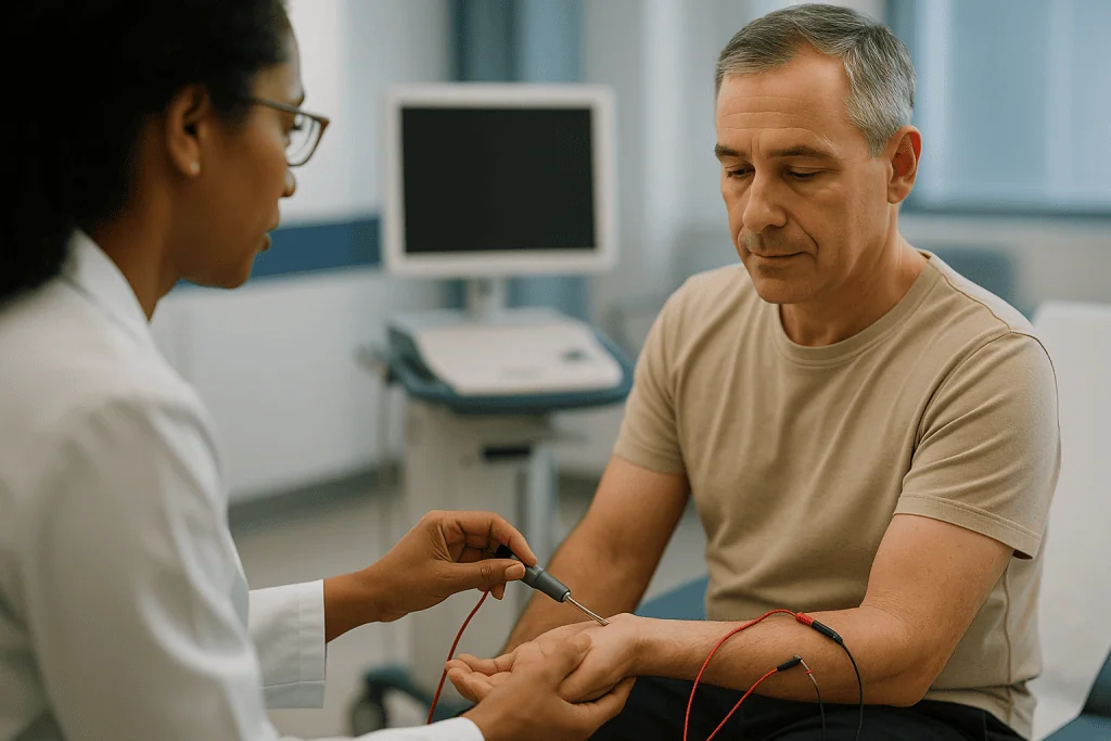
Nerve conduction studies come first in most protocols. They involve surface electrodes taped to the skin and brief electrical pulses. No needles are used. The clinician moves systematically along the nerves suspected of causing the symptoms, often testing the opposite limb for comparison. Two main measurements are taken: conduction velocity, which is the speed of the signal between two points, and amplitude, which reflects how many fibers are responding. Mayo Clinic notes that both nerve conduction studies and needle EMG are typically performed in one session.
Needle EMG follows. A thin recording needle, finer than most injection needles, is inserted into selected muscles. Nothing is injected. The clinician asks you to relax and then to contract the muscle gently while watching and listening to the signal on a screen with a speaker. Different muscles are chosen depending on which nerve roots or peripheral nerves are under suspicion.
Why do both? Because they answer complementary questions. Conduction studies are excellent for localizing a focal nerve lesion, such as compression of the median nerve at the wrist, and for identifying a widespread polyneuropathy. Needle EMG can reach muscles whose nerves are too deep to stimulate on the skin, including the muscles beside the spine supplied directly by nerve roots. It also distinguishes a nerve problem from a primary muscle disease, something conduction studies alone cannot do.
So when someone asks whether you had “the shock test or the needle test,” the honest answer is usually both, and the combination is what makes the result meaningful.
What actually happens during each test, step by step
Knowing the sequence tends to shrink the anxiety. Here is a typical visit, acknowledging that individual labs vary.
For nerve conduction studies you sit or lie on an examination couch with the limb exposed and warm; cold skin slows nerve signals and can produce falsely abnormal readings, so a lab may warm your hand or foot before starting. Electrodes are taped on. Each stimulus feels like a brief static shock or a tap; it is startling more than painful, and it stops the instant the pulse ends. A study of one limb usually involves several nerves and a few dozen pulses.
Needle EMG is quieter but more intimate. The clinician cleans the skin and slides the needle into a muscle. You will feel a pinprick and, in some muscles, a dull ache during the examination. You will be asked to relax completely, then to push gently against the clinician’s hand. The speaker crackles as motor units fire; this is normal and is how the examiner “hears” the pattern. Each muscle takes a minute or two, and the number sampled depends on the question. Mayo Clinic reports that the combined session commonly lasts around 30–60 minutes.
MRI is a different experience altogether. You lie on a motorized table that slides into a tube open at both ends. The machine knocks and buzzes loudly, so ear protection is given. You must stay still; movement blurs the image. The NHS explains that a whole MRI examination usually takes 15–90 minutes depending on how many areas are scanned. Some spine or brain protocols include a contrast dye injected into a vein to highlight inflammation or tumors.
Afterward, you can drive, work and eat normally after any of the three tests. Small bruises at needle sites are the most common aftereffect of EMG; mild soreness fades within a day or so for most people, according to Cleveland Clinic.
Nerve, root, muscle or brain: how doctors decide where to look
The choice of test is driven less by the symptom than by the pattern the clinician finds on examination. A careful history and a hands-on exam often predict the location of the fault with surprising accuracy, and testing then confirms or overturns the hypothesis.
Consider the geography of numbness. Tingling in the thumb, index and middle fingers that wakes you at night suggests the median nerve at the wrist, a peripheral nerve problem that conduction studies handle well. Numbness that runs in a stripe from the neck down the arm into one or two fingers, worsened by turning the head, suggests a nerve root in the neck; here needle EMG of the paraspinal and limb muscles and an MRI of the cervical spine are natural partners. Numbness in both feet that creeps upward in a “stocking” distribution suggests a length-dependent polyneuropathy, and the National Institute of Neurological Disorders and Stroke notes that diabetes is the most common cause of this pattern.
Weakness has its own clues. Weakness with wasting and twitching points toward nerve or motor neuron; weakness that is symmetrical and closest to the trunk, with difficulty rising from a chair, points toward muscle. Weakness with brisk reflexes, spasticity or bladder symptoms points above the nerve root, to the spinal cord or brain, where electrical tests of the limbs are often normal and MRI becomes essential.
There is also the question of time. Electrodiagnostic changes in muscle after a nerve injury develop over days to weeks, so a needle study performed very early after an acute injury may underestimate the damage, and the examiner may suggest waiting. MRI, by contrast, can show a fresh disc herniation or cord lesion immediately.
The examiner at an EMG lab is typically a neurologist or physiatrist who will often extend or shorten the planned study based on what the first few nerves reveal. That flexibility is one of the strengths of electrodiagnosis: the test adapts to you in real time.
EMG test vs MRI vs nerve conduction study: a side-by-side comparison
The table below condenses the practical differences patients ask about most. It reflects general descriptions from MedlinePlus, Mayo Clinic and the NHS rather than any single lab’s protocol.
| Feature | Nerve conduction study | Needle EMG | MRI |
|---|---|---|---|
| What it measures | Speed and size of electrical signals in peripheral nerves | Electrical activity of muscle at rest and during contraction | Detailed anatomy of brain, spinal cord, nerve roots, soft tissue |
| Best at finding | Focal nerve compression, polyneuropathy, myelin vs axon damage | Nerve root injury, motor neuron disease, primary muscle disease | Disc herniation, spinal cord lesions, tumors, inflammation, brain causes |
| Cannot show | Problems above the nerve root; muscle disease | Exact structural cause; sensory-only fibers | How well a nerve or muscle is working |
| Needles | None | Fine recording needle, nothing injected | Only if contrast dye is used |
| Sensation | Brief static-like pulses | Pinprick and dull ache in sampled muscles | Painless; loud and confined |
| Typical duration | About 30–60 minutes combined (Mayo Clinic) | About 15–90 minutes (NHS) | |
| Radiation | None | None | None |
| Main cautions | Pacemakers or stimulators: inform the lab | Blood thinners, infection risk: inform the lab | Metal implants, claustrophobia, kidney function if contrast used |
A few observations follow from this grid. Electrical tests and MRI are rarely competitors; they are more often sequential or complementary. A normal MRI with an abnormal EMG tells a clinician that the problem is functional rather than structural, while an abnormal MRI with a normal EMG raises the possibility that the imaging finding is incidental. Many adults have disc bulges on spine MRI that cause no symptoms at all, which is one reason clinicians resist ordering scans without a matching clinical picture.
Who is usually sent for electrodiagnostic testing first, and who is asked to wait
Electrodiagnostic testing, the umbrella term for NCS and EMG together, tends to lead when the examination points to the peripheral nervous system: a single nerve, a nerve root or a widespread neuropathy. Typical reasons for referral include suspected carpal tunnel syndrome, ulnar nerve compression at the elbow, foot drop, sciatica with weakness, generalized tingling in a person with diabetes, and unexplained muscle wasting. Johns Hopkins Medicine lists these among the common indications for nerve conduction studies.
People are often asked to wait in three situations. The first is timing after an acute injury: because denervation changes in muscle take time to appear, a study performed in the first days after a nerve is stretched or cut may look deceptively mild, and the examiner may schedule it for a few weeks later. The second is when symptoms are purely sensory and intermittent, such as occasional tingling with no numbness on examination; electrical tests may be normal in early, mild compression, and some clinicians prefer a trial of conservative measures before testing. The third is when a structural emergency is suspected, in which case imaging and urgent assessment take priority and electrodiagnosis can follow later.
Certain medical details change the plan rather than cancel it. Anticoagulant medicines increase bruising risk at needle sites, so tell the lab what you take; most people on blood thinners can still be studied with care, and no one should alter a prescribed medicine for the test without speaking to the prescriber. Implanted cardiac devices and spinal cord stimulators are usually compatible with conduction studies, but the lab needs to know in advance. Skin infection or lymphedema in the limb to be tested may lead the examiner to avoid needling that area.
Children and adults with cognitive impairment can be tested, though the examination may be shortened to the most informative muscles. Pregnancy is not a barrier to electrodiagnosis; carpal tunnel symptoms are common in late pregnancy and are frequently evaluated this way.
MRI or EMG for a pinched nerve: when imaging comes first
The phrase “pinched nerve” covers two very different anatomical situations, and the test that fits depends on which one is meant.
If a nerve is compressed at a joint, such as the median nerve at the wrist or the ulnar nerve at the elbow, conduction studies are the usual first test. They can show slowing precisely across the point of compression and estimate how severe it is. MRI of the wrist or elbow is reserved for unusual cases, for example when a mass is suspected or when surgery is being planned and the anatomy is unclear.
If a nerve root is compressed where it leaves the spine, MRI becomes central because it shows the disc, the bony canal and the root itself. Mayo Clinic describes MRI of the spine as the preferred imaging test for suspected disc herniation and spinal cord problems. EMG still earns its place here, since it can confirm that the root seen on the scan is the one actually malfunctioning, and it can date the injury roughly as acute or chronic by the mix of findings.
MRI moves to the front of the queue, often urgently, when the clinical picture suggests the spinal cord or brain rather than a root. Signs that raise that concern include weakness on both sides, a sensory level across the trunk, new difficulty with bladder or bowel control, stiffness and brisk reflexes, or numbness and weakness that came on suddenly over minutes. Electrical tests of the arms and legs may be entirely normal in these conditions because the fault lies upstream of the nerves being measured.
Brain MRI is also the right tool when weakness is accompanied by speech change, facial droop, double vision or loss of coordination. None of those symptoms is evaluated by EMG. The sequencing decision, in other words, is not about which test is “better” but about which part of the chain the examination has implicated.
What does an EMG test show? Reading recruitment, fibrillations and conduction block
EMG reports are written for the referring clinician, and the vocabulary can feel like a foreign language. A few terms carry most of the meaning.
Spontaneous activity is what the muscle does at rest. Healthy muscle is electrically silent apart from brief noise when the needle moves. Fibrillation potentials and positive sharp waves are small, regular discharges from individual muscle fibers that have lost their nerve connection. Their presence indicates active denervation, and their distribution across muscles tells the examiner which nerve or root is involved.
Motor unit potentials appear when you contract the muscle. A motor unit is one nerve cell plus all the muscle fibers it controls. When a nerve is injured and later repairs itself, surviving nerve cells adopt orphaned fibers, so their motor unit potentials become larger and longer. Small, brief, fragmented potentials suggest instead that muscle fibers themselves are sick, the signature of a myopathy.
Reduced recruitment is a phrase patients frequently search. Normally, as you push harder, the nervous system adds more motor units in an orderly way and the screen fills with overlapping signals. When many motor units have been lost, the few that remain fire faster than expected to compensate, yet the screen stays sparse. That pattern, few units firing rapidly, is reduced recruitment, and it points to a neurogenic process: loss of motor axons or a block in conduction. Its opposite, early recruitment, where many small units appear at low effort, points toward muscle disease.
Conduction block, a term from the nerve conduction part of the study, means the signal is large when the nerve is stimulated below a point but much smaller when stimulated above it. The fibers exist but cannot carry the impulse across that segment, usually because myelin is damaged. Conduction block is a hallmark of certain acquired demyelinating neuropathies and is potentially reversible, which is why examiners look for it carefully.
Can EMG diagnose myositis and other muscle diseases?
EMG can strongly suggest myositis, which is inflammation of muscle, but it cannot confirm the diagnosis on its own, and no reputable laboratory would claim otherwise.
In inflammatory muscle disease the needle examination often shows an “irritable myopathy”: fibrillation potentials and positive sharp waves at rest, combined with small, short, polyphasic motor unit potentials and early recruitment during contraction. That combination is distinctive because denervation-type spontaneous activity and myopathic motor units do not usually coexist. The pattern is typically most prominent in muscles close to the trunk, such as the thighs and shoulders, matching the weakness people with myositis describe when rising from a chair or lifting their arms.
EMG also helps in two indirect ways. It can direct a surgeon to a muscle that is clearly abnormal for biopsy, improving the chance that the sample is informative, while the examiner deliberately needles the opposite side so the biopsy site is not disturbed by the needle itself. And it can separate myositis from mimics: the steroid-related muscle weakness that can develop during treatment typically shows myopathic motor units without spontaneous activity, a useful distinction when a patient on long-term anti-inflammatory therapy becomes weaker.
Confirmation, however, rests on other evidence. Blood tests measure creatine kinase, an enzyme released from damaged muscle fibers, and a panel of myositis-specific antibodies. MRI of the thighs can show edema in inflamed muscle and map which groups are affected, which has made it increasingly valuable alongside electrodiagnosis. Muscle biopsy remains the reference standard in many cases, showing inflammatory cells and fiber damage under the microscope.
Other muscle diseases follow a similar logic. Inherited dystrophies and metabolic myopathies produce myopathic EMG patterns, usually without the irritability of inflammation, and are confirmed by genetic testing. Myasthenia gravis, a disorder of the neuromuscular junction, can give a normal routine EMG and requires specialized techniques such as repetitive nerve stimulation or single-fiber EMG to detect the faulty transmission. The treating neurologist chooses the sequence; EMG opens the door rather than closing the case.
What is the most common cause of abnormal EMG results?
There is no single national tally of EMG findings, so any honest answer is about patterns rather than percentages. In general electrodiagnostic laboratories, the most frequent abnormal results fall into three groups: focal entrapment neuropathies, lumbar and cervical radiculopathies, and generalized polyneuropathies.
Entrapment of the median nerve at the wrist, known as carpal tunnel syndrome, is widely regarded as the most common focal neuropathy and one of the most common reasons for referral. Conduction studies show slowing of the median nerve across the wrist while other nerves in the same hand remain normal. Mayo Clinic lists carpal tunnel syndrome among the conditions EMG and nerve conduction studies are routinely used to evaluate.
Radiculopathy, meaning a nerve root compressed or inflamed where it exits the spine, is the second large group. Here conduction studies are frequently normal because the damage is proximal, and the diagnosis rests on needle EMG findings in muscles that share a root but not a peripheral nerve, including the muscles alongside the spine.
Polyneuropathy, damage to many peripheral nerves at once, usually starting in the feet, makes up the third group. The National Institute of Neurological Disorders and Stroke estimates that more than 20 million people in the United States have some form of peripheral neuropathy and identifies diabetes as the leading cause. Other recognized causes include alcohol use, vitamin B12 deficiency, kidney disease, some chemotherapy agents, inherited disorders and autoimmune conditions.
Which group dominates depends heavily on who is referred. A hand-surgery practice sees mostly carpal tunnel; a spine clinic sees radiculopathy; a diabetes service sees polyneuropathy. What matters for an individual is not the statistic but whether the pattern of abnormalities matches the symptoms and the examination. An abnormal finding that does not fit the clinical story is treated with caution and sometimes repeated, because electrodiagnosis, like any test, has false positives, particularly in cold limbs or when technical factors interfere.
What the following days and weeks usually look like
The hours after electrodiagnostic testing are uneventful for most people. You may notice pinpoint marks or small bruises where the needle entered and a dull muscle ache similar to the day after unaccustomed exercise. Cleveland Clinic notes that this soreness typically eases within a day or two, and ordinary activity, including driving home, is fine. No medicine needs to be stopped or started because of the test itself; if you were asked to hold a blood thinner for safety, follow the instructions of the clinician who prescribes it rather than any general rule.
Results arrive on two timelines. The physician who performed the study often sees the pattern in real time and may give you a verbal impression before you leave, though many prefer to review the waveforms formally before committing. A written report usually reaches the referring clinician within days, and a follow-up appointment is where the findings are placed alongside your history, examination and any imaging. Ask at the time of testing how and when you will hear.
MRI results follow a similar path. A radiologist interprets the images and writes a report, which goes to the clinician who ordered the scan. If contrast was used, you may be advised to drink normally to help clear it; most people have no aftereffects.
What comes next depends entirely on the picture that emerges. A clear entrapment may lead to discussion of splinting, activity changes, injections or surgical options. A radiculopathy that matches an MRI finding may lead to a conversation about physiotherapy, time, or referral to a spine specialist. A polyneuropathy opens a search for the cause with blood tests. Normal results are also a result; they narrow the field and often prompt the clinician to reconsider conditions that electrical tests do not detect, such as small-fiber neuropathy or a central cause.
Repeat testing is sometimes scheduled months later to see whether a nerve is recovering, especially after trauma. The treating team decides on that interval case by case.
What people often get wrong about nerve conduction study vs EMG vs MRI
Several durable myths circulate about these tests, and clearing them up changes how people approach the appointment.
Myth one: a normal MRI means nothing is wrong with the nerves. MRI photographs structure; it cannot see a nerve that is electrically failing inside a normal-looking canal, nor a widespread polyneuropathy, nor a disorder of the neuromuscular junction. A normal spine scan in someone with foot numbness simply shifts attention elsewhere.
Myth two: an abnormal MRI explains the symptoms. Degenerative changes and disc bulges are common findings in adults who feel perfectly well. Clinicians look for a match between the level of the disc, the distribution of the symptoms and, often, the EMG pattern before attributing blame to an image.
Myth three: the needle test injects something. Nothing is injected during EMG. The needle is a recording electrode. People who dread injections are frequently surprised by how different the sensation is.
Myth four: the electrical pulses can damage the nerve or the heart. The currents used in conduction studies are small and brief, and MedlinePlus describes the test as having very low risk. Labs do take precautions with implanted devices, which is why they ask.
Myth five: EMG can diagnose everything neurological. It cannot assess the brain or spinal cord, it misses small-fiber neuropathy because those fibers are too thin to record, and it may be normal in the first days after an injury. Its value lies in the peripheral nervous system and muscle, within a window of time.
Myth six: more tests are always better. Ordering all three at once, before examination has focused the question, produces incidental findings and avoidable anxiety. Sequenced testing guided by a clinician who has examined you is both safer and more informative, and it is the approach supported by mainstream guidance from Mayo Clinic and the NHS alike.
Questions to ask your care team before and after testing
A short list of questions, asked at the right moment, turns a confusing referral into a shared plan. You do not need to ask them all; choose the ones that matter to you.
Before the test, it helps to understand the purpose. Ask which part of the nervous system is suspected and what the test is expected to confirm or rule out. Ask whether both nerve conduction studies and needle EMG are planned, and roughly how many muscles may be sampled. If you take an anticoagulant, have an implanted device, or have a skin condition in the limb, ask whether anything should change and who will give that instruction. Ask whether you should avoid lotions on the day, since creams can interfere with electrode contact, and whether to keep the limb warm on the way in.
If MRI is also proposed, ask which region will be scanned and why, whether contrast is expected, and what the radiology department needs to know about implants, prior surgery or kidney function. If enclosed spaces trouble you, raise it early; departments have practical strategies and will discuss options with the ordering clinician.
After the test, the useful questions shift. Ask whether the findings fit the symptoms you reported, and if not, what else is being considered. Ask whether the abnormality looks acute or long-standing, and whether repeat testing is planned to track recovery. Ask what the result changes in terms of the next step, and what you can safely do in the meantime with work, driving and exercise. If a report uses a phrase you do not understand, such as reduced recruitment or conduction block, ask for it to be translated into what it means for you.
Finally, ask who is coordinating the overall picture. When imaging, electrical tests and blood work come from different departments, one clinician should be holding the thread, and knowing who that is prevents results from sitting unread.
When to call your doctor
Most numbness and weakness evolves slowly and can be assessed through planned testing. Some patterns should not wait for a scheduled appointment, because they suggest the spinal cord, brain or a rapidly progressing nerve disorder.
Seek emergency care immediately for sudden weakness or numbness on one side of the body, facial droop, slurred speech, sudden confusion or loss of vision; these are features of a possible stroke, and the American Heart Association and CDC emphasize that treatment is time-critical. Call emergency services rather than driving yourself.
Seek urgent same-day assessment if numbness or weakness is accompanied by new difficulty controlling bladder or bowel, numbness in the area that would touch a saddle, or weakness in both legs; these can indicate compression of the lower spinal cord or nerve roots. Weakness that climbs from the feet upward over hours to days, especially with trouble breathing or swallowing, needs urgent evaluation for an acute inflammatory neuropathy. Weakness or numbness after a significant injury to the neck or back should also be assessed without delay.
Contact your doctor promptly, though not necessarily through an emergency department, if a limb is becoming visibly thinner, if you are dropping objects or tripping more often, if you have persistent muscle twitching with weakness, or if numbness is spreading beyond the area originally affected. People with diabetes should report any new foot numbness, since reduced sensation raises the risk of unnoticed foot injury, a point the National Institute of Neurological Disorders and Stroke highlights.
After testing itself, call the lab or your doctor if a needle site becomes increasingly red, hot, swollen or painful, if there is spreading bruising that worries you, or if you have any reaction after MRI contrast such as rash, swelling or breathing difficulty. Serious complications of these tests are rare, but they should always be reported.
Whatever the result, the interpretation and the next steps belong to the clinician who knows your full history. Use the tests to inform that conversation, not to replace it.
Frequently asked questions
Is an EMG the same thing as a nerve conduction study?
No. A nerve conduction study uses surface electrodes and brief electrical pulses to measure how fast and how strongly nerves carry signals, while needle EMG records electrical activity inside muscle. They are usually done in the same appointment and reported together, which is why the names get blended, but they answer different questions and are performed with different equipment.
What does reduced recruitment mean in EMG?
Reduced recruitment means that when you contract a muscle, fewer motor units than expected are available, and the ones that remain fire faster to compensate. It indicates loss or blockage of motor nerve fibers supplying that muscle, a neurogenic pattern. Its opposite, early recruitment, where many small units appear with little effort, points toward a disease of the muscle itself.
Can EMG diagnose myositis?
EMG can suggest myositis by showing an irritable myopathic pattern: spontaneous discharges at rest plus small, short motor unit potentials with early recruitment. It cannot confirm the diagnosis alone. Blood tests for muscle enzymes and antibodies, MRI showing muscle edema, and in many cases a muscle biopsy are needed, and the treating neurologist or rheumatologist assembles that evidence.
What is the most common cause of abnormal EMG results?
In general laboratories the most frequent abnormal findings are focal nerve entrapments, especially carpal tunnel syndrome, followed by nerve root compression in the neck or lower back, and widespread polyneuropathy. Among polyneuropathies, the National Institute of Neurological Disorders and Stroke names diabetes as the leading cause. The mix depends on which clinic is referring patients.
What does an EMG test show that an MRI cannot?
EMG shows function: whether a nerve is conducting normally, whether muscle has lost its nerve supply, and whether the pattern is neurogenic or myopathic. MRI shows anatomy but not performance. A nerve can look normal on a scan yet conduct poorly, and a disc can look alarming yet cause nothing, so the two tests complement rather than replace each other.
Should I have an MRI or EMG for a pinched nerve?
It depends on where the nerve is pinched. Compression at the wrist or elbow is usually assessed first with nerve conduction studies, which localize the slowing precisely. Compression of a nerve root at the spine usually calls for MRI, often with EMG to confirm which root is malfunctioning. Your examining clinician decides the order based on your symptoms and examination.
Does an EMG test hurt?
Most people describe the nerve conduction pulses as brief static-like shocks that are startling rather than painful. The needle portion causes a pinprick and sometimes a dull ache in the sampled muscle, lasting while the needle is in place. Nothing is injected. Mild soreness or small bruises at needle sites usually settle within a day or two.
How long does an EMG and nerve conduction study take compared with an MRI?
Mayo Clinic reports that a combined nerve conduction study and EMG session commonly lasts about 30–60 minutes, depending on how many nerves and muscles are examined. The NHS states that an MRI examination usually takes 15–90 minutes, depending on the regions scanned. Both allow you to return to normal activity immediately afterward.
Can an EMG be normal even if I have nerve damage?
Yes. EMG and nerve conduction studies do not detect small-fiber neuropathy, because those fibers are too thin to record, and they can be normal in the first days after an acute injury before denervation changes appear. They also do not assess the brain or spinal cord. A normal result narrows the search rather than ending it.
Do I need to stop my medicines before an EMG or MRI?
Do not stop or change any prescribed medicine on your own. Tell the lab about blood thinners, since they raise bruising risk at needle sites, and about any implanted devices or metal before MRI. The clinician who prescribes your medicine, together with the testing team, will decide whether any adjustment is appropriate for your situation.
References
- MedlinePlus: Electromyography (EMG) and Nerve Conduction Studies
- Cleveland Clinic: EMG (Electromyography)
- NHS: MRI scan
- NIH NINDS: Peripheral Neuropathy
This article is for general information only and is not a substitute for professional medical advice. Please consult a qualified doctor about your individual situation.

More from the Blog

Who Benefits From a Comprehensive Check-Up? How Age and Family History Shape the Test List
Most adults benefit from a periodic comprehensive check-up, but the right test list depends on age, sex, family history, and existing conditions rather than…

Before a Comprehensive Check-Up: Fasting Rules, Medical Records and Questions Worth Bringing
To prepare for a health check up, confirm which tests are booked, then follow the fasting instruction you are given, usually 8–12 hours with…
How Long a CT Scan Takes: Preparation, the Scan Itself and the Wait for Results
A CT scan itself usually takes about 10 to 20 minutes, and the moment the X-ray beam is actually on often lasts under a…
CT Coronary Angiogram Explained: What It Shows, Preparation and What Results Mean
A CT coronary angiogram is a non-invasive heart scan that combines an iodine-based contrast dye injected into an arm vein with a fast, heartbeat-timed…

Recovery After a Stereotactic Biopsy: Headache, Pin-Site Soreness, Driving and Results
Stereotactic biopsy recovery is usually short. After a breast biopsy, most people go home the same day and have bruising or soreness for a…

After a PET-CT Scan: Clearing the Tracer, Contact Precautions and When Results Arrive
After a PET-CT scan, most people go home the same day, drink extra fluids and empty their bladder often to help clear the radioactive…