Electromyography
Electromyography (EMG) is a diagnostic test that measures muscle and nerve electrical activity. It helps evaluate numbness, weakness, nerve compression, neuropathy, and neuromuscular disorders.
Quick answer
An EMG test (electromyography) records the electrical activity of muscles and measures how well nerves carry signals to them. It has two parts: a nerve conduction study using skin electrodes and brief pulses, and needle EMG using a fine needle electrode in selected muscles. Physicians use it to investigate numbness, tingling, weakness or pain and to locate nerve or muscle problems. A focused study often takes less than an hour.
What Is an EMG Test?
An EMG test, short for electromyography, is a diagnostic examination that records the electrical activity of your muscles and measures how well your nerves deliver signals to them. It helps your physician work out whether numbness, tingling, weakness, cramping or pain is coming from a nerve, from a muscle, or from the junction where the two meet. It is usually requested when a physical examination, blood tests or imaging have not fully explained your symptoms, and it is often the test that turns a vague clinical picture into a specific diagnosis.
If you are considering this test, your symptoms are probably difficult to put into words. A hand that falls asleep at night. A foot that catches on stairs. Burning in the legs that gets worse in the evening. Weakness that only shows up with certain movements, or pain that seems to travel from the neck or lower back into an arm or leg. These symptoms interfere with sleep, work, walking, grip strength and balance, and they are hard to describe precisely in a consultation. Part of the value of the test is that it does not depend on description: it measures what the nerves and muscles are actually doing.
This is the key distinction from imaging. An MRI or ultrasound shows structure: the shape of a disc, the thickness of a nerve, the anatomy of a joint. Electromyography assesses function: whether electrical signals travel through your nerves at the expected speed and strength, whether your muscles respond normally to those signals, and where along the pathway a problem sits. Structure and function do not always agree, which is why the two kinds of test are so often used together.
In clinical practice, the term EMG usually describes a group of related tests rather than a single measurement. The first is a set of nerve conduction studies, which measure how quickly and how strongly electrical impulses travel along individual nerves. The second is needle electromyography, which records electrical activity inside selected muscles using a very fine needle electrode. Depending on the question your physician needs answered, you may have one component or both, and the plan can change during the appointment as findings emerge.
One thing to be clear about from the start: EMG does not treat anything. It is a diagnostic tool. Its value lies in the decisions it makes possible, whether your symptoms are likely to improve with physiotherapy, whether a compressed nerve needs surgical evaluation, whether medication is appropriate, or whether a different line of investigation should be opened. A carefully performed and carefully interpreted EMG can change the direction of your care.
What is EMG able to show that a scan cannot?
EMG shows how your nerves and muscles are working, while a scan shows how they look, and these are genuinely different pieces of information. A nerve can appear normal on imaging while conducting signals poorly. Equally, imaging can show an abnormality, a bulging disc, a narrowed canal, that is not actually affecting nerve function at all. Many people have imaging findings of this kind without any symptoms. EMG helps determine whether what the scan shows is genuinely the cause of what you feel.
The test can also describe severity and timing in a way imaging cannot. It can suggest whether a nerve problem is mild or significant, whether it appears active and ongoing or old and settled, and, after an injury, whether there are signs that a nerve is regenerating and a muscle is receiving new nerve input. These distinctions matter when the choice is between watching and waiting, structured rehabilitation, or surgery.
What is an EMG compared with a nerve conduction study?
They are two halves of the same examination, and in most clinics they are performed in the same appointment by the same physician. A nerve conduction study measures signals travelling along a nerve, using small electrodes on the skin and brief electrical impulses. Needle EMG records electrical activity inside the muscle itself, using a fine needle electrode, with no medication injected. You may see the first part written as a nerve conduction test on a referral letter: it means the same thing.
Which parts you need depends on the clinical question. Suspected carpal tunnel syndrome may need a focused study of a few nerves at the wrist. Suspected radiculopathy, widespread neuropathy or a possible muscle disease usually needs a broader study, with more nerves measured and more muscles sampled. Your physician decides this, and adjusts it during the test based on what the early measurements show.
Who May Need an EMG Test?
An EMG test may be recommended when your symptoms suggest that nerves or muscles are not functioning normally. Those symptoms can arrive suddenly after an injury, build gradually over months, or come and go with certain positions or activities. In some people they are confined to one hand, arm, foot or leg. In others they are more widespread, affecting both sides or several limbs. Both patterns can be informative, because the distribution of symptoms is one of the first clues to where the problem sits.
Common reasons physicians request the test include numbness, tingling, burning pain, muscle weakness, muscle wasting, cramps, twitching, radiating pain from the spine, and changes in reflexes. The classic examples are familiar: tingling in the thumb, index and middle fingers raises the question of carpal tunnel syndrome at the wrist. Pain travelling from the lower back into the leg raises the question of a lumbar nerve root problem. Weakness that steadily progresses raises the question of a neuromuscular disorder that needs specialist assessment.
The diagnostic process does not start with the machine. It starts with a detailed history and a neurological examination. Expect questions about when the symptoms began, what makes them better or worse, whether they affect one side or both, and whether you have conditions that commonly affect nerves, diabetes, thyroid disease, autoimmune disease, kidney disease, vitamin deficiencies, previous chemotherapy, or prior spine surgery. The examination itself typically covers strength, sensation, reflexes, coordination, gait and muscle bulk. All of this shapes which nerves and muscles are tested.
EMG rarely works alone. MRI is helpful when a disc herniation, spinal stenosis or structural lesion is suspected. Blood tests evaluate metabolic, inflammatory and autoimmune causes. Ultrasound can assist in certain peripheral nerve assessments. What EMG contributes is the functional piece: whether the suspected abnormality is actually affecting nerve or muscle function, and how severe or active that process appears to be.
An EMG is often part of a second opinion rather than a first investigation. If you already have imaging but your symptoms and the scan findings do not fully match, EMG can clarify whether a nerve is truly affected. This matters most before spine or peripheral nerve surgery, because operating on an imaging finding that is not the real cause of symptoms helps no one. Objective functional data makes that mistake less likely.
Conditions an EMG Can Help Evaluate
EMG is used across several specialties because nerve and muscle symptoms arise from many different conditions. The test is at its most useful when the question is one of localisation: where is the problem, how extensive is it, and is it affecting nerves, muscles, or the connection between them? The main groups of conditions are worth understanding in a little detail.
Nerve compression is the most common indication. This includes carpal tunnel syndrome at the wrist, ulnar neuropathy at the elbow, peroneal nerve compression near the knee, and other entrapment neuropathies where a nerve is squeezed at a predictable anatomical point. EMG and nerve conduction studies can pinpoint the location and grade the severity of the compression, information that directly shapes decisions about splinting, injections, physiotherapy, activity changes or surgical consultation.
Radiculopathyoften described as a pinched nerve in the neck or lower back, is another frequent reason for testing. Cervical radiculopathy can cause pain, numbness or weakness in the shoulder, arm or hand; lumbar radiculopathy can cause sciatica-like pain, leg weakness or foot symptoms. EMG helps identify which nerve root is affected and whether the findings suggest active, ongoing irritation or evidence of an older, settled injury. That distinction influences whether treatment is urgent or conservative.
Peripheral neuropathydamage to nerves outside the brain and spinal cord, is a third major indication. Neuropathy may relate to diabetes, alcohol use, vitamin deficiencies, kidney disease, autoimmune disease, infections, medications, chemotherapy or hereditary factors, and in some people no cause is found. Nerve conduction studies characterise the neuropathy: whether it mainly affects the nerve’s insulating covering or the nerve fibre itself, whether sensory nerves, motor nerves or both are involved, and how widespread the process is. That characterisation narrows the list of possible causes considerably. You can read more about how peripheral nerve problems are assessed and treated on our dedicated page.
EMG is also requested for suspected neuromuscular junction disorders such as myasthenia gravis, and for muscle diseases including inflammatory myopathies and certain inherited muscle disorders. Specialised neurophysiological techniques may be added when these conditions are suspected, and the results often direct further testing, antibody studies, muscle enzymes, genetic testing, imaging, or muscle biopsy.
Finally, EMG has an important role after nerve trauma: brachial plexus injury, nerve symptoms after surgery, or recovery after a nerve has been damaged. The test can show whether a nerve is regenerating and whether a muscle is beginning to receive new nerve input. Timing matters here, because some EMG changes only become apparent once the body has had time to respond to the injury: a point your physician will consider when scheduling the study.
What diseases does an EMG rule out?
An EMG helps exclude, or make much less likely, significant damage to the large nerve fibres and to the muscles it samples, and it is often used to distinguish between conditions that look similar from the outside. Weakness and abnormal sensations can come from a compressed nerve root, an entrapped peripheral nerve, a generalised neuropathy, a neuromuscular junction disorder, an inflamed muscle or a motor neuron disease. These require very different management, and EMG is one of the main tools for telling them apart.
It is worth being honest about the limits. A normal EMG does not rule out every condition. Small-fibre neuropathies, which affect the thinnest nerve fibres, can produce burning pain with a normal standard study. Very early nerve injury may not yet show needle EMG changes. And conditions of the brain and spinal cord are generally beyond what EMG measures. A skilled physician interprets a normal result as evidence, often reassuring, sometimes redirecting, rather than as the end of the enquiry.
Is EMG better than MRI?
Neither is better; they answer different questions, and they are frequently used together. MRI shows anatomy in detail, discs, joints, nerve roots, soft tissue, but cannot say whether a structure that looks abnormal is actually disturbing nerve function. EMG measures function directly but cannot show the anatomy causing the problem. In practice, a disc bulge seen on MRI is common even in people with no symptoms at all; an EMG showing that the corresponding nerve root is conducting normally can spare a patient an operation that would not have helped. The reverse is also true: a clearly abnormal EMG can give weight to an imaging finding that might otherwise have been dismissed.
How an EMG Test Is Performed
EMG is usually performed in an outpatient neurophysiology or neurology setting. There is no sedation, no hospital stay and no meaningful recovery period for most patients. A typical appointment moves through a predictable sequence:
- The physician reviews your symptoms, history, medications and any previous imaging or test reports.
- A brief focused examination confirms which nerves and muscles should be studied.
- Nerve conduction studies are performed first, using surface electrodes and brief electrical impulses.
- If needed, needle EMG follows, sampling selected muscles at rest and during gentle contraction.
- The physician may extend or adjust the study based on what the early measurements show.
- Findings are summarised, and a formal report is prepared for your treating physician.
Preparing for the test
Before the study, your care team reviews your medical background in detail. Tell your physician if you take blood-thinning medication, have a bleeding disorder, have a pacemaker or another implanted electrical device, have lymphoedema, or have an active skin infection in the area to be tested. None of these automatically prevents an EMG, but each can influence how the test is planned, which nerves are stimulated, which muscles are sampled, and what precautions are taken.
On the day of the study, keep the skin clean and avoid lotions, oils or heavy creams, because these interfere with surface electrode contact. Wear comfortable clothing that allows access to the arm, leg, neck, back or shoulder region, depending on where your symptoms are. In most cases you can eat normally and continue your usual medications unless your physician has given you different instructions.
Temperature matters more than most people expect. Cool skin slows nerve conduction and can make a healthy nerve look borderline, so the team may warm your hands or feet before measuring: a small step that noticeably improves accuracy. It also helps to bring a list of your current medications, any previous EMG or nerve conduction reports, and recent imaging, so the physician can plan the study around what has already been examined rather than starting from zero.
The nerve conduction study
The nerve conduction portion begins with small electrodes placed on the skin. One electrode records the response of the nerve or muscle; another delivers a brief electrical impulse to stimulate the nerve. Patients describe the sensation as a quick tap, a pulse, or a mild shock. It can be startling the first time and momentarily uncomfortable, but each impulse is brief and carefully controlled, and the intensity is adjusted to what is needed for a clear recording.
The equipment measures two main things: how fast the signal travels along the nerve, and how strong the response is when it arrives. A nerve that is compressed, damaged or affected by neuropathy produces signals that are slower, smaller, delayed or absent. The physician compares your measurements against expected values and against the pattern of your symptoms, and will often test the same nerve on both sides of the body, because your own unaffected side is a useful benchmark.
Depending on the question, the physician may also record late responses such as F-waves and H-reflexes. These signals travel up towards the spinal cord and back again, so they carry information about the innermost portion of the nerve pathway, the segment closest to the spine, which routine conduction measurements along the arm or leg cannot reach. They are particularly helpful when a nerve root problem or an early generalised neuropathy is suspected.
Needle EMG
If needle EMG is needed, the physician inserts a very fine sterile needle electrode into selected muscles. Nothing is injected: the needle is a recording device, not a syringe. It listens to the muscle’s electrical activity in two states: at complete rest, when a healthy muscle should be electrically quiet, and during gentle contraction, when the pattern of activity shows how well the nerve is driving the muscle. You may be asked to lift your foot, bend your wrist, straighten your arm or tense a specific muscle in a controlled way while the physician watches and listens to the signal.
The number of muscles tested depends entirely on the clinical question. Suspected carpal tunnel syndrome may need only a focused sample. Suspected radiculopathy or a widespread neuropathy usually needs several muscles across a limb, because the pattern across muscles, which are affected and which are spared, is what localises the problem.
When a neuromuscular junction disorder such as myasthenia gravis is suspected, the physician may add repetitive nerve stimulation, in which a nerve is stimulated several times in quick succession to see whether the muscle response holds steady or fades. In selected cases, single-fibre EMG, a highly sensitive technique that records from individual muscle fibres, is used to examine the transmission between nerve and muscle in fine detail.
Does an EMG test hurt?
An EMG test causes brief discomfort for most people, but it is generally well tolerated and no anaesthesia is needed. The nerve conduction impulses feel like short, sharp taps, over in a fraction of a second, and easier to tolerate once the first few have passed. The needle portion can cause a brief sting on insertion and a cramping or aching sensation while the muscle is sampled; the discomfort stops when the needle is withdrawn from each site. Experience varies from person to person and from muscle to muscle, some sites are more sensitive than others, and you can tell the physician at any point if you need a pause. Some muscle soreness afterwards is normal and settles quickly.
How long does an electromyography take?
A focused electromyography study may take less than an hour, while more complex evaluations take longer. The main drivers of duration are how many limbs need to be assessed, how many diagnostic questions are on the table, and what the early findings show: a study can reasonably expand mid-appointment if the first measurements point somewhere unexpected. If your referral involves several possible diagnoses, or comparison of both sides of the body, plan for a longer session and allow additional time for registration and discussion afterwards.
The equipment and what it records
Modern EMG systems combine sensitive amplifiers, surface electrodes, needle electrodes, digital signal processing and specialised software. They convert the very small electrical signals produced by nerves and muscles into waveforms on a screen and, during needle EMG, into sound, experienced physicians read both the shape and the sound of the signal. From these recordings the physician evaluates conduction speed, response amplitude, spontaneous activity in resting muscle, recruitment patterns during contraction, and other features that point towards specific diagnoses.
What the technology cannot do is think. EMG is a physician-directed examination, not a machine-generated printout. The specialist decides which nerves and muscles to test, adapts the study in real time as findings emerge, and integrates the results with your symptoms, examination and history. This clinical judgment matters most in exactly the situations that are hardest to untangle: complex cases, previous operations, mixed nerve and spine findings, or symptoms that refuse to follow a textbook pattern.
After the test
Most patients return to normal activities immediately. Mild soreness, tenderness or small bruises can appear at needle sites and usually resolve quickly. Serious complications are uncommon when the test is performed with appropriate technique and patient screening; if you take blood thinners or have other relevant conditions, your physician will explain any specific precautions that apply to you.
Results may be discussed shortly after the test or delivered in a formal report, depending on the clinical workflow and whether other consultations are pending. The report typically lists the nerves and muscles tested, describes any abnormalities, and gives the likely diagnostic interpretation. Your treating physician then uses it to decide the next step, conservative treatment, medication, rehabilitation, further imaging, injections, surgical evaluation or additional laboratory testing.
Recovery After an EMG Test
There is no formal recovery process after an EMG: the timeline below reflects what most patients can expect in the days and weeks that follow.
| Time Period | What Patients Can Expect |
|---|---|
| Day 1 | You may return to routine activities immediately unless your physician advises otherwise. Mild soreness, brief tenderness or small bruising may occur at needle sites. |
| First Week | Any minor discomfort usually improves. Your physician may review the EMG report with you and discuss how it relates to your symptoms and examination findings. |
| First Month | Treatment planning may begin or be adjusted based on the results. This may include rehabilitation, medication, further imaging, specialist consultation or surgical evaluation where appropriate. |
| Longer Term | Follow-up depends on the diagnosis. Some patients need monitoring of nerve recovery; others begin a structured treatment plan for compression, neuropathy or neuromuscular disease. |
Understanding Your EMG Results
An EMG report is a technical document, but its purpose is practical: to answer the question your physician asked when requesting the study. A good report does more than label findings normal or abnormal: it describes where any problem sits, how severe it appears, and whether it looks active or long-standing, so that your treating physician can turn it into a concrete next step.
What happens when EMG is positive?
A positive EMG test result means the study found an abnormality, and the report will say what kind, where, and how significant it appears. From there, the path depends entirely on the diagnosis. A clearly localised nerve compression might lead to splinting, injections or a surgical opinion. A generalised neuropathy usually triggers a search for the underlying cause through blood tests and metabolic assessment. Findings suggesting a neuromuscular disorder lead to specialist referral and further targeted testing. An abnormal result is the beginning of a plan, not a verdict: it tells your medical team what they are dealing with, which is precisely what makes effective treatment possible.
What does a normal EMG mean?
A normal EMG is genuinely useful information, not a wasted test. It makes significant damage to the large nerve fibres and sampled muscles much less likely, which can rule out some of the more worrying explanations for your symptoms and redirect attention towards other causes, joint disease, vascular problems, central nervous system conditions or pain syndromes. Two caveats apply. Small-fibre neuropathy can cause real symptoms with a normal standard study, and testing performed very early after a nerve injury may precede the changes the needle examination looks for. Your physician weighs both when interpreting a normal result against a symptomatic patient.
Why Acting Early Matters
Many people wait when nerve symptoms begin, especially when numbness or weakness comes and goes. That is understandable, but persistent or progressive nerve and muscle symptoms deserve careful evaluation, because several of the conditions EMG detects respond best when identified before nerve damage becomes advanced.
The pattern is consistent across conditions. A compressed nerve that initially causes intermittent tingling can progress to constant numbness, weakness and muscle wasting. In carpal tunnel syndrome, delayed diagnosis allows hand weakness and loss of fine motor control to develop. In cervical or lumbar radiculopathy, untreated compression can affect walking, grip strength and daily function. In peripheral neuropathy, identifying a treatable contributor, diabetes control, a vitamin deficiency, a medication effect, autoimmune activity, gives the best chance of slowing progression.
Some neuromuscular disorders also benefit from timely specialist evaluation, because early treatment can influence symptom control and long-term planning. EMG earns its place here by distinguishing between conditions that look alike on the surface but need entirely different management: muscle fatigue, weakness and abnormal reflexes can each arise from nerve root compression, generalised neuropathy, neuromuscular junction disease, muscle inflammation or motor neuron disease. Clarifying which one is present reduces uncertainty and prevents treatments aimed at the wrong target.
Delay carries costs beyond the diagnosis itself. Pain can become chronic. Compensation patterns strain other muscles and joints. Mobility can decline. And a nerve injured for long enough may not recover completely even after the underlying cause is addressed. Not every symptom signals a serious condition, but physicians generally treat progressive weakness, loss of balance, foot drop, wasting of the hand muscles, severe radiating pain, or numbness on both sides as findings that justify earlier rather than later assessment.
Benefits of Electromyography
The practical value of the test lies in the functional information it adds, information no other routine investigation provides in the same way.
| Benefit | What It Means for You |
|---|---|
| Clarifies the source of symptoms | Helps determine whether numbness, tingling, pain or weakness is more likely related to nerve compression, neuropathy, muscle disease or another neuromuscular condition. |
| Localises nerve problems | Can identify whether the issue is at the wrist, elbow, neck, lower back, a peripheral nerve or another location: the foundation of accurate treatment planning. |
| Assesses severity and activity | Provides information about how significantly a nerve or muscle is affected and whether findings suggest ongoing irritation, chronic change or recovery. |
| Supports treatment decisions | May guide choices such as physiotherapy, medication, injections, surgical consultation, lifestyle changes or further diagnostic testing. |
| Adds value when imaging is unclear | Helps determine whether an abnormality seen on MRI or other imaging is actually affecting nerve function and matches your symptoms. |
| Useful for second opinions | Provides objective functional data that helps a specialist team review your diagnosis and recommend an appropriate care pathway. |
Factors That Influence How Useful the Results Are
A good EMG result is not simply a normal or abnormal report. The most useful result is one that answers the right clinical question and clarifies the next decision in your care. Several factors determine whether the study achieves that.
Timing. After an acute nerve injury, certain EMG changes take time to appear. A study performed very early may still yield useful nerve conduction information, but some needle EMG findings become clearer later. Your physician sets the timing based on whether your symptoms are sudden, progressive, traumatic or long-standing, and sometimes a repeat study weeks later tells the fuller story.
Clinical accuracy. The study must be planned around your actual symptoms and examination findings. A focused study suits suspected carpal tunnel syndrome; leg weakness, suspected neuropathy or possible motor neuron disease needs a broader one. Testing the wrong nerves and muscles produces a technically flawless report that answers nothing.
Patient factors. Age, body temperature, diabetes, previous surgery, old injuries, swelling, medications and other medical conditions can all influence nerve conduction measurements. Cold limbs, for instance, slow conduction and can mimic abnormality. This is why EMG interpretation requires clinical context rather than isolated numbers.
Technical quality. Accurate electrode placement, appropriate stimulation, careful muscle selection and clean signal recording all contribute to reliable results. Modern neurophysiology equipment captures detailed signals, but reducing artefacts and reading the patterns correctly still depends on the person holding the electrode.
Integration with other findings. This is usually where the meaningful conclusion emerges. EMG may confirm a diagnosis, narrow the possibilities, or show that your symptoms are not explained by the suspected condition. A normal study that excludes significant nerve damage and redirects attention elsewhere can be exactly as valuable as an abnormal one.
Ultimately, the outcome depends on what the EMG reveals and how that information is used. A treatable nerve compression identified in time can be managed before function is lost. A generalised neuropathy prompts a systematic search for causes. A suspected neuromuscular disorder reaches specialist hands earlier and with greater clarity. The test is a means; the decision it enables is the end.
How an EMG Fits Into Care at Acibadem
Patients rarely need an EMG in isolation. More often they arrive with prior scans, incomplete reports, persistent symptoms or conflicting recommendations, and what they need is a reliable diagnosis and coordinated guidance. In that setting, the quality of interpretation and the organisation around the test matter as much as the test itself.
At Acibadem, EMG is performed within a multidisciplinary clinical environment. Depending on the suspected condition, neurology, neurosurgery, orthopaedics, physical medicine and rehabilitation, endocrinology, rheumatology, oncology and pain medicine teams may all contribute. For a patient with complex symptoms, previous operations, a cancer history, autoimmune disease, diabetes-related complications or possible neuromuscular disease, this coordination means the EMG findings are read in the full medical context rather than as a standalone report.
In practice, that looks like this: for spine and peripheral nerve conditions, EMG findings are reviewed alongside MRI and other imaging to establish whether a compression is functionally significant. For suspected neuropathy, physicians combine the study with laboratory testing, metabolic evaluation, medication history and neurological examination. For suspected neuromuscular disorders, additional specialised testing is arranged when clinically necessary. Sometimes earlier tests are repeated when their quality, timing or completeness leaves a question open, so that decisions rest on current, directly comparable measurements: a separate guide describes what happens between your last test and the start of treatment.
The EMG result is never treated as the end of the process; it informs the next step. Some patients need reassurance and conservative management. Others need physiotherapy, medication, ergonomic adjustments, metabolic optimisation, injection therapy, surgical evaluation or further neurological testing. Where a second opinion is the purpose of the visit, Acibadem physicians review existing records to establish whether an EMG is needed at all, whether previous findings should be repeated or expanded, and how the results bear on the decision in front of you: a review that carries particular weight before spine surgery, carpal tunnel surgery, nerve decompression procedures or complex neuromuscular treatment decisions.
Moving Forward with a Clearer Diagnosis
Living with unexplained numbness, weakness, tingling or radiating pain is unsettling, and the questions it raises are hard ones. Is this a spine problem? Is a nerve compressed? Is neuropathy developing? Would surgery help, or would rehabilitation be the better path?
Electromyography helps answer these questions by measuring how your nerves and muscles are actually functioning. It identifies patterns that imaging alone cannot show, distinguishes between conditions that feel identical from the inside, and gives your physician the functional evidence needed to choose the right next step. For many patients, the real value of an EMG is not just finding an abnormality: it is replacing uncertainty with a structured, specific plan.
Preparation
- Your doctor may ask about medications, bleeding disorders, pacemakers, or implanted devices before the test. Avoid applying lotions, oils, or creams to the skin on the day of electromyography. Bring previous imaging, blood tests, and nerve study results if available.
Aftercare
- Most patients can return to daily activities immediately after electromyography. Mild muscle tenderness, bruising, or tingling may occur at needle insertion sites and usually resolves quickly. Contact your doctor if you develop increasing pain, swelling, redness, or fever.
Frequently Asked Questions
What affects the cost of electromyography?
The final cost depends on whether EMG is performed alone or with nerve conduction studies, how extensive the assessment needs to be, the specialist and hospital setting, and whether consultation, reporting, translation, or follow-up are included.
How can I get a personalised EMG quote from Acibadem?
You can request a free consultation by sharing your symptoms, previous test results, medical reports, and any referral notes. The medical team can review your case and provide a personalised plan and quote based on the recommended diagnostic pathway.
Is EMG usually included in a package for international patients?
It may be offered as part of a diagnostic package that can include neurology consultation, EMG or nerve conduction studies, report preparation, and patient coordination. Inclusions should always be confirmed before travel.
Will I need nerve conduction studies as well as EMG?
Many patients have both tests because they provide complementary information about nerves and muscles. A specialist decides which tests are appropriate after reviewing your symptoms and examination findings.
Can the EMG result change the overall treatment cost?
Yes. EMG is a diagnostic test, and its findings may indicate whether further imaging, laboratory tests, medication, physiotherapy, injections, or surgery should be considered. This can affect the overall care plan and cost.
Is this comparison medical or financial advice?
No. This information is general and educational. Diagnosis, test selection, and cost estimates should be discussed with a qualified specialist, and a personalised quote is recommended before making travel or treatment decisions.
Medically reviewed by the Acıbadem International Medical Board September 1, 2026
See our medical review board →
Update history
- PublishedJune 8, 2026
- Medical review approvedSeptember 1, 2026
- Last content updateSeptember 1, 2026
References1
- Electromyography medlineplus.gov
Trusted care for international patients
From the Acibadem Blog
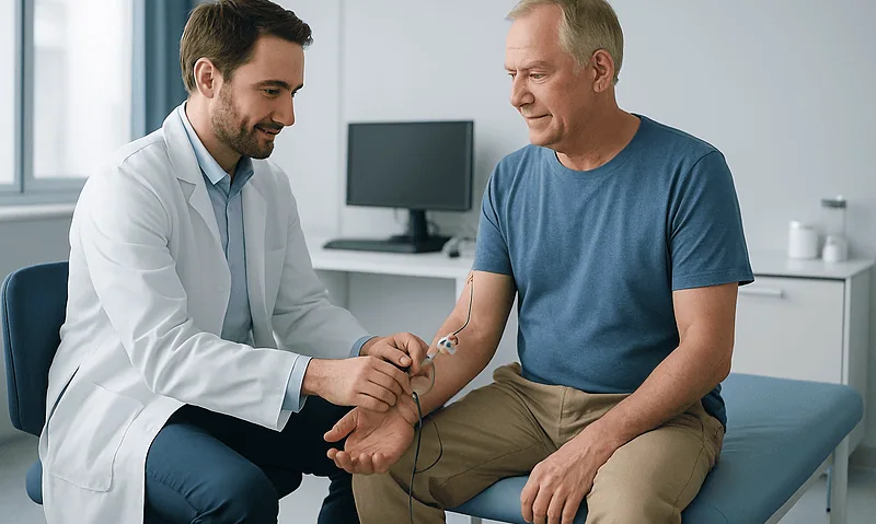
What Happens During an EMG Test? Nerve Conduction Study, Needle Electrodes and Timing
During an EMG test, a clinician first performs a nerve conduction study, placing sticky surface electrodes on the skin and delivering brief electrical pulses…

EMG Test Preparation: Lotions, Pacemakers, Blood Thinners and What to Wear on the Day
To prepare for an EMG test, arrive with clean, lotion-free skin, wear loose clothing that lets the examiner reach the arms and legs, and…