

What Happens During an EMG Test? Nerve Conduction Study, Needle Electrodes and Timing
Key Takeaways
- An EMG appointment almost always has two parts: surface-electrode nerve conduction testing first, then a solid needle electrode in selected muscles that records but never injects.
- Mayo Clinic puts a typical study at about 30 to 60 minutes, with combined studies stretching toward 90 minutes when many nerves and muscles are sampled.
- Cold skin slows nerve conduction enough to mimic disease, so warming the limb before measurement is a standard part of the test rather than a delay.
- Lotion, oil or sunscreen on the skin changes surface measurements, which is why labs ask you to shower and skip moisturizer on the day.
- Radiculopathy from a pinched spinal nerve root often shows a normal nerve conduction study and an abnormal needle exam, because the compression sits upstream of where surface tests can reach.
- Small fiber neuropathy and many pain conditions test normal on EMG, so a normal result narrows the search without ending it.
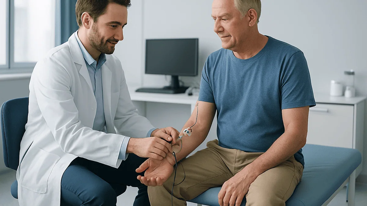
During an EMG test, a clinician first performs a nerve conduction study, placing sticky surface electrodes on the skin and delivering brief electrical pulses to measure how fast and how strongly nerves carry signals. Then a thin, solid needle electrode is inserted into selected muscles to record their electrical activity at rest and during gentle contraction. Nothing is injected. The combined study usually takes about 30 to 90 minutes, and a neurologist interprets the results.
The referral letter says ‘EMG/NCS’, the appointment is at eight in the morning, and the person holding it has spent the evening searching one question: what happens during an EMG test? The tingling in two fingers has been there for months. Now someone is proposing needles in the forearm, and the internet is split between ‘barely noticed it’ and ‘worst hour of my life’.
Both accounts describe the same procedure, which tells you how much depends on expectation. An electromyography (EMG) test measures the electrical chatter of muscles, and it almost always travels with a nerve conduction study, which measures how well nerves carry signals. Together they answer a question that a scan cannot: is the wiring or the motor at fault?
This explainer walks through the room, the electrodes, the small shocks, the needle, the clock and the report, with the evidence behind each step and none of the folklore.
What happens during an EMG test, step by step
You will be asked to sit or lie on an examination table with the arm or leg exposed. The clinician, often a neurologist or a physiatrist (a physician specializing in physical medicine and rehabilitation), or a trained technologist working alongside one, begins by checking skin temperature, because cold slows nerve signals and can mimic disease. A warm pack or a few minutes under a heated blanket is routine, not a sign that something is wrong.
The first half is the nerve conduction study. Small sticky pads go over a muscle and along the path of a nerve. A handheld stimulator delivers brief electrical pulses, each lasting a fraction of a second, and a computer records how long the signal takes to arrive and how large it is. The stimulator moves to several spots, and the intensity climbs step by step until the response stops getting bigger. That maximal response is the point of the exercise.
Then comes the needle portion, the electromyography itself. A fine, solid needle electrode, similar in thickness to an acupuncture needle, slides into a muscle. Nothing is injected. A speaker crackles as the muscle fibers fire, and a screen draws the waveforms. You will be asked to relax completely, then to push gently against the examiner’s hand, then to push harder. The needle comes out, the next muscle is chosen, and the cycle repeats.
The order matters. Nerve conduction data tell the examiner which muscles are worth sampling, so the needle map is drawn in real time rather than from a fixed list. That is why two people with the same referral can have very different numbers of needle sites, and why an experienced examiner sometimes stops early because the answer is already clear.
Nerve conduction study vs EMG: what is the difference?
People use ‘EMG’ as shorthand for the whole appointment, but the two halves test different things and feel different.
A nerve conduction study, sometimes written NCS or NCV (for nerve conduction velocity), measures the nerve alone. Think of it as a timing gate on a highway: a pulse enters at one point, a sensor records when it arrives at another, and the distance between them divided by the delay gives the speed. Healthy nerve fibers conduct faster than highway traffic, and a nerve squeezed at the wrist or elbow shows up as a delay at exactly that segment. The size of the arriving signal, the amplitude, tells a second story: how many nerve fibers are still carrying traffic.
Electromyography measures the muscle. Muscles fire only when a nerve tells them to, so a muscle that crackles when it should be silent, or that recruits too few fibers when asked to work, is a readout of both its own health and that of its nerve supply. The needle electrode sees changes that surface pads cannot, including the tiny spontaneous discharges that appear when muscle fibers have lost their nerve connection.
Most labs run the nerve conduction study first because its numbers are quantitative and reproducible, while the needle exam is interpretive and guided by what the first half found. According to the Mayo Clinic, both are usually performed in the same visit and interpreted together, which is why the report you eventually see reads as a single opinion rather than two separate results.
One practical consequence: if you are told you are having ‘just a nerve conduction study’, there may be no needle at all. If you are told ‘an EMG’, ask whether both parts are planned. The answer shapes what the hour will feel like.
What does the needle electrode actually record?
The needle listens at three moments, and each one answers a different question.
First, insertion. A healthy muscle produces a short burst of electrical noise as the needle passes through it, then falls quiet. If the burst is prolonged, or the muscle keeps firing on its own, that is called abnormal spontaneous activity, and it is one of the clearest signs that muscle fibers have lost their nerve input.
Second, rest. You will be asked to let the limb go completely limp, which is harder than it sounds when someone is holding a needle in it. Examiners have tricks: they may position the limb so gravity does the relaxing, or ask you to tense the opposite muscle. In a normal muscle the speaker goes silent. Fibrillation potentials, which are tiny rhythmic discharges from individual muscle fibers, and positive sharp waves, a related pattern, are the classic findings of denervation, meaning the nerve fibers to that muscle have been damaged. These changes take time to appear after an injury, which is one reason a study performed very early can look normal.
Third, contraction. During a gentle push the examiner watches the shape and size of motor unit potentials, the combined electrical signature of one nerve fiber and all the muscle fibers it controls. Large, long, complex units suggest a nerve problem in which surviving fibers have adopted orphaned muscle fibers. Small, short, crowded units point toward a disease of the muscle itself, a myopathy. Then you push harder, and the examiner judges recruitment: whether the muscle brings in extra units smoothly or leaves gaps because units are missing.
None of this is guesswork, but all of it is interpretation. The waveforms are read live, in context, by a physician who also has your history and examination in mind. Cleveland Clinic notes that the same equipment is used to assess disorders of muscle, of nerve and of the junction between them.
How long does an EMG take?
Plan for an hour, and do not be surprised if it is shorter. Mayo Clinic puts a typical study at about 30 to 60 minutes, and Cleveland Clinic gives a range that stretches toward 90 minutes when both the nerve conduction and needle portions are extensive. A single-nerve question, such as a suspected pinched nerve at the wrist, sits at the short end. A whole-body question, such as unexplained weakness in both arms and legs, sits at the long end because more nerves and more muscles have to be sampled.
Several things stretch the clock that have nothing to do with severity. Cold hands or feet must be warmed before the numbers mean anything, and a limb that arrives chilled from a winter parking lot adds noticeably to the visit on its own. Difficulty relaxing prolongs the resting portion of the needle exam. Comparison with the unaffected side, which examiners often do to establish your personal baseline, doubles some measurements.
Other things shorten it. When the referral question is narrow and the first few measurements are clearly normal or clearly abnormal, an experienced examiner may stop, because more data would not change the report. That is efficiency, not corner-cutting.
Build in time on both ends. Check-in and a brief history and physical examination come first; the examining physician needs to know where the symptoms are before deciding where to look. Afterward there is usually a short conversation, though not always a result. Most people walk out and drive home, and no sedation is involved, so no one needs to accompany you unless you prefer company. Johns Hopkins notes that fasting is generally not required, so eat and drink as usual before you arrive, unless your own instructions say otherwise.
How painful is an EMG nerve test? The honest EMG test pain level
Honest answer: it is uncomfortable, most people describe it as tolerable, and it is briefer than the anticipation.
The nerve conduction pulses feel like the static shock from a carpeted floor, repeated. Cleveland Clinic uses almost exactly that comparison. Each pulse lasts a fraction of a second, the muscle jumps involuntarily, and the sensation is over before you have decided how to react. The pulses build in intensity, so the last few at each site are the ones people remember. Anxiety amplifies them; a slow exhale timed to the click of the stimulator genuinely helps.
The needle portion is where reviews diverge. Insertion feels like a quick pinprick or a firm pinch. What follows, a dull ache while the needle sits in the muscle and is repositioned, is the part people find unpleasant, and it varies by muscle. Small muscles of the hand and foot, and muscles close to bone, tend to be more sensitive than the fleshy bulk of the thigh or upper arm. Mayo Clinic acknowledges that you may feel some pain or discomfort during needle insertion and that it should ease once the needle is removed.
Guarding makes it worse: tensing the muscle around the needle. Telling the examiner where it hurts makes it better, because they can often adjust the angle, and asking how many muscles are planned lets you count down. Numbing cream is rarely used, since it would not reach the muscle and could alter surface measurements, and needle sites are usually kept to what the clinical question requires.
A useful reframing: the needle records, it does not inject. There is no medicine going in and no fluid stretching the tissue, which is why the ache fades quickly. Soreness afterward is common and mild; the recovery section covers it.
What should you not do before an EMG?
The list is short, and most of it is about your skin.
Do not apply lotion, oil, sunscreen or powder to the arms or legs on the day of the test. Surface electrodes need clean, dry skin to make a reliable electrical contact, and a greasy film changes the numbers. Mayo Clinic suggests showering shortly before the appointment specifically to remove natural oils. Skip the moisturizer afterward.
Do not smoke or drink caffeinated beverages in the hours before the test if your lab asks you to abstain. Johns Hopkins notes that cigarettes and caffeinated drinks may be restricted for about two to three hours beforehand at some centers, because stimulants can affect muscle activity. Not every lab requires this, so follow your own instructions.
Do not stop, skip or double any prescribed medicine on your own. Some medicines matter to the examiner: blood thinners raise the chance of bruising at needle sites, and Mayo Clinic advises telling the team about them, though the test can usually still be done. Muscle relaxants and medicines for myasthenia gravis, a condition in which nerve signals fail to reach muscle properly, can alter results, and the neurologist may give specific instructions about timing. Those instructions come from the prescribing clinician and the testing physician, never from an article.
Do not forget to mention implanted devices. A pacemaker or implantable defibrillator is not an automatic barrier, but the team needs to know so they can choose where and how to stimulate, according to Cleveland Clinic. The same goes for a bleeding disorder, lymphedema in the limb being tested, or a skin infection at a likely needle site.
Do wear loose clothing that can be pushed above the elbow or knee, or expect a gown. Do eat normally. Do arrive warm; cold hands mean a slower start.
Who is usually referred for an EMG, and who is asked to wait
The referral almost always follows a pattern of symptoms that a clinical exam alone cannot localize: numbness or tingling in a distribution that follows one nerve, such as the thumb and first two fingers; weakness without pain; muscle wasting; cramps, twitching or fasciculations, which are visible ripples under the skin; or pain radiating from the neck or low back into a limb. MedlinePlus lists conditions commonly evaluated this way, including carpal tunnel syndrome, peripheral neuropathy, radiculopathy from a pinched spinal nerve root, muscular dystrophy, myasthenia gravis and amyotrophic lateral sclerosis, or ALS.
The test is also used to plan and to monitor. Surgeons may want objective evidence of nerve compression before operating on a wrist or elbow. Neurologists may repeat a study months apart to see whether a nerve is recovering after injury.
Some people are asked to wait, and the reasons are practical rather than alarming. After an acute nerve injury, the needle findings that confirm damage take time to develop, so a study done within days can miss what a later one would show; the neurologist chooses the timing. An active skin infection over the muscles of interest usually postpones needle sampling until it clears. Significant swelling from lymphedema in the limb may shift the plan to the other side or to surface recordings only. A recent change to anticoagulant therapy may prompt a conversation with the prescriber first.
A few people are steered toward other tests instead. When the question is clearly about the brain or spinal cord rather than the peripheral nerves, imaging or other studies may be more useful. When symptoms are purely sensory and fleeting, the referring doctor may decide the test would not change management. Those are judgment calls, and they sit with the treating team.
What happens during an EMG test for carpal tunnel, sciatica or neuropathy
The choreography changes with the question, and it helps to know which version you are booked for.
For suspected carpal tunnel syndrome, compression of the median nerve at the wrist, most of the appointment is nerve conduction. Electrodes sit over the palm and fingers while the stimulator works along the forearm and wrist. The examiner is looking for a delay specifically across the wrist compared with the segment above it, and often compares the median nerve with the neighboring ulnar nerve in the same hand as a built-in control. Needle sampling, when done, is usually limited to a thumb muscle and a few forearm muscles to check whether the compression has begun to affect muscle. The NHS lists nerve conduction tests among the investigations used when carpal tunnel is suspected.
For sciatica-type symptoms or a suspected pinched nerve in the neck, the emphasis flips toward the needle. Nerve conduction is often normal in radiculopathy because the compression is close to the spine, upstream of where surface tests can reach. So the examiner samples muscles supplied by different spinal levels, sometimes including the paraspinal muscles beside the spine, and asks which levels show denervation. Expect more needle sites than in a wrist study.
For a generalized neuropathy, the study is broad by design. Several nerves in the arms and legs are tested, usually starting with the legs because polyneuropathy affects the longest nerves first. Sensory and motor responses are compared, and the pattern, slowed speed versus shrunken amplitude, separates nerves whose insulation is damaged from nerves whose fibers are lost. The NIH describes this distinction as part of how the underlying cause is narrowed down.
Whatever the target, your job stays the same throughout: stay warm, relax on request, push when asked, and say so if something hurts more than expected.
EMG and nerve conduction study side by side
Seen together, the two halves of the appointment stop looking like one long ordeal and start looking like a pair of complementary measurements.
| Feature | Nerve conduction study | Needle EMG |
|---|---|---|
| What it measures | Speed and size of signals traveling along a nerve | Electrical activity of muscle fibers at rest and during effort |
| Electrodes | Adhesive pads on the skin plus a handheld stimulator | Fine solid needle inserted into muscle; nothing injected |
| Sensation | Brief static-like shocks that make the muscle twitch | Pinprick on entry, dull ache while recording |
| Best at detecting | Entrapments such as carpal tunnel; widespread neuropathy | Radiculopathy, motor neuron disease, myopathy |
| Output | Numbers compared with laboratory normal ranges | Waveforms and sounds interpreted live by the physician |
| Place in the visit | Usually first; length depends on nerves tested | Usually second; length depends on muscles sampled |
| Afterward | Nothing to care for | Possible small bruises and soreness for a few days |
Two rows deserve a second look. The ‘best at detecting’ row explains why some people leave with almost no needle sites and others with many: the examiner is choosing the tool that can actually see the problem. A nerve pinched at the wrist lives within reach of surface stimulation, so pads do most of the work. A nerve pinched where it leaves the spine does not, so the needle has to look at the muscles downstream for indirect evidence.
The ‘output’ row explains why results are not instant. Nerve conduction numbers land in the computer immediately and are compared with norms adjusted for age and height. Needle findings are judgments about waveform shape and firing pattern, made by a physician who then has to reconcile them with the first half and with your history. That synthesis is the report, and it is what your referring doctor is waiting for. Neither half stands alone; Mayo Clinic describes them as commonly performed and interpreted together.
Do you get EMG results immediately?
Sometimes a first impression, rarely a full answer.
The physician performing the study sees the nerve conduction numbers as they appear and hears the needle findings in real time, so they often know the broad shape of the result before you have put your shoes back on. Many will share it: ‘the median nerve is slowed at the wrist’, or ‘I did not find evidence of nerve damage in the muscles I tested’. Some prefer not to, especially when the referring doctor has asked to deliver results, or when the findings are subtle and need to be weighed against imaging and blood tests.
The formal report takes longer. Mayo Clinic describes the neurologist interpreting the results and preparing a written report that goes to the doctor who ordered the test, who then discusses it with you at a follow-up visit. Cleveland Clinic gives a similar picture. Ask at check-out how and when the report will reach you, and whether you should book the follow-up now.
The report contains tables of conduction values for each nerve tested, with normal ranges beside them; a summary of needle findings for each muscle; and an impression paragraph that pulls the two together. The impression is written for your doctor, so expect terms like ‘demyelinating’, meaning the nerve’s insulating layer is affected, ‘axonal’, meaning the fibers themselves are lost, ‘chronic’ and ‘active’. Ask for it to be translated. A finding described as ‘mild’ electrically does not always match how symptoms feel, and a normal study does not mean symptoms are imagined; some conditions, especially those affecting the smallest nerve fibers, are invisible to this test.
The result is one input into a decision, never the decision itself. What happens next, whether that is a splint, a referral, a repeat study or nothing at all, is worked out between you and the treating team.
What is the most common cause of abnormal EMG results?
It depends on which half is abnormal and where the person was referred from, but a few conditions account for most abnormal reports.
Carpal tunnel syndrome is the single most common entrapment neuropathy and, in a typical outpatient laboratory, one of the most frequent reasons a nerve conduction study is abnormal. The median nerve, running through a tight tunnel of bone and ligament at the wrist, is squeezed, and conduction slows precisely across that segment while the rest of the nerve tests normal. The NHS notes that pregnancy, repetitive hand work and a family history make it more likely.
Peripheral polyneuropathy is the other heavyweight. Here many nerves are affected in a length-dependent pattern, feet before hands, and the abnormality shows up as slowed or shrunken responses in several nerves rather than one. The NIH’s National Institute of Neurological Disorders and Stroke identifies diabetes as the most common cause of peripheral neuropathy in the United States, with other causes including alcohol misuse, vitamin deficiencies, autoimmune conditions, certain chemotherapies and inherited disorders.
Radiculopathy, irritation of a nerve root where it exits the spine, usually from a herniated disc or narrowing of the spinal canal, is the most common reason the needle portion is abnormal while conduction studies are normal. Ulnar neuropathy at the elbow, the ‘funny bone’ nerve, is the next most frequent entrapment.
Less common but critical are the diseases the test is uniquely placed to find: motor neuron disease such as ALS, disorders of the nerve-muscle junction such as myasthenia gravis, and primary muscle diseases. These are not frequent findings, and an abnormal study is not a diagnosis of any of them; the electrical pattern is combined with examination, imaging and blood tests before a name is attached. When something is unclear, the honest phrasing in a report is ‘consistent with’, not ‘proves’.
What the days after an EMG usually look like
Most people are back to ordinary life the moment they leave the room. There is no sedation to wear off and no dressing to change. Driving is fine. Work is fine. The sticky residue from the surface electrodes washes off with soap.
Needle sites are the only thing that lingers. Mayo Clinic describes temporary minor bruising that should fade within several days, and some muscle soreness, similar to the ache after unaccustomed exercise, is common for a day or two. Muscles sampled near bone, such as those beside the shin or the small muscles of the hand, tend to complain most. A cool pack applied over clothing for short periods is reasonable; ask the team what they suggest for soreness rather than reaching for something on your own, especially if you take blood thinners.
Exercise the same day is not prohibited, though a heavy session using the sampled muscles will feel worse than usual. Let the ache guide you.
Beyond the first couple of days, the waiting is about the report. Depending on the clinic’s process, the written interpretation reaches the referring doctor, and a follow-up appointment turns the numbers into a plan. If you have not heard anything by the time you were told to expect it, call the office that ordered the test. Reports do occasionally sit in an inbox.
Repeat studies, if any, are usually months apart, because nerves recover slowly, over months rather than days. A follow-up EMG is asking whether the picture has changed, not re-checking the same day’s answer, so a second study sooner than that would rarely add information. If your symptoms change substantially in the meantime, that is a reason to contact the referring doctor rather than to wait for the scheduled repeat.
What people often get wrong about EMG
Myth: the needle injects something. It does not. The EMG needle is a solid recording electrode, and nothing goes into the muscle. People who have had steroid injections or nerve blocks expect the same swelling ache and are usually relieved.
Myth: the electric shocks can damage nerves. The pulses are brief, low-energy and designed to trigger a response a nerve is built to carry. MedlinePlus describes the test as carrying very little risk. The twitch you feel is your own muscle doing its normal job on command.
Myth: a normal EMG means nothing is wrong. The test reads the large nerve fibers and the muscles. Small fiber neuropathy, which produces burning pain and altered temperature sensation, often tests normal because those fibers are too small to be measured this way. So do many pain conditions that do not involve nerve damage at all. A normal result narrows the search; it does not end it.
Myth: an abnormal EMG is a diagnosis. It is a pattern. ‘Chronic denervation at two spinal levels’ describes what the muscles are doing electrically; whether the cause is a disc, arthritis or something else requires imaging and examination. Reports typically say ‘consistent with’ rather than naming a disease outright.
Myth: you have to stop your medicines. Only if your neurologist specifically says so, and only after checking with whoever prescribed them. Stopping a blood thinner without instruction carries its own risk.
Myth: more needle sites mean something serious was found. Sometimes it means the question was broad from the start. Sometimes it means findings needed confirmation in a second muscle. The number of sites reflects the question, not the verdict.
Myth: the test is the same everywhere. Protocols vary by lab and physician, and normal values are specific to each laboratory. That is why reports are compared with the lab’s own reference ranges and why bringing a copy of any earlier study helps the examiner judge change over time.
Questions to ask your care team
Arrive with a few questions, and leave with the answers written down. These are the ones that most often change how the day feels and what happens afterward.
Before the study:
- Which nerves and roughly how many muscles do you expect to test, and can that change as you go?
- Is there anything about my medicines, including blood thinners or medicines for a muscle or nerve condition, that you need to know or would want to adjust after checking with my prescriber?
- Does my implanted device, bleeding history or skin condition change how you will do this?
During the study:
- Can you tell me before each needle site so I can relax the right muscle?
- If a site is especially painful, is there another muscle that would answer the same question?
After the study:
- Can you tell me what you saw today, even if it is preliminary?
- Who will receive the formal report, and how will I get a copy?
- Is a follow-up appointment needed, and should I book it now?
- Would a repeat study be useful later, and if so, what would you be looking for?
One more question belongs to your referring doctor rather than the lab: how will this result change the plan? A test that would not alter what happens next is worth reconsidering, and a good clinician will say so. If the answer is ‘it will tell us whether surgery, a splint or watchful waiting makes sense’, you know exactly what the hour of electrodes is buying. If the answer is vague, ask again. The people who find the test least stressful are usually the ones who understood beforehand why it was ordered and what each half was for.
When to call your doctor
Complications after EMG are uncommon, and MedlinePlus rates the overall risk as very low, but the needle does pass through skin and muscle, so a short watch list is sensible.
Call the office that performed the test, or your own doctor, if a needle site becomes increasingly red, warm or swollen, or begins to drain in the days afterward; those are signs of infection, which Mayo Clinic lists among the rare risks. Call if bruising spreads well beyond the site or a lump forms and keeps growing, particularly if you take a blood thinner, since that may indicate bleeding into the muscle. Call if you develop new numbness, weakness or persistent sharp pain in the tested limb that was not there before the study, because nerve irritation from the needle, though rare, should be assessed.
Two situations deserve emergency care rather than a phone call. If the needle was placed in muscles of the chest wall, which is occasionally done, sudden shortness of breath or sharp chest pain afterward could signal a collapsed lung, a rare complication that Mayo Clinic specifically mentions. And regardless of the test, sudden weakness on one side of the body, facial drooping, slurred speech, loss of bladder or bowel control with back pain, or rapidly spreading weakness are red flags for conditions that cannot wait for a report.
Separately, if the symptoms that prompted the test are worsening quickly while you wait for results, let the referring doctor know. A study booked for gradual tingling should be re-thought if the hand has gone weak within a week.
Every decision about what to do with the findings, including whether to treat, watch or test again, belongs with the clinicians who examined you and read the study. This article explains the room; they know your case.
Frequently asked questions
How painful is an EMG nerve test?
Most people describe it as uncomfortable but tolerable. The nerve conduction pulses feel like repeated static shocks that make the muscle twitch, and each lasts a fraction of a second. The needle portion feels like a pinprick on entry followed by a dull ache while the electrode records, which Mayo Clinic notes should ease once the needle is removed. Relaxing the muscle rather than tensing against the needle makes a real difference, and you can ask the examiner to warn you before each site.
What not to do before an EMG: lotions, coffee, medicines?
Skip lotion, oil, sunscreen or powder on the limbs being tested, and shower beforehand to remove natural skin oils, as Mayo Clinic advises. Johns Hopkins notes some labs restrict smoking and caffeinated drinks for about two to three hours before the test. Do not stop or skip any prescribed medicine on your own; tell the team about blood thinners, muscle relaxants and medicines for nerve or muscle conditions, and let the neurologist and your prescriber decide together whether anything needs adjusting.
Do you get EMG results immediately?
Often you get a preliminary impression, not a final answer. The physician performing the study sees the nerve conduction numbers and hears the needle findings in real time and may share the broad picture before you leave. The formal written report, which Mayo Clinic describes the neurologist preparing for the doctor who ordered the test, comes later and is usually discussed at a follow-up visit. Ask at check-out how and when the report will reach you.
What is the most common cause of abnormal EMG results?
Carpal tunnel syndrome, compression of the median nerve at the wrist, is the most common entrapment neuropathy and one of the most frequent reasons a nerve conduction study is abnormal. Peripheral polyneuropathy is the other major cause, and the NIH identifies diabetes as the most common cause of peripheral neuropathy in the United States. Radiculopathy from a pinched spinal nerve root is the usual reason the needle portion is abnormal while conduction studies are normal.
How long does an EMG take from start to finish?
Mayo Clinic puts a typical study at about 30 to 60 minutes, and Cleveland Clinic gives a range that can extend toward 90 minutes when both nerve conduction and needle portions are extensive. A narrow question, such as a suspected pinched nerve at the wrist, sits at the short end; a search for widespread neuropathy sits at the long end. Add time for check-in, a brief history and examination, and warming cold hands or feet before measurements begin.
Nerve conduction study vs EMG: do I need both?
Usually, yes, though not always. The nerve conduction study measures how fast and how strongly a nerve carries a signal using surface electrodes and small electrical pulses. The needle EMG measures the electrical activity of the muscle itself. Mayo Clinic describes them as commonly performed together and interpreted as one study, because each sees problems the other cannot. Some narrow questions, such as suspected carpal tunnel, can occasionally be answered with nerve conduction alone; the examining physician decides during the visit.
Can I drive home after an EMG?
Yes, in almost all cases. No sedation or anesthesia is used, so there is nothing to wear off, and the test does not impair coordination. You may notice mild soreness or small bruises at needle sites, which Mayo Clinic says should fade within several days, but these do not affect driving. If you feel anxious about the test or expect a long study of the legs, bringing a companion is a comfort choice rather than a medical requirement.
Can an EMG be done if I take blood thinners or have a pacemaker?
Often, yes, but the team must know in advance. Mayo Clinic notes that blood thinners increase the chance of bruising at needle sites and asks patients to disclose them, while the test can usually still proceed. Cleveland Clinic advises telling the team about a pacemaker or implantable defibrillator so they can plan where and how to deliver stimulation. Never stop an anticoagulant on your own; any adjustment is a decision for the prescribing clinician and the testing physician together.
Can a normal EMG miss nerve damage?
Yes. EMG and nerve conduction studies read the large nerve fibers and the muscles they supply. Small fiber neuropathy, which causes burning pain and altered temperature sensation, often tests normal because those fibers are too small to measure this way. A study done within days of an acute injury can also look normal because the needle changes that confirm damage take time to develop. A normal result narrows the search, and your treating team decides what, if anything, comes next.
Does the EMG test pain level differ between arms and legs?
It varies more by muscle than by limb. Small muscles of the hand and foot, and muscles lying close to bone such as those beside the shin, tend to be more sensitive to the needle than the fleshy bulk of the thigh or upper arm. Nerve conduction pulses feel similar wherever they are applied, though stronger stimulation is sometimes needed in the legs. Telling the examiner where it hurts lets them adjust the angle or choose an alternative muscle.
References
- MedlinePlus: Electromyography (EMG) and Nerve Conduction Studies
- Cleveland Clinic: EMG (Electromyography)
- NHS: Carpal tunnel syndrome
- NIH NINDS: Peripheral Neuropathy
This article is for general information only and is not a substitute for professional medical advice. Please consult a qualified doctor about your individual situation.

More from the Blog

What a CT Scan Shows, and Why Doctors Order One
A CT scan shows detailed cross-sectional images of bones, organs, blood vessels and soft tissue by combining many X-ray views into slices. Doctors most…

Can an Ultrasound Detect Cancer? What It Can Show, Its Limits and the Next Steps
An ultrasound can detect abnormal lumps, masses and changes in organs that may be cancer, and it is especially good at telling a fluid-filled…
What a PET-CT Scan Shows That a CT Alone Cannot: Metabolic Activity in Cancer Staging
A PET-CT scan shows how actively tissues are using energy, mapped onto a detailed anatomical picture. The PET part tracks a mildly radioactive sugar…
When Does a Brain Lesion Need a Stereotactic Biopsy Instead of Open Surgery?
A brain lesion is usually sampled with a stereotactic needle biopsy when the tissue diagnosis will change treatment but removing the lesion is not…

Stereotactic Brain Biopsy Step by Step: Planning Scans, Coordinates and the Needle Path
A stereotactic brain biopsy uses a detailed MRI or CT scan, a three-dimensional coordinate system and a thin needle to remove a tiny sample…
How an Echocardiogram Is Done, How Long It Takes, and Echo vs ECG
A standard echocardiogram is an ultrasound scan of the heart. You undress from the waist up, wear a gown, lie on your left side,…