

Recovery After a Stereotactic Biopsy: Headache, Pin-Site Soreness, Driving and Results
Key Takeaways
- "Stereotactic" describes an imaging-guided coordinate method, not a body part, and the breast and brain versions of the procedure have very different recoveries.
- After a stereotactic breast biopsy, most people go home the same day and are advised to avoid strenuous activity for about 24 hours, according to MedlinePlus.
- Headache after a brain biopsy comes from the scalp, bone membrane, and pin sites rather than the brain itself, and the expected pattern is one that fades rather than builds.
- The breast marker clip is smaller than a sesame seed, MRI-compatible, does not trigger airport scanners, and is designed to stay permanently.
- Driving yourself home may be possible after a breast biopsy without sedation, but never after a brain biopsy, where clearance depends on both medical judgment and licensing rules.
- Biopsy results generally take a few days to a couple of weeks per the NHS, and brain samples often take longer because of molecular testing, not because of bad news.
Stereotactic biopsy recovery is usually short. After a breast biopsy, most people go home the same day and have bruising or soreness for a few days. After a brain needle biopsy, a mild headache and tenderness at the frame pin sites are common for several days, with a longer wait before driving. Results typically take days to a couple of weeks. Your treating team sets the specifics.
The bandage on her forehead was smaller than she expected. Four neat dots where the frame had been pinned, a shaved patch behind the hairline the size of a coin, and a headache that felt like she had slept in the wrong position. Nobody had told her that the most unsettling part of a stereotactic biopsy would be the quiet afternoon afterward, when the drama is over and the questions begin.
Across town, someone else is peeling back a strip of surgical tape on her breast, looking at a bruise and wondering whether the tiny metal clip they mentioned is really staying in there forever. Two very different procedures share one name, and the first thing an honest guide to stereotactic biopsy recovery has to do is tell them apart.
What follows is the practical middle ground: what the headache means, why the pin sites ache, when the car keys come back, and what the wait for results actually involves.
Why 'stereotactic biopsy' means two different procedures
Stereotactic simply describes a method, not a body part. It means using a three-dimensional coordinate system, built from imaging, to guide a needle to a precise target. The word gets attached to two common procedures that feel nothing alike to the patient.
The first is the stereotactic breast biopsy. Here, mammography images taken from two angles let a computer calculate the exact position of a spot that showed up on a screening mammogram, most often a cluster of calcifications too small to feel. A hollow needle samples that spot through the skin, usually under local anesthetic, and most people go home the same day, according to MedlinePlus.
The second is the stereotactic brain biopsy. A neurosurgeon uses a CT or MRI scan, plus either a rigid frame fixed to the skull with pins or a frameless navigation system, to pass a fine needle through a small opening in the bone and sample tissue deep in the brain. Mayo Clinic describes this as the usual way to obtain tissue when a lesion is hard to reach or when open surgery is not planned.
The title of this article mentions headache and pin-site soreness, which belong to the brain procedure, alongside driving and results, which matter to both. So the guide covers both, side by side, and flags clearly which advice applies where. When your own paperwork just says “stereotactic biopsy,” the body part on the consent form settles the question.
How a stereotactic biopsy actually works, step by step
Picture a map with a pin in it. The imaging is the map; the biopsy needle is the pin; the stereotactic system is the set of coordinates that makes sure the two line up.
For the breast, you lie either face down on a special table with the breast lowered through an opening, or sit upright at a mammography unit. The breast is compressed, two images are taken from different angles, and the computer works out the depth and position of the target. After numbing the skin, the radiologist inserts a needle and removes several small cores of tissue. A tiny marker clip, usually made of titanium or a similar material, is often left at the site so the spot can be found again on future mammograms. A final image confirms the clip’s position, per MedlinePlus.
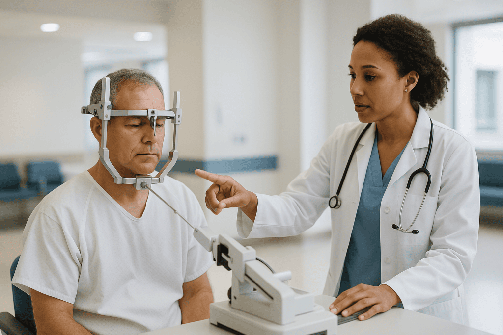
For the brain, the sequence is longer. With a frame-based method, the frame is fixed to the skull under local anesthetic at four points, a scan is done with the frame in place, and the coordinates are calculated from that scan. In the operating room, often under general anesthesia and sometimes under sedation, the surgeon makes a small incision, drills an opening a little wider than a pencil, and advances the needle along the calculated path to take samples. Frameless systems skip the pins and instead track the needle against a scan using skin markers or optical cameras. Either way, a pathologist may look at a preliminary sample during the procedure to confirm the needle reached abnormal tissue.
The whole point of the precision is to take a small amount of tissue from exactly the right place while disturbing as little healthy tissue as possible. That precision is also why recovery is generally quicker than after open surgery.
Who is usually offered a stereotactic biopsy, and who is asked to wait
A biopsy is only ordered when imaging alone cannot answer the question. For the breast, that typically means a mammogram has shown calcifications, a small mass, or a distortion of the tissue that cannot be felt and cannot be confidently called benign. Mayo Clinic notes that a biopsy is the only way to know for sure whether a suspicious area is cancer. If the same area shows up clearly on ultrasound, an ultrasound-guided biopsy is often chosen instead because it is quicker and needs no compression.
For the brain, the decision is weightier. A stereotactic needle biopsy is usually considered when a lesion sits deep, near critical structures, or in more than one location, and when the treatment plan depends on knowing the exact tissue type. When a lesion is accessible and removal is already the plan, the surgeon may skip a separate biopsy and obtain tissue during the operation itself, as Mayo Clinic outlines.
Some people are asked to wait or to prepare first. Blood-thinning medicines, including anticoagulants and antiplatelet drugs, raise the risk of bleeding at the biopsy site, so the team may ask the prescribing clinician whether a pause is safe and for how long. That is never a decision for the patient to make alone. People with a current infection near the site, uncontrolled blood pressure, or a bleeding disorder may also be rescheduled. For the breast procedure, very thin breast tissue or a target lying close to the chest wall can make the standard approach difficult, and the radiologist may suggest a different guidance method.
The general rule holds across both: the team chooses the least invasive route that still gets a reliable answer.
What the first 24 hours of stereotactic biopsy recovery look like
The first day sets the tone, and it differs sharply between the two procedures.
After a breast biopsy, you usually leave within an hour or two. MedlinePlus advises keeping the dressing on as instructed, using a cold pack to limit swelling, and avoiding strenuous activity for about a day. A snug bra worn around the clock for the first day or two helps keep the tissue still and reduces bruising. Some oozing onto the dressing is expected; soaking through it is not.
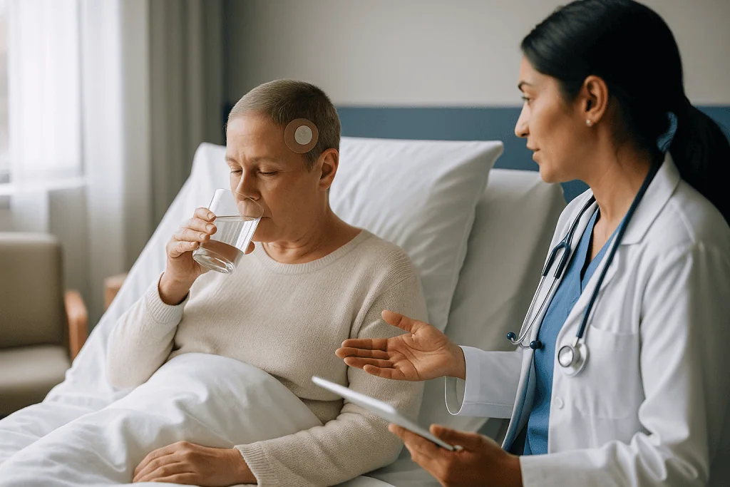
After a brain biopsy, plan on staying in the hospital at least overnight. Nurses check your neurological status at regular intervals: are you awake, oriented, moving both sides normally, speaking clearly? A follow-up CT scan is commonly done within the first hours to check for bleeding at the needle track, which is the main early complication the team watches for. Fatigue is nearly universal, partly from anesthesia and partly from the emotional weight of the day. The pin sites will feel tender and may be slightly swollen; the incision is closed with a few stitches or staples under a small dressing.
Both groups share one piece of advice: have someone with you. Even a breast biopsy done under local anesthetic leaves many people shakier than they expected, and after a brain biopsy you will not be driving yourself anywhere. Eat lightly, drink normally, and rest. The urge to “get on with things” is understandable, but the tissue you have just had sampled benefits from a quiet day.
Headache after a stereotactic brain biopsy: what is normal
The headache surprises people because the brain itself has no pain receptors. What hurts is everything around it: the scalp where the incision was made, the periosteum (the sensitive membrane covering the bone) where the frame pins pressed, the small drill opening, and the muscles of the scalp and neck that tense up during a long procedure. Air that enters the skull during the biopsy can also cause a dull, positional ache that eases as the body absorbs it over days.
A typical post-biopsy headache is mild to moderate, worse when you first sit or stand, better lying flat, and steadily improving. It should respond to the pain relief your team prescribes, which is usually a simple analgesic rather than anything strong. Your surgical team will explain what to take and how often; that is their call, not this article’s.
What matters is the pattern rather than the presence. A headache that fades across several days is the expected course. One that builds instead of fading, that arrives suddenly and severely, or that comes with vomiting, drowsiness, confusion, weakness on one side, or a new seizure is a different story. Those signs can indicate bleeding or swelling at the biopsy site and belong in the emergency category described later in this article. MedlinePlus lists worsening headache, seizures, and new weakness among the symptoms that should always prompt medical review in someone with a brain lesion.
Small things help. Keep your head raised on a couple of pillows for the first nights. Avoid straining, heavy lifting, or bending your head below your heart for as long as your surgeon advises. Stay hydrated. Caffeine withdrawal is an underrated headache cause after hospital stays, so if you normally drink coffee, ask whether you can resume it.
Pin-site soreness and wound care: stereotactic brain biopsy aftercare
Pin sites are the four small marks, usually two on the forehead and two at the back of the head, where a frame was fixed to the skull. They look trivial and can hurt out of proportion to their size, because the pins press on the periosteum, which is densely supplied with nerves. Expect tenderness, a little swelling, and sometimes a small bruise around each one. Some people notice a temporary numb or tingling patch on the forehead if a small sensory nerve was pressed; this usually settles over weeks. If your procedure was frameless, you will have no pin sites at all, just the skin markers’ adhesive residue and the single incision.
Care is simple. Keep the sites clean and dry for the period your team specifies, then wash gently with mild soap and pat dry. Do not pick at scabs. Watch for the signs that distinguish normal healing from infection: growing redness spreading outward, warmth, thick or discolored drainage, and increasing rather than decreasing pain.
The incision behind the hairline is closed with stitches or staples. Your discharge instructions will say when they come out and who removes them; this is usually done at a follow-up visit rather than at home. Hair around the site can be washed once you are cleared, but avoid hair dye, harsh products, or heat styling directly over the wound until it has fully closed.
Sleep is often the hardest part. Lying on the pin sites at the back of the head hurts, so many people sleep propped up or on their side for the first week. A soft, folded towel under the neck rather than a firm pillow against the sore spots can make a surprising difference.
Stereotactic breast biopsy recovery: bruising, the marker clip and the ice pack
The breast version of stereotactic biopsy recovery is measured in days rather than weeks. MedlinePlus describes soreness and bruising at the site for a few days, with a small adhesive strip or dressing over the needle entry point. The needle used for breast biopsy is wider than a blood-draw needle because it must remove intact cores of tissue, so a bruise the size of a plum is not unusual, especially if several samples were taken or the target sat deep.
Cold helps more than heat in the first day. A wrapped ice pack applied for short periods reduces swelling and dulls the ache. After that, most people manage with the pain relief their team suggests and a supportive bra. Bruising can look dramatic for a week and then fade through the familiar green-yellow stages.
The marker clip prompts more questions than anything else. It is smaller than a sesame seed, sits where the tissue was sampled, does not set off airport scanners, and is compatible with MRI, per Mayo Clinic. Its job is to let future imaging identify the exact spot, whether the result is benign and simply needs watching, or abnormal and needs further treatment. It is not a tracking device and does not need removal.
A firm lump under the bruise in the days afterward is usually a hematoma, a collection of blood in the tissue, and it typically softens and shrinks on its own. If it keeps growing, feels hot, or the skin over it turns red and tight, that needs a call. The same applies to any bleeding that does not stop with firm, direct pressure held steadily for a set period your team specified.
How painful is a stereotactic biopsy, really?
People search this question because they want honesty, not reassurance. So here it is, drawn from what the major sources describe and what patients consistently report.
For the breast procedure, the sharpest moment is the local anesthetic injection, a brief sting and burn that lasts seconds. After that, most people feel pressure and a pushing or tugging sensation as the needle is positioned and fired, sometimes with a clicking or vacuum sound from the device, rather than pain. Mayo Clinic notes that discomfort is generally mild. The compression of the breast during imaging and the awkward position on the table for the duration of the procedure are, for many, more uncomfortable than the biopsy itself. Afterward, the ache is comparable to a bruise from a knock.
For the brain procedure, the frame placement is the part people remember. Local anesthetic is injected at each pin site before the pins are tightened, and the sensation is usually described as intense pressure rather than sharp pain, lasting a minute or two. If the biopsy itself is done under general anesthesia, you feel nothing during it. If it is done under sedation with local anesthetic, you may sense pressure and hear the drill but should not feel pain. Afterward, the headache and pin-site soreness described above dominate, typically manageable with simple pain relief prescribed by your team.
Anxiety amplifies pain, which is not a moral failing but a physiological fact. Ask what you will feel and when. Ask whether you can listen to music. Tell the team if numbing does not seem to have taken effect; more local anesthetic can almost always be given.
Driving after a biopsy: when can you get behind the wheel?
This is where the two procedures diverge most sharply, and where reading the wrong web page could genuinely cause harm.
After a stereotactic breast biopsy done under local anesthetic alone, many people are able to drive themselves home, and MedlinePlus lists this among the routine possibilities. The caveats: if you received any sedation, you must not drive that day; if you feel faint, shaky, or in enough pain that turning the wheel or braking hard would be a problem, arrange a ride. Seat belts cross the chest, and a fresh biopsy site under the belt can be sore, so a folded towel between belt and skin helps.
After a stereotactic brain biopsy, you will not drive yourself home, full stop. You have had a procedure inside the skull, usually under anesthesia, and the team needs to be confident there is no bleeding, swelling, or seizure risk before you are safe behind the wheel. How long that takes depends on the diagnosis, whether you have had any seizure, whether you are taking medicines that affect alertness, and the driving rules of the state or country where you live. Many licensing authorities impose a mandatory period off the road after any seizure or after intracranial surgery, and those rules are legal requirements rather than suggestions. Your neurosurgical team will tell you when it is medically reasonable to drive, and you may need to check the licensing rules yourself or through your primary care clinician.
A practical point: do not treat the first drive as a test. Start with a short daytime trip on familiar roads with a passenger, once you have been cleared. Fatigue and slowed reaction time can linger longer than headache.
How long does a brain or breast biopsy take to heal? Side by side
Healing is not one event but several: skin closing, bruising fading, energy returning, and the emotional dust settling. The table lays out typical patterns described by the cited sources and by standard postoperative practice. Ranges are typical, not promises; your team’s instructions override anything here.
| Recovery milestone | Stereotactic breast biopsy | Stereotactic brain biopsy |
|---|---|---|
| Leaving the hospital | Usually same day (MedlinePlus) | Usually after at least one night of observation |
| Strenuous activity | Avoid for about 24 hours (MedlinePlus) | Avoid lifting and straining until surgeon clears you, often a week or more |
| Soreness or headache | A few days | Several days, gradually fading |
| Bruising visible | Around one to two weeks | Pin-site bruises fade over one to two weeks |
| Stitches or staples | Usually none; adhesive strip falls off | Removed at follow-up, timing set by surgeon |
| Driving | Often same day if no sedation | Not until medically cleared and legally permitted |
| Results | Days to about two weeks (NHS) | Days to about two weeks; some tests longer (NHS) |
Two things the table cannot show. First, energy lags behind wound healing after the brain procedure; feeling tired for a couple of weeks is common and does not mean something has gone wrong. Second, the skin over a breast biopsy site heals quickly, but a firm area or small dent where tissue was removed can persist for months and is usually harmless. Tell your team if it grows or changes.
The NHS describes biopsy results generally as taking from a few days to a couple of weeks, depending on the tests the pathologist needs to run. Molecular or genetic analyses of brain tissue can extend that further.
Waiting for results: what the '2 week biopsy rule' really means
Search for “2 week biopsy rule” and you will find two quite different ideas tangled together. Untangling them saves needless worry.
The first meaning is a healthcare access standard, best known from the United Kingdom, where an urgent referral for suspected cancer is meant to lead to a specialist appointment within two weeks. It is a target for how quickly you are seen, not a rule about how long a biopsy takes to heal or how long results take. It says nothing about whether your result is good or bad.
The second meaning is the informal expectation that results arrive within about two weeks. That lines up with what the NHS describes: results generally return within a few days to a couple of weeks, depending on what the pathologist needs to do. A simple benign breast core may be read within days. A brain sample often requires additional stains and sometimes molecular testing to classify a tumor precisely, because modern brain tumor classification depends on genetic features as well as appearance under the microscope, as Mayo Clinic describes. Those extra tests are the usual reason a brain biopsy result takes longer, not a sign of bad news.
A third, unhelpful myth sometimes hides inside the phrase: that a delay beyond two weeks means the result is cancer. It does not. Delays reflect laboratory workload, the need for a second pathologist’s opinion, or samples being sent to a specialist center. Ask at discharge when results are expected and how you will receive them, whether at an appointment, by phone, or through a patient portal. Then ask whom to call if that date passes with no word.
What percentage of stereotactic biopsies are cancer?
The honest answer is that no single percentage applies to everyone, and this article will not invent one. What the mainstream sources do say is consistent and worth holding onto.
For breast biopsies, Mayo Clinic is clear that most breast changes turn out not to be cancer, and a biopsy is often ordered precisely because imaging cannot distinguish a harmless finding from a worrying one. Screening programs deliberately set their thresholds so that they catch early cancers at the cost of biopsying many benign lesions. The proportion that proves malignant depends heavily on what was seen: a cluster of calcifications with a low-suspicion appearance carries a very different likelihood from a spiculated mass. Your radiologist’s report will often include an assessment category that reflects this, and asking what category your finding was given is a fair question.
For brain biopsies, the arithmetic runs the other way. A stereotactic brain biopsy is rarely done unless imaging has already shown a lesion that needs a tissue diagnosis, so the question is usually not “is it something” but “what exactly is it.” The possibilities include tumors of several grades, infections, inflammatory conditions, and lymphoma, each with a different treatment path. MedlinePlus notes that even brain tumors that are not cancerous can cause serious problems because of where they sit, which is why tissue diagnosis matters regardless of the label.
If a number would help you cope, ask your team for the likelihood in your specific case. They have seen your imaging; a search engine has not. And whatever the result, it is the start of a plan rather than a verdict delivered by a statistic.
What people often get wrong about stereotactic biopsy recovery
Some myths are harmless. A few can lead to real trouble.
“The needle can spread cancer.” This is the most persistent worry, and the mainstream evidence does not support it as a meaningful risk for either breast or brain biopsy. Mayo Clinic does not list tumor spread among the risks of breast biopsy; the recognized risks are bruising, bleeding, infection, and occasionally needing a repeat sample. Biopsy remains the standard way to diagnose because the information it gives outweighs any theoretical concern.
“A small incision means a small procedure.” True for the breast. Misleading for the brain. The scalp wound after a stereotactic brain biopsy is tiny, but a needle has passed through brain tissue, and the observation period, driving restrictions, and activity limits reflect that, not the size of the dressing.
“If I feel fine, I can skip the follow-up.” The follow-up is where stitches come out, where results are explained in context, and where the next step is agreed. Feeling well is the expected state after a biopsy, not a reason to miss the appointment.
“The marker clip needs to come out.” It does not. It is designed to stay, is MRI-compatible, and helps future imaging.
“I can restart my blood thinner when I feel like it.” If your prescribing clinician paused an anticoagulant or antiplatelet medicine for the biopsy, the timing of restarting it is a medical decision balancing bleeding risk at the biopsy site against the clotting risk the medicine was protecting you from. Confirm the plan before you leave, and follow it exactly.
“A headache after a brain biopsy means the biopsy went wrong.” A fading headache is expected. A worsening one is the signal to act, as the next sections explain.
Questions to ask your care team before you go home
The best time to ask is before discharge, when the people who did the procedure are still within reach. Write the answers down, or have someone with you who will.
- Which type of stereotactic biopsy did I have, and what exactly was sampled?
- When will results be ready, how will I receive them, and whom do I call if that date passes?
- What kind of headache or soreness is expected, and what pattern would worry you enough to want a call?
- When can I shower, wash my hair, or get the site wet?
- When do the stitches or staples come out, and who removes them?
- When may I drive, and are there licensing rules I need to check myself?
- Which of my regular medicines were paused, and exactly when and how do I restart them?
- What activity limits apply, for how long, and does that include lifting children, exercise, or work?
- If the sample was not adequate, would I need a repeat biopsy or a different procedure?
- Who is my point of contact over a weekend or at night?
Two questions people rarely ask but should. First: “If the result changes the plan, what would the next steps look like?” Knowing the possible branches in advance takes some of the fear out of the waiting period. Second: “Is there anything I should stop doing right now that I have not thought of?” This catches things like hot tubs, swimming, hair treatments, contact sports, and flying, each of which teams may restrict for different lengths of time after a cranial procedure.
The NHS encourages people to ask their care team about aftercare specifics, because instructions vary with the site and the technique used. There is no such thing as a silly question at this stage; there is only the question you wish you had asked at two in the morning.
When to call your doctor: red-flag signs after a stereotactic biopsy
Most recoveries are uneventful. The point of knowing the warning signs is not to worry about them but to recognize them instantly if they appear, so you can act without hesitation.
After a stereotactic brain biopsy, seek emergency care immediately for:
- A headache that suddenly becomes severe, or that steadily worsens instead of easing
- New weakness, numbness, or clumsiness on one side of the body or face
- Difficulty speaking, understanding speech, or new vision changes
- Confusion, unusual drowsiness, or difficulty waking
- A seizure of any kind, even brief
- Repeated vomiting
- Clear or blood-tinged fluid leaking from the wound or from the nose
These can signal bleeding or swelling at the biopsy site, and MedlinePlus lists new or worsening neurological symptoms among the signs that always warrant urgent assessment.
After a stereotactic breast biopsy, call your team the same day for:
- Bleeding that soaks through the dressing or does not stop with steady, firm pressure
- A lump under the skin that keeps enlarging
- Spreading redness, warmth, or thick discharge at the site
- Fever or chills
- Pain that increases after the first day rather than settling
For either procedure, contact the team about fever, wound redness that spreads, or anything that simply feels wrong to you. You know your body; a clinician would rather take an unnecessary call than miss a necessary one. If you cannot reach your team and a brain-related red flag is present, go to the nearest emergency department or call emergency services rather than waiting.
Every decision about treatment, medicines, and next steps rests with the team who performed your biopsy and know your imaging. This article can describe what usually happens; only they can tell you what is happening to you.
Frequently asked questions
How long does it take to heal after a stereotactic biopsy?
Skin healing is quick for both procedures, but the timelines differ. After a stereotactic breast biopsy, soreness and bruising typically last a few days, with strenuous activity avoided for about 24 hours, per MedlinePlus. After a stereotactic brain biopsy, headache and pin-site tenderness usually fade over several days, stitches come out at follow-up, and fatigue can linger for a couple of weeks. Your team’s instructions set your specific limits.
What percentage of stereotactic biopsies are cancer?
There is no single reliable percentage, because it depends on what the imaging showed. For breast biopsies, Mayo Clinic notes that most breast changes are not cancer, and screening thresholds are deliberately set to biopsy many benign findings. Brain biopsies are usually done on lesions already known to be abnormal, so the question there is what type of tissue it is. Ask your team for the likelihood in your specific case.
How painful is a stereotactic biopsy?
For the breast, the local anesthetic sting is usually the sharpest moment, followed by pressure and tugging rather than pain, and Mayo Clinic describes discomfort as generally mild. For the brain, frame pin placement causes intense pressure for a minute or two, and the biopsy itself is done under anesthesia or sedation so it is not felt. Afterward, a bruise-like ache or a moderate headache is typical and manageable with prescribed pain relief.
What is the 2 week biopsy rule?
It usually refers to one of two things. In the United Kingdom, the two-week standard is about how quickly someone with suspected cancer is seen by a specialist after urgent referral, not about the biopsy itself. Informally, people also use it to mean results arriving within about two weeks, which matches the NHS description of results taking a few days to a couple of weeks. A delay beyond that does not indicate cancer.
Why do I have a headache after a stereotactic brain biopsy?
The brain has no pain receptors, so the headache comes from surrounding structures: the scalp incision, the sensitive membrane over the bone where frame pins pressed, the small drill opening, and tense scalp and neck muscles. Air that enters during the procedure can add a dull positional ache. A headache that steadily fades is expected; one that worsens, arrives suddenly, or comes with vomiting, weakness, or confusion needs emergency assessment.
What is stereotactic brain biopsy aftercare for the pin sites?
Keep the pin sites clean and dry for the period your team specifies, then wash gently with mild soap and pat dry without picking at scabs. Tenderness, mild swelling, and small bruises are normal and fade over one to two weeks. Watch for spreading redness, warmth, thick discharge, or pain that increases rather than settles, which can signal infection. Sleeping propped up or on your side eases pressure on sore spots at the back of the head.
Can I drive myself home after a stereotactic biopsy?
After a stereotactic breast biopsy done with local anesthetic only, many people can drive themselves home, as MedlinePlus notes, provided they had no sedation and feel steady. After a stereotactic brain biopsy, you must not drive yourself home, and driving after that depends on medical clearance from your neurosurgical team plus the licensing rules where you live, which may impose mandatory periods after seizures or intracranial procedures.
What does stereotactic breast biopsy recovery involve day by day?
Day one is rest, a cold pack, a supportive bra, and no heavy activity, per MedlinePlus. Over the next few days, soreness settles while bruising may look worse before it fades over one to two weeks. The adhesive strip usually falls off on its own. A firm lump under the bruise is often a small hematoma that shrinks by itself; a lump that keeps growing, bleeding that will not stop, or fever warrants a call.
Does the marker clip left after a breast biopsy need to be removed?
No. The clip is designed to stay permanently and marks the exact spot that was sampled so future mammograms or MRI can find it, whether the result was benign and needs monitoring or abnormal and needs treatment. Mayo Clinic notes it is MRI-compatible and does not set off airport security. It is smaller than a sesame seed and cannot be felt. If the area is later removed surgically, the clip comes out with the tissue.
How long do stereotactic biopsy results take, and why might they be delayed?
The NHS describes biopsy results generally taking from a few days to a couple of weeks. Breast cores with clear findings may be read within days. Brain samples often take longer because pathologists run additional stains and, increasingly, molecular tests to classify tumors precisely, as Mayo Clinic describes. Delays also come from laboratory workload or second opinions. A longer wait is not a coded message about the result; ask your team when to expect news.
References
This article is for general information only and is not a substitute for professional medical advice. Please consult a qualified doctor about your individual situation.

More from the Blog
After a PET-CT Scan: Clearing the Tracer, Contact Precautions and When Results Arrive
After a PET-CT scan, most people go home the same day, drink extra fluids and empty their bladder often to help clear the radioactive…
Preparing for an MRI: Eating, Metal, What to Wear
For most MRI scans you can eat and drink normally beforehand, because the magnet images tissue, not stomach contents. The exceptions are scans of…

What Does a Comprehensive Check-Up Include? Blood Work, Imaging and the Follow-Up Plan
A comprehensive check-up usually includes a medical and family history, a physical examination with blood pressure and body measurements, and blood and urine tests…
What a CT Scan Shows, and Why Doctors Order One
A CT scan shows detailed cross-sectional images of bones, organs, blood vessels and soft tissue by combining many X-ray views into slices. Doctors most…
Can an Ultrasound Detect Cancer? What It Can Show, Its Limits and the Next Steps
An ultrasound can detect abnormal lumps, masses and changes in organs that may be cancer, and it is especially good at telling a fluid-filled…
What a PET-CT Scan Shows That a CT Alone Cannot: Metabolic Activity in Cancer Staging
A PET-CT scan shows how actively tissues are using energy, mapped onto a detailed anatomical picture. The PET part tracks a mildly radioactive sugar…