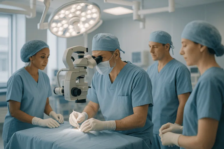
Stereotactic Biopsy
Stereotactic biopsy is a diagnostic procedure that uses a three-dimensional coordinate system derived from CT or MRI scans to guide a thin needle to a precise target, most commonly a lesion in…

Quick answer
A stereotactic biopsy is an image-guided procedure in which a thin needle is directed to a precise location, most often in the brain, using three-dimensional coordinates from CT or MRI scans, to remove a small tissue sample for diagnosis. It is used for deep or hard-to-reach lesions and typically requires only a small skull opening and a short hospital stay.
What is stereotactic biopsy?
A stereotactic biopsy is a way of taking a small sample of tissue from a precise location inside the body using a three-dimensional map made from imaging scans. The word “stereotactic” refers to a system of coordinates that lets a surgeon or radiologist guide a thin needle to a target that cannot be seen or felt from the outside. A biopsy is simply the removal of a small piece of tissue so that it can be examined under a microscope by a pathologist, a doctor who specializes in diagnosing disease from tissue samples.
The technique is most often discussed in two settings. In the brain, a stereotactic biopsy is used to sample a lesion (an abnormal area) found on a computed tomography (CT) or magnetic resonance imaging (MRI) scan when it is deep, small, or close to areas that control movement, speech, or vision. In the breast, a similar approach uses mammography or MRI to guide a needle to a suspicious area that can only be seen on imaging. This page focuses mainly on stereotactic brain biopsy, which is managed by neurosurgery teams, while noting where the principles apply more broadly.
Conditions in which a stereotactic brain biopsy may be considered include:
- Suspected brain tumors, whether they appear to have started in the brain or spread from elsewhere in the body
- Lesions that could be infection, inflammation, or a tumor, where the scan alone cannot tell them apart
- Suspected lymphoma of the central nervous system, a cancer of immune cells that may be treated without open surgery
- Lesions in people who are not well enough for a larger operation
The goal is diagnostic rather than curative: the procedure aims to answer the question “what is this?” so that the right treatment can be planned.
Who is a candidate
Understanding who needs stereotactic biopsy starts with the imaging findings and the person’s overall health. Your doctor may suggest this procedure when:
- An abnormal area has been found on a brain scan and a tissue diagnosis is needed before deciding on treatment
- The lesion is located deep in the brain or in a region where a larger open operation would carry a high risk of damaging important functions
- There are several lesions and the team needs to confirm the diagnosis of one of them
- A larger operation is not advisable because of age, other medical conditions, or the likely type of disease
- The results are expected to change management, for example by distinguishing a tumor from an infection or inflammatory condition
A stereotactic biopsy may not be suitable in some situations. If the lesion is large and causing dangerous pressure inside the skull, a surgeon may prefer an open operation that both removes tissue and relieves the pressure. If the imaging appearance is already very characteristic and a biopsy would not change the treatment, the risks may not be justified. Lesions with a very rich blood supply, or those located in areas where even a needle track carries a high risk of bleeding, may call for a different approach. People who take blood-thinning medication or who have bleeding disorders need careful assessment first, because bleeding is the main risk of the procedure. A multidisciplinary team, often including neurosurgery, neuroradiology (imaging of the nervous system), and pathology specialists, usually weighs these factors together.
How the procedure works
The stereotactic biopsy procedure is generally divided into three stages: planning, the biopsy itself, and the immediate period afterward. Details vary between hospitals and between the frame-based and frameless techniques described below.
Before the procedure
- A high-resolution MRI or CT scan is taken, sometimes on the day of the biopsy. Small stickers called fiducial markers may be placed on the scalp so the scan can be matched to the head during surgery.
- In the frame-based method, a lightweight metal frame is attached to the head with pins after numbing the skin. The frame provides fixed reference points. In the frameless method, a computer navigation system tracks the position of the head and instruments without a rigid frame.
- The surgeon uses planning software to choose the safest path from the scalp to the target, avoiding major blood vessels and critical brain regions.
During the procedure
- Anesthesia may be general (fully asleep) or local with sedation (awake but relaxed and pain-free). The choice depends on the person, the location of the lesion, and the team’s practice.
- A small area of hair is usually shaved and the scalp is cleaned. A short incision is made and a small hole, often described as the width of a pencil, is drilled in the skull. This is called a burr hole.
- Guided by the coordinates, a thin biopsy needle is advanced to the target. One or more small tissue samples are taken. In many cases a pathologist examines a sample immediately to confirm that it contains abnormal tissue; if not, another sample may be taken.
- The needle is withdrawn, the incision is closed with stitches or staples, and the frame, if used, is removed.
After the procedure
- You are monitored in a recovery area, and a CT scan is often performed within the first hours to check for bleeding.
- Most people are observed overnight, although some centers discharge selected patients the same day and others keep people longer if there are concerns.
The biopsy itself typically takes about one to two hours, though the total time including scanning, planning, and recovery is longer.
Preparation for a stereotactic biopsy
Preparation focuses on safety and on making the imaging as accurate as possible. Your team will give you specific instructions, which may include the following:
- Medication review. Tell your doctors about every medicine and supplement you take. Blood thinners such as warfarin, clopidogrel, and newer anticoagulants, as well as aspirin and some anti-inflammatory drugs, usually need to be stopped for a set period beforehand. Never stop a prescribed medicine without instructions from the prescribing doctor.
- Blood tests. Tests of blood clotting, blood count, and kidney function are commonly requested, especially if contrast dye will be used for the scan.
- Fasting. If general anesthesia or sedation is planned, you will typically be asked not to eat or drink for several hours before the procedure.
- Allergies. Report any previous reactions to contrast dye, anesthesia, latex, or antibiotics.
- Practical arrangements. Arrange for someone to accompany you home and stay with you for the first night, and avoid driving until your team confirms it is safe.
- Hair and skin. Wash your hair as instructed; avoid hair products on the day. Only a small area is usually shaved.
Steroid medication is sometimes prescribed before the procedure to reduce swelling around the lesion, and anti-seizure medication may be given in certain cases. These decisions are individual and will be explained by your team.
Recovery and aftercare
Stereotactic biopsy recovery time is generally shorter than recovery from open brain surgery, because the incision is small and the brain is disturbed as little as possible. Even so, the experience varies from person to person.
- First 24 hours. Many patients feel tired and have a mild headache or soreness at the incision and, if a frame was used, at the pin sites. Nurses check your alertness, strength, speech, and pupils regularly. A follow-up CT scan is commonly performed.
- Going home. Most people go home within one to two days if the post-procedure scan is reassuring and they are stable. Simple pain relief is usually enough for discomfort.
- First week. Fatigue is common. Light activity around the house is typically encouraged, while heavy lifting, strenuous exercise, and driving are usually avoided until your team clears them. Keep the incision clean and dry according to instructions.
- Stitches or staples are often removed at about seven to fourteen days, or dissolve on their own.
- Return to work for desk-based jobs is often possible within one to two weeks, though this depends on how you feel, the underlying diagnosis, and any further treatment that is planned.
Pathology results usually take several days to a couple of weeks, because some tests, such as genetic or molecular analysis of tumor tissue, take longer than a standard microscope examination. Your team will arrange a follow-up appointment to discuss the findings and the next steps.
Risks and side effects
Weighing stereotactic biopsy risks and benefits is an important part of the decision. The main benefit is a tissue diagnosis obtained through a very small opening, which allows treatment to be planned with much greater confidence than imaging alone. The procedure is widely regarded as having a lower complication rate than open surgery, but it is still an operation on the brain and carries real risks.
- Bleeding. The most important risk. Small amounts of bleeding along the needle track are common and usually cause no symptoms; less often, a larger bleed can cause new weakness, speech problems, or reduced alertness and may require urgent treatment.
- New neurological symptoms. Temporary or, rarely, permanent changes such as weakness, numbness, or difficulty speaking, depending on the area sampled.
- Seizures. Irritation of the brain surface can occasionally trigger a seizure during or after the procedure.
- Infection. Uncommon but possible at the wound or, rarely, deeper in the brain or its coverings.
- Non-diagnostic sample. Sometimes the tissue taken does not give a clear answer, and a repeat biopsy or a different type of operation may be needed.
- Anesthesia-related effects. Nausea, sore throat, or, rarely, more serious reactions.
- Frame-related discomfort. Pin-site soreness, small scabs, or minor bruising where the frame was attached.
Your surgeon can explain how these general risks apply to your particular lesion and health, since location, size, and blood supply all influence the level of risk.
Results and outlook
In most cases, a stereotactic biopsy provides enough tissue for a diagnosis, and the addition of an immediate pathology check during the procedure has generally improved the chance of a useful sample. The diagnosis then guides the plan, which might be surgery to remove a tumor, radiation therapy, chemotherapy or other drug treatment, antibiotics or antiviral medication for an infection, immune-modulating treatment for an inflammatory condition, or simply monitoring.
The outlook after the biopsy itself is usually determined far more by the underlying diagnosis than by the procedure. The biopsy does not treat the lesion; it explains it. Modern tissue analysis often includes molecular and genetic testing, which can refine the diagnosis and, for some tumors, influence which treatments are likely to help. Because these tests take time, final results may arrive in stages.
Occasionally a biopsy is inconclusive, either because the sample came from the edge of the lesion, because the tissue was distorted by prior steroid treatment, or because the condition is difficult to identify from a small sample. In these cases your team may discuss repeat sampling, a different surgical approach, or a period of watchful observation with repeat scans.
Cost considerations
The cost of a stereotactic biopsy varies considerably between countries, hospitals, and individual cases. Rather than a single price, it is helpful to understand the elements that usually contribute:
- Imaging. The planning MRI or CT, contrast dye, and any post-procedure scans.
- Operating room and equipment. The use of a stereotactic frame or a computer navigation system, disposable biopsy needles, and operating theater time.
- Professional fees. The neurosurgeon, anesthesiologist, radiologist, and pathologist.
- Laboratory work. Standard microscope examination, immediate intraoperative review, and any additional molecular or genetic tests, which can add significantly to the total.
- Hospital stay. The length of observation, which depends on how quickly you recover and whether any complication occurs.
- Follow-up. Clinic visits, wound checks, and repeat imaging.
Insurance coverage, referral pathways, and whether the biopsy is bundled with subsequent treatment all affect what a patient ultimately pays. Hospitals such as Acibadem typically provide an individualized estimate after reviewing the imaging and the proposed plan. Asking in advance which items are included in a quoted figure can help avoid surprises.
Frequently asked questions
Is a stereotactic biopsy painful?
Most people describe the procedure as uncomfortable rather than painful. If you are awake with sedation, the scalp is numbed with local anesthetic, and you may feel pressure or a brief pushing sensation rather than sharp pain. Afterward, headache and soreness at the incision or pin sites are common for a few days and usually respond to simple pain relief. Severe or worsening pain is not expected and should be reported.
How long does the stereotactic biopsy procedure take?
The biopsy itself commonly takes about one to two hours. The whole process, including the planning scan, attaching a frame if one is used, anesthesia, and recovery-room monitoring, usually takes most of a day. Times vary depending on the technique, the location of the lesion, and whether the pathologist needs additional samples during the procedure.
What is the typical stereotactic biopsy recovery time?
Many patients go home within one to two days and feel largely back to normal within one to two weeks, although fatigue can linger. Return to driving, work, and strenuous activity depends on your team’s advice, your diagnosis, and any planned treatment. Recovery from the biopsy is generally quicker than from open brain surgery because the incision and the disturbance to the brain are small.
Who needs a stereotactic biopsy rather than open surgery?
A stereotactic biopsy is often chosen when a lesion is deep, small, or in a region where open surgery would be risky, when several lesions are present, when the person is not well enough for a larger operation, or when the suspected diagnosis, such as lymphoma, is usually treated without surgical removal. Open surgery may be preferred when a lesion is causing dangerous pressure or when removing it is likely to be part of the treatment anyway.
What are the main stereotactic biopsy risks and benefits?
The main benefit is a reliable tissue diagnosis obtained through a very small opening, which allows treatment to be tailored to the actual disease. The main risks are bleeding, new or worsened neurological symptoms, seizures, infection, and the possibility that the sample does not give a clear answer. For most people these risks are considered lower than those of open surgery, but they are not zero, and they vary with the lesion’s location.
Will I need to stay in the hospital?
An overnight stay for observation is common, mainly to watch for delayed bleeding and to repeat a scan if needed. Some centers discharge selected patients the same day after a normal scan, while others keep people longer if there are concerns. Your surgeon will explain what is planned in your case.
Can a stereotactic biopsy miss the diagnosis?
Yes, occasionally. A sample may come from tissue at the edge of the lesion, may be too small for certain tests, or may be altered by steroid medication given beforehand. Immediate pathology review during the procedure reduces this risk but does not eliminate it. If the result is unclear, your team may discuss repeat sampling, a different surgical approach, or careful monitoring with follow-up imaging.
When to see a doctor
Anyone who has been told that a scan shows an abnormal area in the brain should be assessed by a specialist, usually a neurosurgeon or neurologist, who can discuss whether a stereotactic biopsy or another approach is appropriate. Symptoms that commonly lead to such scans include new or worsening headaches, especially those that are worse in the morning or with lying down; new seizures; persistent nausea or vomiting without another explanation; gradual weakness or numbness on one side of the body; difficulty speaking or understanding speech; changes in vision; or changes in personality, memory, or alertness. These symptoms do not mean a tumor is present, but they warrant evaluation.
After a stereotactic biopsy, seek urgent medical care, through emergency services if necessary, if you experience any of the following:
- Sudden severe headache, or a headache that steadily worsens despite pain relief
- New weakness, numbness, or clumsiness in the face, arm, or leg
- New difficulty speaking, understanding, or finding words
- Confusion, unusual drowsiness, or difficulty waking
- A seizure, or a seizure that is different from any you have had before
- Repeated vomiting
- Fever, chills, or increasing redness, swelling, warmth, or discharge at the incision
- Clear fluid leaking from the wound
- Sudden changes in vision or a stiff neck with light sensitivity
Less urgent concerns, such as mild pin-site soreness, small amounts of dried blood on the dressing, or questions about medication and activity, can usually be raised with your care team at the scheduled follow-up or by contacting the ward as instructed at discharge.
Preparation
- Tell your team about all medicines, particularly blood thinners and aspirin, which usually need to be paused under medical guidance. Blood tests and a planning MRI or CT scan are typically arranged beforehand. Fast as instructed if sedation or general anesthesia is planned, and arrange for someone to take you home and stay with you the first night.
Aftercare
- Expect mild headache and soreness at the incision or pin sites for a few days, usually eased by simple pain relief. Keep the wound clean and dry, avoid heavy lifting, strenuous exercise, and driving until cleared by your team, and attend the follow-up visit for wound checks and pathology results. Seek urgent care for sudden severe headache, new weakness, speech difficulty, seizures, confusion, or fever.
Medically reviewed by the Acıbadem International Medical Board September 13, 2026
See our medical review board →
Update history
- PublishedSeptember 13, 2026
- Medical review approvedSeptember 13, 2026
- Last content updateSeptember 13, 2026
References3
From the Acibadem Blog

When Does a Brain Lesion Need a Stereotactic Biopsy Instead of Open Surgery?
A brain lesion is usually sampled with a stereotactic needle biopsy when the tissue diagnosis will change treatment but removing the lesion is not…

Stereotactic Brain Biopsy Step by Step: Planning Scans, Coordinates and the Needle Path
A stereotactic brain biopsy uses a detailed MRI or CT scan, a three-dimensional coordinate system and a thin needle to remove a tiny sample…