

When Does a Brain Lesion Need a Stereotactic Biopsy Instead of Open Surgery?
Key Takeaways
- The deciding question is not needle versus opening but whether the tissue result will change treatment; if removing the lesion is the treatment, open surgery usually supplies the diagnosis as a byproduct.
- Deep-seated, multiple, diffuse or eloquent-area lesions, and suspected lymphoma or infection, are the classic reasons a team chooses a stereotactic needle over a craniotomy.
- Modern glioma classification uses World Health Organization grades 1 to 4 plus molecular markers that can only be read from tissue, which is why imaging alone rarely settles a suspected tumor.
- Bleeding along the needle track is the main serious risk of stereotactic biopsy, and a non-diagnostic sample is its main practical limitation; frozen section and multiple cores reduce the second.
- Corticosteroids can shrink lymphoma so fast that a later biopsy shows only dead tissue, so teams coordinate steroid timing with the procedure; never adjust these medicines yourself.
- Hospital stay after a stereotactic biopsy is often a single night, compared with several days to about a week after a craniotomy, though the final molecular report can take a week or more either way.
A brain lesion is usually sampled with a stereotactic needle biopsy when the tissue diagnosis will change treatment but removing the lesion is not the goal or is not safe: deep-seated or multiple lesions, suspected lymphoma or infection, lesions in speech or movement areas, or people too unwell for a long operation. Open surgery is favored when the lesion is accessible, is pressing on the brain, and removing it is itself part of the treatment plan.
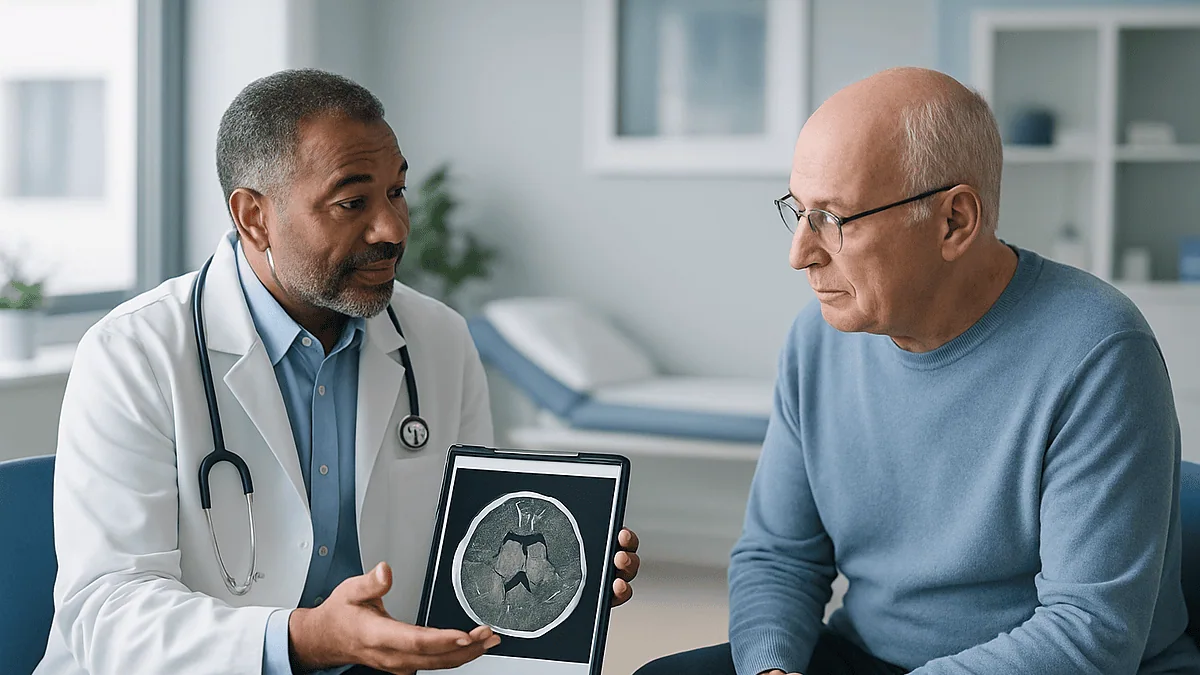
The radiologist’s report uses a word nobody wants to read at a kitchen table: lesion. It sits in the left thalamus, deep in the middle of the brain, and it lights up with contrast. Two neurosurgeons have looked at the same MRI. One talks about a needle and a night in the hospital. The other mentions a craniotomy. The scan has not changed; the question has: when is a brain biopsy needed, and when does the lesion call for opening the skull instead?
That question is less about courage and more about geometry, probability, and what the answer will be used for. Surgeons weigh where the lesion sits, what it most likely is, whether taking it out would help or harm, and whether a rice-grain sample of tissue will be enough for the pathologist and the molecular lab.
This explainer walks through that reasoning in plain language, including the risks, the trade-offs, what recovery looks like, and the questions worth asking before anyone signs a consent form.
When is a brain biopsy needed at all?
A biopsy is the removal of a small piece of tissue so a pathologist can examine it under a microscope and run molecular tests. Imaging, even excellent imaging, describes shape, size, blood flow and water content. It cannot reliably say which cells are present. That gap is the whole reason biopsies exist.
The honest test for whether a brain biopsy is needed is a single question: will the answer change what happens next? If a scan shows a mass that could be a glioma (a tumor arising from the brain’s supporting cells), a lymphoma (a cancer of immune cells), a metastasis (cancer spread from elsewhere), an abscess (a pocket of infection), or an inflammatory lesion, each of those leads to a completely different treatment. Radiation planning, chemotherapy choice, antibiotics, immune-suppressing drugs, or careful observation all hinge on what the tissue shows.
Modern tumor classification raises the stakes further. The World Health Organization system grades brain tumors from 1 to 4 and now folds in molecular markers such as specific gene mutations and chromosome changes, as the Mayo Clinic and National Cancer Institute both describe. Those markers cannot be read off an MRI. They come from tissue.
A biopsy is usually not needed when the imaging appearance is so characteristic that the team is confident of the diagnosis and treatment would not differ, or when the lesion is going to be removed anyway. In that second case the operation itself supplies the tissue, and a separate needle procedure would add risk without adding information.
So the first fork in the road is not “needle or open” at all. It is “do we need tissue, and if so, what is the least harmful way to get enough of it?”
What actually happens during a stereotactic brain biopsy
Stereotactic means “guided in three dimensions.” The surgeon uses a recent MRI or CT scan to map the exact coordinates of the lesion, then steers a thin needle to that point through a small opening in the skull. Think of it as using satellite navigation for the inside of the head rather than looking with the eyes.
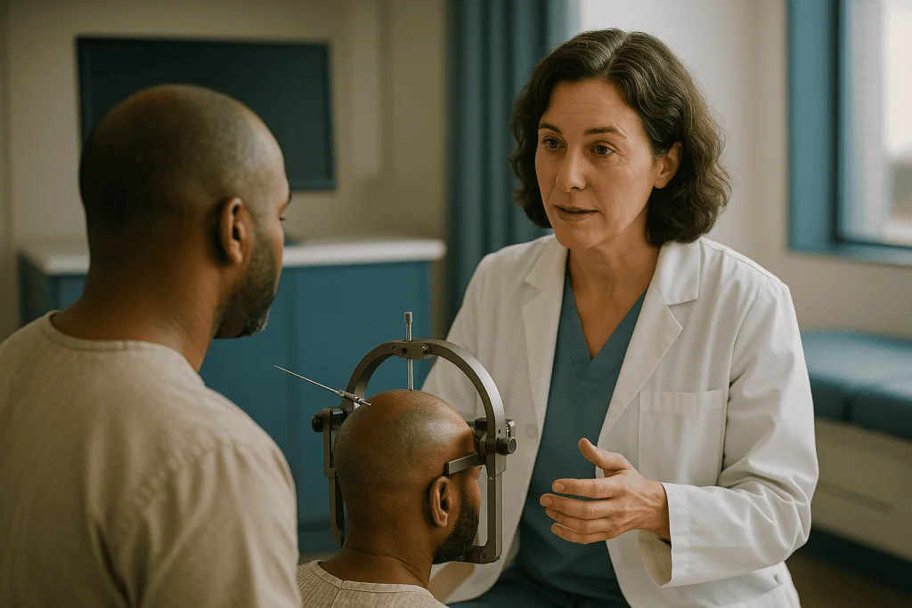
Two broad approaches exist. Frame-based biopsy uses a lightweight ring fixed to the skull under local anesthetic that acts as a fixed reference grid. Frameless biopsy uses skin markers or facial contours registered to a computer navigation system, with the needle tracked by cameras. Both aim at the same thing: reaching a chosen spot within a few millimeters.
Once the target is planned, the scalp is numbed and a short incision made. A burr hole, a small opening drilled through the skull roughly the width of a fingertip, gives the needle a path. The tough membrane covering the brain is opened, and the needle advances along the planned trajectory. Several small cores of tissue are taken, often from more than one depth, so the pathologist sees the lesion’s center and its edge.
Many centers send one sample for an immediate “frozen section,” a rapid look while the patient is still on the table, to confirm that the needle has hit abnormal tissue. If it has not, another pass can be made. The remaining tissue goes for formal processing and molecular testing.
The procedure can be done under general anesthesia or, in selected people, under sedation with local anesthetic. It is usually measured in an hour or two of operating time, and a CT scan afterward checks for bleeding before the person moves to a ward or observation unit, a sequence described in patient information from Johns Hopkins Medicine.
Brain biopsy vs craniotomy: what "open surgery" really means here
A craniotomy is an operation in which a section of skull bone is temporarily removed so the surgeon can see and work on the brain directly, then the bone is fixed back in place. When people say “open surgery” for a brain lesion they usually mean a craniotomy done to remove as much of the lesion as safely possible, which surgeons call a resection.
The distinction with a stereotactic biopsy is not simply small versus large. It is about purpose. A needle biopsy has one job: to obtain a diagnosis. A craniotomy typically has two: to obtain a diagnosis and to treat by removing tissue, relieving pressure, or both. That double purpose is why open surgery is often preferred for an accessible mass that is causing symptoms. The tissue arrives in the pathology lab as a byproduct of an operation that was going to happen anyway.
There is also a middle option, the open biopsy. Here the surgeon makes a small craniotomy over a lesion near the surface, takes a generous sample under direct vision, and closes without attempting full removal. It offers more tissue than a needle and better control of bleeding, at the cost of a larger opening.
MedlinePlus describes hospital stays after brain surgery as commonly running several days to about a week, with recovery at home measured in weeks. Needle biopsy stays are far shorter, often one night. Those differences matter to a person who is frail, who lives alone, or who needs to start other treatment quickly.
Neither route is “better” in the abstract. The right one depends on what the lesion is likely to be, where it sits, and what the team intends to do with the answer.
Which lesions usually point toward a needle rather than an opening
Location comes first. Lesions buried in the thalamus, basal ganglia, brainstem or deep white matter sit beneath structures that control movement, sensation, alertness and speech. Reaching them with a craniotomy means traveling through healthy brain; a needle along a carefully planned path disturbs far less tissue. Surgeons often describe these as deep-seated or eloquent-area lesions, meaning the surrounding brain does something you would notice losing.

Pattern comes next. Multiple lesions scattered through both hemispheres are unlikely to be removed one by one. Whether they turn out to be metastases, lymphoma, infection or inflammation, the treatment will be medical or radiation-based, and the team needs one representative sample, not a series of operations.
Suspected lymphoma is a classic needle case. Primary central nervous system lymphoma responds to chemotherapy and radiation rather than surgical removal, so cutting it out offers little benefit and carries the usual risks of open surgery. A small, well-targeted sample is what the hematology and oncology teams actually need.
Diffuse, infiltrating lesions that spread through brain tissue without a clear edge on MRI are another example. There may be no discrete mass to resect, yet the grade and molecular profile still steer treatment.
The person matters as much as the lesion. Someone with serious heart or lung disease, someone with advanced cancer elsewhere, or someone whose overall condition makes a multi-hour operation unwise may be offered a biopsy specifically because it is the shorter, lighter procedure. Age alone is not the deciding factor; overall health and goals of care are.
Finally, when non-surgical treatment is already the plan, the biopsy simply confirms the target. Radiation oncologists and medical oncologists cannot responsibly treat a shadow on a scan without knowing what it is.
Who is asked to wait: when is a brain biopsy needed later rather than now?
Not every lesion earns a procedure on first sight. Neurosurgeons and neuro-oncologists regularly recommend a period of watching with repeat MRI, and it helps to understand why that is not the same as doing nothing.
Some appearances are reassuring enough to monitor. A small, smooth, contrast-enhancing lump attached to the brain’s outer covering, found incidentally on a scan done for another reason, is very often a meningioma, a usually slow-growing tumor of the membranes. The NHS notes that many such tumors are low grade and may only need monitoring. A repeat scan in a few months tells the team whether it is growing; a biopsy would tell them what it is called but not necessarily change the plan.
Other situations call for the opposite: acting, but not with a biopsy first. A large mass with swelling that is shifting the brain and causing drowsiness, weakness or worsening headache generally needs pressure relieved. Open surgery to remove it is both the diagnostic and the therapeutic step, so a separate needle procedure would only delay things. A suspected abscess may be drained through a burr hole, which both treats the infection and yields a sample for culture.
Some people are asked to wait because something else needs sorting first. Blood-thinning medicines increase bleeding risk, and the surgical team and prescribing clinician will decide together how to manage them around the procedure; that is never a decision to make alone at home. A known cancer elsewhere may be biopsied at a safer site, such as a lung or lymph node, before anyone touches the brain.
When is a brain biopsy needed later? Typically when a watched lesion grows, changes character on imaging, or starts producing symptoms. The waiting period is a form of information gathering, with a defined trigger for action.
Can you diagnose a brain tumor without a biopsy?
Sometimes, and it is worth being precise about when. Imaging can be confident enough to guide treatment in a few circumstances. A person with widespread lung cancer who develops several ring-enhancing brain lesions almost certainly has metastases, and teams often treat on that basis. A classic meningioma appearance, as above, is frequently managed without tissue. Certain diffuse brainstem tumors in children have historically been treated on imaging alone because the biopsy risk in that location was judged too high, although practice varies and tissue is increasingly sought for molecular testing.
Advanced MRI adds clues rather than answers. Perfusion imaging measures blood flow through a lesion; spectroscopy measures chemical signatures; diffusion imaging measures how freely water moves. Each nudges the probability toward tumor, abscess or inflammation. PET scanning, which tracks a radioactive tracer taken up by active cells, can help separate tumor recurrence from radiation injury. The Mayo Clinic lists these among the tools used alongside, not instead of, tissue diagnosis.
A lumbar puncture, sampling the fluid around the spinal cord, can occasionally make the diagnosis without brain tissue. Lymphoma cells, infection, or inflammatory markers may be found there. When it works, it spares a brain procedure; when it is negative, it does not rule anything out.
Blood-based “liquid biopsy” tests that look for tumor DNA are an active research area. For brain tumors specifically, the blood-brain barrier limits how much tumor material reaches the bloodstream, and these tests are not an established replacement for tissue in routine care. Any claim that a blood test can do the biopsy’s job today should be read as promising research, not current standard practice.
The molecular classification that now drives glioma treatment still requires tissue. That single fact keeps the biopsy central.
Stereotactic brain biopsy risks: how risky is it, honestly
Bleeding is the complication surgeons worry about most. The needle passes through brain tissue and through small blood vessels. Most bleeding is tiny and silent, visible only on the routine CT afterward. Occasionally a bleed is large enough to cause new weakness, speech difficulty, drowsiness or seizure, and rarely it is life-threatening. Johns Hopkins Medicine and the Cleveland Clinic both list bleeding, infection, seizure, stroke-like deficits, and reactions to anesthesia as the recognized risks. The chance rises with deep targets, lesions rich in blood vessels, and impaired blood clotting.
A second risk is not medical but informational: the sample may not answer the question. Brain lesions are not uniform. A needle can land in dead tissue at the center, in swollen but normal brain at the edge, or in a low-grade corner of a tumor that is high grade elsewhere. Pathologists call this sampling error. Frozen section during the procedure reduces it, and taking cores from several depths helps, but it cannot be eliminated. A “non-diagnostic” result sometimes means a second procedure.
Infection at the wound site, headache, and scalp numbness around the incision are the more common, less serious problems. Seizure can occur because any irritation of the brain surface can trigger one, which is why some teams use anti-seizure medicine around the time of surgery; that choice sits with the anesthetic and surgical team.
How does this compare with open surgery? A craniotomy carries every risk above plus those of a longer operation, a larger wound, more blood loss, and more brain manipulation, balanced against the benefit of removing the lesion. The comparison is not needle-safe versus open-dangerous. Both are serious brain procedures; one is simply smaller and does less.
Asking the team how they estimate risk for your particular lesion, given its location and your health, is more useful than any general figure.
Stereotactic biopsy and open surgery side by side
The table below is a summary of typical differences, not a scoring sheet. Individual cases move between columns depending on location, imaging appearance and the person’s health, and the treating team’s judgment overrides any generalization here.
| Feature | Stereotactic needle biopsy | Open surgery (craniotomy) |
|---|---|---|
| Main purpose | Diagnosis only | Diagnosis plus removal or pressure relief |
| Skull opening | Small burr hole | Section of bone temporarily removed |
| Typical lesions | Deep, multiple, diffuse, suspected lymphoma or infection, eloquent-area lesions | Accessible single masses causing symptoms; lesions where removal is the treatment |
| Amount of tissue | Several small cores | Large specimen, often the whole lesion |
| Risk of non-diagnostic sample | Present, reduced by frozen section and multiple cores | Low, because tissue is abundant |
| Bleeding control | Indirect; relies on planning and post-procedure imaging | Direct, under the surgeon’s view |
| Anesthesia | General or sedation with local | General, occasionally awake for mapping |
| Usual hospital stay | Often one night (Johns Hopkins Medicine) | Several days to about a week (MedlinePlus) |
| Recovery at home | Days to a couple of weeks | Weeks, with rehabilitation if needed |
| Who tends to be steered here | People for whom removal would not help, or who could not tolerate a long operation | People with a removable lesion where surgery itself treats symptoms |
Two rows deserve emphasis. “Main purpose” explains most decisions: if removing the lesion is the treatment, the team will almost always favor open surgery. “Risk of non-diagnostic sample” explains why some surgeons choose an open biopsy for a lesion near the surface even when they do not plan to remove it; more tissue means a more confident pathology report and a full molecular panel.
Why steroids and timing matter: the lymphoma trap
People with a newly discovered brain lesion are often started on corticosteroids, medicines that reduce swelling around the lesion and can quickly ease headache, weakness or confusion. They work by calming the leaky blood vessels that cause the surrounding brain to swell. For most tumors this is helpful and does not interfere with diagnosis.
Lymphoma is the exception, and it is one of the more important practical reasons a surgeon may want to move quickly or, alternatively, ask for a pause. Lymphoma cells are exquisitely sensitive to steroids. Within days the lesion can shrink dramatically on MRI and the cells within it can die or change shape. A biopsy taken after that may show only inflammation or dead tissue, leaving the team without a diagnosis and the person facing a repeat procedure once the lesion regrows.
This is why, when imaging raises suspicion of lymphoma, the neurosurgical and oncology teams may coordinate the timing of any steroid treatment with the biopsy. The decision is a balance: a person who is drowsy from swelling may need steroids regardless, and safety comes first. The point for readers is simply that this coordination exists, and that changing or stopping any prescribed medicine before a procedure is a decision for the prescribing clinician and surgical team, never something to do on your own.
A related timing issue involves blood thinners and antiplatelet medicines, which reduce clotting and increase the chance of bleeding along the needle track. The team will give specific instructions about these, often in consultation with the doctor who prescribed them, because the reason the medicine was started (a heart valve, a stent, a previous clot) carries its own risks if it is interrupted.
Ask directly: does the timing of my current medicines affect this biopsy, and who is coordinating that?
Non-cancer reasons for a brain biopsy: infection, inflammation and unexplained disease
The word biopsy is so tightly linked to cancer that many people assume a recommended brain biopsy means a tumor is already suspected. Often it is. Frequently, though, the differential includes conditions that are treatable in entirely different ways, and that is exactly why tissue is needed.
Infection is a common one. In a person whose immune system is suppressed, whether by HIV, transplant medicines or chemotherapy, a ring-enhancing lesion could be a bacterial abscess, a fungal infection, toxoplasmosis (a parasitic infection), or a viral condition called progressive multifocal leukoencephalopathy. Each has a different treatment, and some are dangerous to treat blindly. Where a lesion looks like an abscess, a needle procedure both drains it and captures material for culture.
Inflammatory and autoimmune conditions form another group: vasculitis (inflammation of blood vessel walls), sarcoidosis, and unusual forms of demyelination, where the insulating coating of nerve fibers is damaged. Treatment usually involves suppressing the immune system, which would be harmful if the lesion were actually an infection or a lymphoma. The biopsy protects against that error.
Rarely, a biopsy is considered for rapidly progressive neurological decline of unknown cause, including suspected prion disease, where standard tests have not given an answer. These are careful, multidisciplinary decisions because the diagnostic yield may be modest and the risks are real.
Sometimes the tissue can come from somewhere safer. Intravascular lymphoma, a rare cancer growing inside small blood vessels, is occasionally diagnosed from random skin biopsies. A suspected systemic disease with brain involvement may be confirmed from a lymph node, lung or skin. Good teams look for the least invasive source of a reliable answer before proposing a brain procedure, and it is reasonable to ask whether such an option exists.
Brain biopsy recovery time: what the following days and weeks usually look like
The first hours are about watching. After the procedure a CT scan checks for bleeding, and nurses perform regular neurological checks: asking your name and the date, testing grip strength, shining a light in the eyes. Headache at the wound site is common and usually mild to moderate; nausea from anesthesia can occur. Most people are eating and walking the same day or the next morning.
Discharge is often the following day when the CT is clear and the neurological checks are stable, according to patient information from Johns Hopkins Medicine. Some people stay longer, particularly if they were unwell before the procedure, live far away, or had a target the surgeon considered higher risk.
At home, the wound is usually closed with stitches or staples that are removed or dissolve within one to two weeks. The scalp may feel numb or tender. Fatigue is normal and tends to ease over a few days. Desk-based work, driving and heavier activity return on the team’s advice rather than a fixed calendar; most people are back to routine within days to a couple of weeks, unless the underlying condition itself limits them. Anyone who has had a seizure will have specific driving restrictions set by their doctor and local regulations.
Results take longer than the wound. A preliminary pathology report may arrive within a few days, but the molecular tests that now define many tumor types can take a week or more, as the Mayo Clinic notes about tissue analysis. Waiting for that final report is frequently the hardest part of the whole process, and it is fair to ask in advance when and how results will be shared.
Recovery after a craniotomy follows a longer arc: several days to about a week in hospital and weeks of gradual return, sometimes with physical, speech or occupational therapy, per MedlinePlus.
What people often get wrong about brain biopsies
“A biopsy will spread the tumor.” This fear comes up constantly. In standard stereotactic practice there is no good evidence that a needle biopsy causes a brain tumor to spread, and the procedure remains a routine step in guideline-based care described by the National Cancer Institute and others. The theoretical concern is real enough that surgeons plan trajectories carefully, but it does not outweigh the value of knowing what the lesion is.
“If they recommend a biopsy, it must be cancer.” Not so. As the earlier section shows, infection, inflammation and benign tumors are all reasons for sampling. The biopsy is proposed precisely because the team does not yet know.
“Open surgery is always more thorough, so it must be better.” Thoroughness is only a virtue when removal helps. For lymphoma, multiple metastases or diffuse disease, a large operation adds risk without changing the treatment path. Choosing a needle in those cases is not cutting corners; it is matching the procedure to the purpose.
“A negative or benign biopsy means everything is fine.” A small sample can miss the most aggressive part of a lesion. Teams interpret biopsy results alongside imaging, and if the two disagree they may repeat the biopsy or recommend removal. A reassuring report is good news, not a guarantee.
“If they can reach it with a needle, they can take it out.” A needle track a few millimeters wide through deep brain is a very different proposition from an operative corridor wide enough to remove a mass. Reachability and resectability are separate judgments.
“You wake up a different person.” Most people leave a stereotactic biopsy with a small dressing, a headache, and their personality intact. Serious deficits are the uncommon complication, not the expected outcome, and the team will describe your individual risk.
Questions to ask your care team
Bringing written questions to a neurosurgical consultation is not being difficult; it is how good decisions get made. The following are the ones that most reliably clarify why a particular route is being proposed.
- What do you think this lesion most likely is, and what else is on your list? Understanding the range of possibilities explains why tissue is or is not needed.
- How will the biopsy result change my treatment? If the honest answer is “it might not,” that is worth knowing before accepting any risk.
- Why a needle rather than open surgery in my case, or the reverse? Ask about location, size, the number of lesions and your general health.
- Is there a safer place in the body to get this diagnosis, such as a lymph node, lung lesion, skin, or spinal fluid?
- What is the chance the sample will not give an answer, and what would happen next if it does not?
- Do any of my current medicines, especially blood thinners or steroids, need to be timed or adjusted, and who is coordinating that with my other doctors?
- Will I be asleep or sedated, and how long is the procedure expected to take?
- How long do you expect me to stay in hospital, and what would extend that?
- When will the preliminary and final results be ready, and how will I receive them?
- If this is a tumor, will the sample be large enough for full molecular testing?
- What symptoms after discharge should make me call, and what number do I use out of hours?
- If I chose to wait and rescan instead, what would you be watching for, and how long would you wait?
A team that welcomes these questions is a team you can work with. Write the answers down, or bring someone who will; consultations about the brain are hard to remember in full.
When to call your doctor
Before any procedure, a brain lesion can declare itself with changes that need prompt medical assessment rather than a scheduled appointment. Seek urgent care, calling emergency services if severe, for a first-ever seizure; a sudden severe headache unlike any before; new weakness, numbness or drooping on one side of the face or body; new difficulty speaking, understanding or seeing; sudden confusion or unusual drowsiness; or repeated vomiting with headache, particularly on waking. The NHS and Mayo Clinic list these among the symptoms that warrant same-day evaluation.
After a stereotactic biopsy, the same list applies with added weight, because any of these could signal bleeding at the biopsy site. Contact the neurosurgical team or emergency services without delay for worsening headache not eased by the medicines you were sent home with; new or worsening weakness, clumsiness, speech or vision problems; a seizure; increasing sleepiness or difficulty waking; or repeated vomiting.
Call the surgical team the same day, without waiting for a routine check, if the wound becomes increasingly red, swollen, hot or painful; if it leaks pus or clear fluid; if you develop a fever, neck stiffness or sensitivity to light, which can indicate infection around the brain; or if a calf becomes swollen and painful, or you develop chest pain or breathlessness, which can indicate a blood clot after any period of reduced mobility.
Between those extremes, ordinary recovery includes mild headache, scalp tenderness, tiredness and a bruised feeling around the wound. When in doubt, ring the number you were given; teams expect these calls and would rather hear from you early. Every decision about further imaging, treatment or a repeat procedure stays with your treating team, informed by what they find.
Frequently asked questions
How risky is a brain biopsy?
A stereotactic brain biopsy is a serious procedure whose most important risk is bleeding along the needle path, which can occasionally cause new weakness, speech problems, seizure or drowsiness. Infection, seizure and anesthetic reactions are also recognized. Serious complications are uncommon, and risk rises with deep targets, blood-vessel-rich lesions and impaired clotting. Your surgeon can estimate risk for your specific lesion far more meaningfully than any general figure.
What are the 7 warning signs of brain cancer?
There is no validated list of exactly seven signs; that number comes from headlines, not guidelines. Brain tumors can cause persistent or worsening headache, seizures, new weakness or numbness, speech or vision changes, unusual drowsiness, personality change, or vomiting with headache. Every one of these has far more common causes, so the point is not self-diagnosis but knowing that new, persistent or sudden neurological symptoms deserve a doctor’s assessment.
Can you diagnose brain cancer without a biopsy?
Sometimes, but only in limited situations. Imaging can be confident enough when a person with known widespread cancer develops typical metastases, or when a lesion has the classic look of a meningioma. Advanced MRI, PET and spinal fluid tests add clues. Blood-based tumor DNA tests remain research tools for brain tumors. The molecular classification that now guides glioma treatment still requires tissue, so a biopsy or resection usually remains necessary.
How long do you stay in hospital after a brain biopsy?
Often one night. After a stereotactic biopsy a CT scan checks for bleeding and nurses monitor neurological function; if both are reassuring, discharge the next day is common, according to Johns Hopkins Medicine patient information. Stays are longer for people who were unwell beforehand or had a higher-risk target. Open surgery is different: MedlinePlus describes several days to about a week in hospital after a craniotomy.
What is the difference between a brain biopsy and a craniotomy?
A brain biopsy removes a small tissue sample for diagnosis, usually through a fingertip-sized burr hole guided by imaging coordinates. A craniotomy temporarily removes a section of skull so the surgeon can see the brain directly, most often to remove a lesion as well as diagnose it. One is a diagnostic procedure; the other is typically both diagnostic and therapeutic, which is why the choice depends on whether removal would help.
What is the typical brain biopsy recovery time?
Most people are walking and eating within a day, go home the next morning, and return to light routine within days to a couple of weeks, depending on the team’s advice and the underlying condition. Wound closures come out or dissolve within one to two weeks. Fatigue and scalp tenderness are normal early on. Full recovery after a craniotomy takes weeks rather than days, sometimes with rehabilitation therapy.
Can a stereotactic brain biopsy be wrong or inconclusive?
Yes. Because the needle samples only a small part of the lesion, it can miss the most abnormal area or land in dead or swollen tissue, giving a non-diagnostic or misleadingly mild result. Surgeons reduce this with frozen-section checks during the procedure and cores from several depths. If the pathology does not match the imaging, the team may repeat the biopsy or recommend open surgery.
Are you awake during a stereotactic brain biopsy?
It depends on the center and the person. Many stereotactic biopsies are done under general anesthesia. Some are performed with sedation and local anesthetic numbing the scalp, since the brain itself has no pain receptors. Awake techniques are more often used in open surgery when the surgeon needs to map speech or movement. Your anesthetic team will explain which approach they recommend and why.
Why do surgeons sometimes want a brain biopsy done before steroids are given?
Because lymphoma cells are extremely sensitive to corticosteroids. Steroids can shrink a lymphoma within days and change or kill the cells, so a later biopsy may show only inflammation, forcing a repeat procedure once the lesion regrows. When imaging raises suspicion of lymphoma, teams coordinate timing. If swelling is dangerous, steroids come first regardless. Never start, stop or change these medicines without the prescribing clinician’s instruction.
Who decides between a stereotactic biopsy and open surgery?
The treating team, ideally at a multidisciplinary meeting that includes neurosurgeons, neuroradiologists, neuro-oncologists and pathologists, with you and your family part of the final decision. They weigh the lesion’s location and likely nature, whether removal would relieve symptoms, your overall health, and what treatment each possible diagnosis would lead to. Asking how the result will change your plan is the most useful question you can bring.
References
This article is for general information only and is not a substitute for professional medical advice. Please consult a qualified doctor about your individual situation.

More from the Blog

What a CT Scan Shows, and Why Doctors Order One
A CT scan shows detailed cross-sectional images of bones, organs, blood vessels and soft tissue by combining many X-ray views into slices. Doctors most…

Can an Ultrasound Detect Cancer? What It Can Show, Its Limits and the Next Steps
An ultrasound can detect abnormal lumps, masses and changes in organs that may be cancer, and it is especially good at telling a fluid-filled…
What a PET-CT Scan Shows That a CT Alone Cannot: Metabolic Activity in Cancer Staging
A PET-CT scan shows how actively tissues are using energy, mapped onto a detailed anatomical picture. The PET part tracks a mildly radioactive sugar…

Stereotactic Brain Biopsy Step by Step: Planning Scans, Coordinates and the Needle Path
A stereotactic brain biopsy uses a detailed MRI or CT scan, a three-dimensional coordinate system and a thin needle to remove a tiny sample…
What Happens During an EMG Test? Nerve Conduction Study, Needle Electrodes and Timing
During an EMG test, a clinician first performs a nerve conduction study, placing sticky surface electrodes on the skin and delivering brief electrical pulses…
How an Echocardiogram Is Done, How Long It Takes, and Echo vs ECG
A standard echocardiogram is an ultrasound scan of the heart. You undress from the waist up, wear a gown, lie on your left side,…