

CT Coronary Angiogram Explained: What It Shows, Preparation and What Results Mean
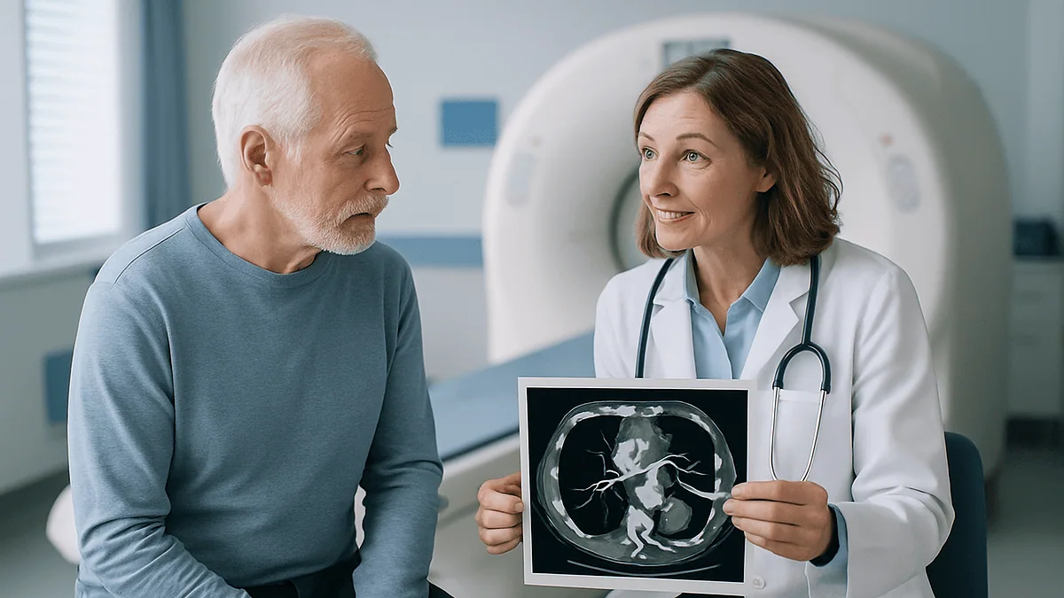
Key Takeaways
- The actual image capture in a CT coronary angiogram takes only a few seconds, though the whole appointment, including preparation and observation, lasts around an hour.
- Image quality depends on a slow heart rate, ideally in the low sixties or below, which is why many people are given a short-acting heart-slowing medicine solely as a photography aid.
- Avoiding caffeine for about 12 hours and food for about 4 hours before the scan reduces the chance of a blurred, fast-heart-rate study that has to be repeated.
- A coronary calcium score of zero suggests very little hardened plaque, but soft, non-calcified plaque does not show on a calcium scan and requires the contrast angiogram to detect.
- Narrowing of 50 to 69 percent on CT is considered moderate and often needs a functional test to determine whether it actually limits blood flow, because percentages on a still image do not always predict that.
- The contrast dye is cleared by the kidneys within a day, so drinking extra water after the scan and having a recent kidney function blood test beforehand both matter more than most people expect.
A CT coronary angiogram is a non-invasive heart scan that combines an iodine-based contrast dye injected into an arm vein with a fast, heartbeat-timed CT scanner to produce detailed images of the coronary arteries. It shows whether plaque is narrowing those arteries and by roughly how much. The scan itself takes seconds, the visit about an hour, and most people go home the same day; results are interpreted alongside symptoms and risk factors by the treating team.
The strangest part, people often say afterward, is the warmth. A nurse warns you it is coming, the dye goes in, and for about thirty seconds you feel as though you have wet yourself or swallowed a mouthful of hot tea. Then it passes, the table slides back out, and the whole thing is over before you have finished wondering whether it has started.
That small anticlimax hides a genuinely remarkable piece of engineering. In the space of a few heartbeats, a modern scanner captures hundreds of thin slices of a moving organ and reassembles them into a three-dimensional map of arteries no wider than a drinking straw. Cardiologists can then scroll through those vessels the way you might scroll through a street view, looking for the fatty deposits that cause angina and heart attacks.
What follows is an honest account of the test: who it helps, what it cannot tell you, how to prepare, and what the numbers on the report actually mean.
What is a CT angiogram, and how is a coronary one different?
Any CT angiogram (often shortened to CTA) is a computed tomography scan performed while an iodine-based contrast agent flows through the blood vessels. Ordinary CT distinguishes bone from soft tissue well but struggles to separate blood from the vessel wall. The contrast fixes that: it makes flowing blood appear bright white, so the inside of an artery stands out sharply against anything that narrows or blocks it. According to Mayo Clinic, CT angiography can be used to examine vessels almost anywhere in the body, from the brain and neck to the lungs, kidneys and legs.
A CT coronary angiogram (CCTA) is the version aimed specifically at the arteries that feed the heart muscle. Those vessels are small, they curve around a surface that is constantly contracting, and a blurred image is a useless one. Cardiac scanners therefore synchronize image capture with the electrocardiogram, taking their pictures in the brief pause between beats when the heart is nearly still. The NIH StatPearls chapter on coronary CT angiography describes this ECG gating as the technical step that turned the test from a research curiosity into routine clinical practice.
You will sometimes see the phrase cardiac CT used loosely to cover several related scans. A coronary calcium score is a cardiac CT without contrast that counts calcified plaque. A CT angiogram of the heart is the contrast version that shows the artery lumen itself. Both can be done in the same appointment, and many centers do exactly that.
Why would a doctor order a CT coronary angiogram?
The most common reason is chest pain that does not yet have a clear explanation. Someone in their fifties notices tightness when climbing stairs, or a person with diabetes reports breathlessness that could be heart-related or could be nothing of the kind. The clinician wants to know whether coronary artery disease is present before deciding what to do next, and the CT angiogram answers that question without threading a catheter into the body.
Both the NHS and Cleveland Clinic list several other situations in which the scan is useful:
- Checking a congenital abnormality in how the coronary arteries branch or where they arise
- Assessing bypass grafts or stents in people who have already had heart procedures, where clinically appropriate
- Investigating an equivocal or inconclusive result from a stress test
- Planning certain heart valve or structural procedures, where the anatomy needs to be mapped in advance
The scan is particularly good at one thing: ruling disease out. A clean, plaque-free set of coronary arteries on CT is strongly reassuring, and it spares a patient the small but real risks of an invasive angiogram. That strength shapes who is referred. Guideline bodies generally position CCTA as a first-line test for people at low to intermediate likelihood of coronary disease, rather than for those whose story already makes a severe blockage very likely. In that second group, going straight to invasive angiography, which can treat a narrowing as well as show it, may make more sense. The decision belongs with the treating team, who weigh symptoms, risk factors and previous tests together.
How does the scanner actually see inside a beating heart?
Picture a doughnut-shaped machine with an X-ray tube on one side and a ring of detectors on the other, spinning around you several times a second. Each rotation captures a thin cross-sectional slice; a computer stacks those slices into a volume you can rotate and slice in any direction. That is basic CT.
Three additions make the coronary version work. First, timing. Small electrodes on the chest feed the heart rhythm into the scanner, which either fires only during the quiet diastolic phase or records continuously and later keeps just the frames from that phase. Second, speed. Coronary studies rely on multidetector scanners capable of covering the whole heart within a few heartbeats, sometimes a single one, which is why an older or slower machine cannot simply be repurposed for the job. Third, a slow, steady heart rate. StatPearls notes that image quality improves markedly when the pulse is held around the low sixties or below, so many people are given a short-acting medicine that slows the heart before the scan. That medicine is not a treatment; it is a photography aid, and it wears off within hours.
Immediately before imaging, a second medicine may be sprayed or dissolved under the tongue to widen the coronary arteries for a few minutes. Wider vessels are simply easier to measure. The contrast agent, timed to arrive in the coronary arteries at the exact moment the images are taken, then makes the blood glow. The result is a picture in which the artery lumen, the fatty or calcified plaque within the wall, and the muscle beyond can each be told apart.
CT angiogram vs calcium score vs invasive angiogram: what is the difference?
Patients frequently arrive with these three tests tangled together, and the differences matter.
A coronary calcium scan is the simplest. There is no contrast and no medicine to slow the heart. The scanner counts flecks of calcium in the artery walls and produces a score. Mayo Clinic explains that calcium is a marker of plaque that has been present long enough to harden, so a score of zero suggests very little disease, while higher numbers indicate a greater plaque burden. What the calcium scan cannot show is soft, non-calcified plaque or how much any deposit narrows the vessel.
The CT coronary angiogram adds contrast and answers those questions. It shows both calcified and soft plaque, estimates how much of the lumen is lost, and maps the anatomy of every major branch. It is still non-invasive: one cannula in an arm vein and nothing threaded toward the heart.
Invasive coronary angiography, sometimes called cardiac catheterization, is the traditional gold standard. A thin tube is guided from the wrist or groin up to the heart, contrast is squirted directly into each coronary artery, and X-ray movies are recorded. The NHS notes that the same procedure allows a blocked artery to be opened with a balloon and stent in the same sitting. That treatment capability is its great advantage; its disadvantages are a small risk of bleeding, vessel injury or stroke, a longer recovery, and greater cost and staffing.
Put simply: the calcium scan asks whether plaque exists, the CT angiogram asks how much and where, and the invasive angiogram can do something about it.
How do you prepare for a CT coronary angiogram?
Preparation is light compared with most hospital procedures, but a few details genuinely change the quality of the images.
Caffeine is the first. Coffee, tea, energy drinks, cola and even chocolate raise the heart rate, and Mayo Clinic advises avoiding them for about 12 hours before the scan. The same source suggests not eating for roughly 4 hours beforehand; plain water is usually encouraged, because being well hydrated helps the kidneys clear the contrast afterward. Individual centers vary, so the instructions on your appointment letter override anything written here.
Medicines are the second detail. Bring a list of everything you take. Most can continue as normal, though some diabetes medicines are occasionally paused for a short period around contrast injection, and anyone who takes a daily medicine to slow the heart will be asked whether they have taken it. Do not stop or start anything on your own; the imaging team or prescribing clinician will tell you what to do.
Third, disclose anything relevant to contrast safety. Tell the team if you have had a previous reaction to contrast dye, have kidney disease, have asthma, are pregnant or might be, are breastfeeding, or have a thyroid condition. None of these automatically rules out the scan, but each changes how it is planned.
Finally, the practical things. Wear a top that is easy to remove; you will change into a gown. Leave necklaces at home, as metal near the chest can streak the images. Ask whether you may drive afterward, because the heart-slowing medicine occasionally causes light-headedness.
What happens on the day, step by step?
Expect to be in the department for around an hour, although the scanning itself takes seconds. Mayo Clinic puts the actual image acquisition at a few seconds and the full appointment, including preparation, at roughly an hour.
It usually unfolds like this. A nurse or radiographer checks your identity, allergies, kidney function results and pregnancy status, then places a small cannula, most often in the crook of the elbow. Electrodes go on your chest so the scanner can read your rhythm. Your pulse is checked; if it is on the fast side, you may be given the heart-slowing medicine as a tablet or through the cannula and asked to rest for a while until it takes effect.
You then lie on your back on the scanner table with your arms above your head. The table moves you into the ring. A staff member will ask you to practice holding your breath, because chest movement blurs the arteries as surely as a fast heartbeat does; the hold is typically short, around 5 to 10 seconds. A vessel-widening medicine may be given under the tongue at this point.
The contrast is injected by an automatic pump. You feel the warm flush and sometimes a metallic taste. The scanner completes its rotations while you hold your breath, and the sequence is done. Staff check the images for quality while you are still on the table in case a repeat is needed. The cannula comes out, and after a brief observation period you are free to go.
Is a CT angiogram painful?
Not in any meaningful sense, and it helps to know exactly what the odd sensations are so they do not alarm you.
The cannula insertion is a brief, sharp pinch, the same as having blood drawn. Some people find lying with their arms overhead for several minutes uncomfortable in the shoulders. The scanner itself makes a steady whirring or humming sound and is open at both ends, so it is far less enclosing than an MRI tunnel; claustrophobia is rarely a problem.
Then there is the contrast. Cleveland Clinic describes the classic trio: a wave of warmth spreading through the body, a metallic taste in the mouth, and a distinct sensation of having passed urine. You have not. The warmth comes from the iodine solution dilating small blood vessels and stimulating nerve endings as it circulates, and it fades within a minute or so. A few people feel briefly nauseated.
The vessel-widening medicine can cause a short-lived headache or a pounding feeling in the head. The heart-slowing medicine occasionally leaves people feeling a little flat or dizzy on standing. Neither effect is painful, and both wear off within hours.
Anything beyond this is worth mentioning at once. Itching, a rash, swelling of the lips or throat, wheezing or a feeling of faintness during or shortly after the injection can indicate a contrast reaction. The NHS describes serious reactions as rare, and the imaging team is trained and equipped to deal with them, which is one reason you are asked to wait a short time before leaving.
How long does it take to recover from a CT angiogram?
Recovery, in the sense most people mean it, is almost immediate. There is no incision, no puncture of an artery and no sedation, so you are not confined to a bed afterward. Mayo Clinic advises that people can generally return to normal activities the same day, with one practical exception: if you were given a medicine to slow your heart, the department may ask you to wait a short while and to have someone else drive.
The main task in the first day is hydration. Drinking more water than usual helps the kidneys flush the contrast agent, which is excreted in urine. You may notice you pass urine a little more often; that is the contrast leaving, not a problem.
Breastfeeding parents often ask whether they must pump and discard milk. Current guidance from major radiology bodies, summarized on the Cleveland Clinic page, is that only a tiny fraction of iodinated contrast reaches breast milk and even less is absorbed by the infant, so most people can continue feeding as normal. Ask your own team if you prefer a specific instruction.
Watch the cannula site for a day or two. A small bruise is common. Increasing redness, swelling or pain at the site, or a rash, hives or breathing difficulty appearing hours later, should prompt a call to the department or an urgent care service. Delayed contrast reactions, usually mild skin rashes, can occur up to a week later and are worth reporting so they are recorded for future scans.
Results typically take a few days, because a radiologist or cardiologist must reconstruct and review the images vessel by vessel before writing the report.
What does a CT angiogram show, and how do you read the report?
Reports have become more standardized in recent years, which is good news for patients trying to understand them. Most describe two things: how much calcified plaque is present, if a calcium score was performed, and how severely each artery is narrowed.
Calcium scores are grouped into bands. Mayo Clinic summarizes them as follows: zero means no detectable calcified plaque; 1 to 99 is a small amount; 100 to 399 is moderate; 400 or more is extensive. Bear in mind that soft plaque does not calcify, so a low score does not guarantee a completely clear artery.
Narrowing, or stenosis, is usually reported as a percentage of the artery diameter lost. StatPearls describes the widely used reporting categories, which the table below simplifies.
| Report wording | Approximate narrowing | What it generally means |
|---|---|---|
| No plaque | 0% | No visible coronary disease |
| Minimal | 1 to 24% | Plaque present, flow essentially unaffected |
| Mild | 25 to 49% | Non-obstructive disease |
| Moderate | 50 to 69% | May or may not limit blood flow; further testing sometimes needed |
| Severe | 70 to 99% | Likely to limit flow, especially with exertion |
| Occluded | 100% | Vessel completely blocked |
The report may also comment on plaque character, on the anatomy of each vessel, and on incidental findings in the lungs or elsewhere that happened to be in the picture. None of these numbers is a diagnosis on its own. A 55 percent narrowing in someone with no symptoms and a 55 percent narrowing in someone with classic exertional chest pain are read very differently.
What happens if they find a blockage during a CT angiogram?
Nothing dramatic happens in the scanner room. Unlike an invasive angiogram, a CT cannot treat what it finds, so the immediate result is a conversation rather than a procedure.
What follows depends on how severe the narrowing is and how it lines up with your symptoms. For minimal or mild plaque, the usual response is preventive: the finding confirms that atherosclerosis has begun, and the focus shifts to slowing it down through blood pressure control, cholesterol management, stopping smoking, physical activity and, where the clinician judges appropriate, long-term cholesterol-lowering and blood-thinning medicines. Those decisions rest with the prescribing clinician and depend on your overall risk, not the scan alone.
For moderate narrowing, the question is whether the plaque actually restricts blood flow. Percentages measured on a still image do not always predict that. A stress test, a specialized CT perfusion or flow analysis, or an invasive assessment during catheterization may be recommended to find out.
For severe narrowing or a blocked vessel, particularly with symptoms, referral to a cardiologist for invasive angiography is common. There, the same narrowing can be measured directly and, if appropriate, treated with a balloon and stent. Some patterns of disease, such as narrowing in several vessels or in the main left coronary artery, may lead to a discussion of bypass surgery instead.
Two reassurances. A finding of plaque is common and does not mean a heart attack is imminent. The point of the scan is to catch disease early enough that its course can be changed, and the choice among options is always made with you, weighing benefits, risks and what matters to you.
What are the risks: radiation, contrast dye and your kidneys?
Three risks are worth understanding honestly.
Radiation is the one people ask about most. CT uses X-rays, and the coronary version historically delivered a relatively high dose because of the need for many rapid images. Newer scanners and lower-dose protocols have reduced this substantially; StatPearls notes that modern techniques can bring the effective dose of a coronary CT angiogram down to a level comparable to or lower than an invasive angiogram in many cases. The NHS describes the added lifetime cancer risk from a single CT scan as small, and the amount you receive should be recorded and available on request. Pregnancy is the situation in which radiation is weighed most carefully, which is why you will be asked about it.
Contrast reactions are the second. Mild effects, such as nausea, a rash or hives, are uncommon and usually settle without treatment. Severe allergic-type reactions are rare, according to the NHS, and imaging departments keep the means to treat them immediately at hand. A previous reaction is the strongest predictor of another one, so always mention it.
The kidneys are the third. Iodinated contrast can temporarily reduce kidney function, particularly in people whose kidneys are already impaired, who are dehydrated, or who have diabetes. This is why a recent blood test of kidney function is usually required and why you are encouraged to drink water before and after. In most people with normal kidney function the risk is very low.
The two short-acting medicines used for image quality have their own small risks, mainly a temporary drop in heart rate or blood pressure, and there are people for whom they are unsuitable. The team screens for this before giving them.
Who might not be a good candidate, and what are the alternatives?
The test is powerful but not universal, and knowing its limits helps explain why a clinician might suggest something else.
Image quality suffers when the heart rate cannot be brought down, as in atrial fibrillation or other irregular rhythms, or when someone cannot hold their breath for several seconds. Heavy calcification is another problem: dense calcium scatters X-rays and can make a vessel look more narrowed than it is, which is one reason the scan is aimed at people at lower likelihood of advanced disease. Very small stents can be hard to assess for the same reason. Body size affects image noise, and severe kidney impairment or a serious previous contrast reaction may make the injection inadvisable.
Alternatives fall into two groups. Functional tests ask whether the heart muscle receives enough blood under stress, rather than mapping the arteries directly. These include exercise ECG testing, stress echocardiography, nuclear perfusion imaging and stress cardiac MRI, the last of which involves no radiation. The American Heart Association describes these as complementary approaches, each suited to different questions and different patients.
The other alternative is to go straight to invasive coronary angiography, especially when the likelihood of significant disease is already high or when treatment during the same procedure is anticipated.
None of these is universally better. A good referral matches the test to the question being asked and to the individual in front of the clinician, and it is entirely reasonable to ask why a particular test has been chosen over another.
When should you see a doctor about chest symptoms?
A CT angiogram is a planned test for people whose symptoms are stable. It is not the right pathway for an emergency, and recognizing the difference can save a life.
Call emergency services immediately if you or someone near you has chest pain or pressure lasting more than a few minutes, or that comes and goes, especially if it spreads to the arm, jaw, neck or back, or is accompanied by breathlessness, cold sweat, nausea, light-headedness or a sense of dread. The American Heart Association stresses that symptoms in women, older adults and people with diabetes are more often atypical, presenting as unusual fatigue, indigestion-like discomfort or shortness of breath rather than crushing pain. Sudden severe breathlessness, fainting, or a rapid irregular heartbeat with chest discomfort are also emergency signs. Do not drive yourself.
Arrange a non-urgent appointment if you notice chest tightness, pressure or breathlessness that reliably appears with exertion and settles with rest, particularly if you smoke, have high blood pressure, high cholesterol, diabetes or a close relative who developed heart disease before age 60. That pattern is exactly what the scan was designed to investigate.
After the scan itself, contact the imaging department or seek urgent care for a spreading rash, swelling of the face or throat, wheezing, or increasing pain and redness at the cannula site. Report any reaction, however mild, so it is documented for the future.
What questions should you ask before and after the scan?
Patients who ask good questions tend to leave with a clearer sense of what the result means for them. Consider these before the appointment:
- Why this test rather than a stress test or an invasive angiogram in my case?
- Will a calcium score be done at the same time?
- Do I need a kidney blood test first, and has it been arranged?
- Should I take my usual medicines on the morning of the scan?
- Will I be given a heart-slowing medicine, and can I drive afterward?
Once the report is back, a different set of questions becomes useful:
- Was the image quality good enough to answer the question, or were some segments unclear?
- How much plaque is there, and is it mostly calcified or soft?
- Does any narrowing appear severe enough to limit blood flow, and how do you know?
- Were there any incidental findings outside the heart, and do they need follow-up?
- What would change about my treatment if we did nothing further?
The last question is the one most worth asking. A CT angiogram is a snapshot of anatomy at one moment. Its value comes from how it alters the plan: reassuring someone whose arteries are clear, sharpening prevention in someone with early plaque, or pointing toward further tests and treatment in someone with a significant narrowing. Understanding which of those three situations you are in, and why, is the real result of the scan. The pictures are just the means of getting there.
Frequently asked questions
What is a CT angiogram?
A CT angiogram is a CT scan performed while an iodine-based contrast dye flows through the blood vessels, making the blood appear bright so the inside of arteries can be seen clearly. When aimed at the heart, it is called a CT coronary angiogram and uses heartbeat-timed imaging to map the coronary arteries and detect plaque or narrowing without inserting a catheter.
Why would a doctor order a CT coronary angiogram?
Most often to investigate chest pain or breathlessness when coronary artery disease is possible but not certain, particularly in people at low to intermediate likelihood of disease. It is also used to check the anatomy of unusual coronary arteries, assess grafts or stents in selected cases, clarify an inconclusive stress test, and plan certain heart procedures where the vessel layout must be known in advance.
Is a CT angiogram painful?
No. The cannula placement feels like a blood test, and the scan itself is silent apart from a whirring sound. The contrast produces a brief warm flush, a metallic taste and a sensation of having passed urine, all of which fade within about a minute. The vessel-widening medicine can cause a short headache. Anything more, such as a rash, wheezing or faintness, should be reported to staff immediately.
How long does it take to recover from a CT angiogram?
Almost no recovery is needed. There is no incision or sedation, and most people resume normal activities the same day. If you were given a heart-slowing medicine, you may be asked to wait briefly and to avoid driving until it wears off. Drinking extra water for the rest of the day helps clear the contrast through the kidneys. Written results usually follow within a few days.
What happens if they find a blockage during a CT angiogram?
The scan cannot treat a blockage, so the finding leads to a discussion rather than an immediate procedure. Mild plaque usually prompts a stronger focus on prevention and risk-factor control. Moderate narrowing may need a functional test to see whether it restricts blood flow. Severe narrowing or a blocked vessel, especially with symptoms, often leads to referral for invasive angiography, where a stent can be placed if appropriate.
How long does a CT coronary angiogram take?
The image acquisition itself lasts only a few seconds, during a short breath-hold. The full appointment typically takes about an hour, which includes checking your history, placing a cannula, attaching ECG electrodes, waiting for any heart-slowing medicine to take effect, the scan, and a brief observation period afterward to make sure you have had no reaction to the contrast.
How much radiation is in a CT angiogram of the heart?
More than a chest X-ray but, with modern scanners and low-dose protocols, often comparable to or lower than an invasive angiogram. The added lifetime cancer risk from a single scan is described by the NHS as small. Your department records the dose delivered and can share it on request. Pregnancy is the situation in which radiation exposure is weighed most carefully, so always mention it.
What is the difference between a cardiac CT and an angiogram?
Cardiac CT is an umbrella term that includes a non-contrast calcium score and the contrast-enhanced CT coronary angiogram, both performed non-invasively from an arm vein. An invasive angiogram, or cardiac catheterization, threads a thin tube from the wrist or groin to the heart to inject contrast directly. The invasive test carries slightly more risk but can treat a narrowing with a stent during the same procedure.
Can you eat or drink coffee before a CT angiogram?
Most centers ask you to avoid caffeine for around 12 hours and to stop eating about 4 hours before the scan, because caffeine speeds the heart and food can worsen nausea from the contrast. Plain water is usually encouraged to support kidney clearance of the dye. Follow the exact instructions on your appointment letter, since protocols vary between departments.
What does a calcium score of zero mean?
It means the scan detected no calcified plaque in the coronary arteries, which is associated with a low near-term risk of heart events. It does not prove the arteries are entirely clear, because early, soft plaque has not yet hardened and is invisible on a calcium scan. That is why a contrast CT angiogram is sometimes performed alongside it when symptoms are present.
References
This article is for general information only and is not a substitute for professional medical advice. Please consult a qualified doctor about your individual situation.

More from the Blog

Recovery After a Stereotactic Biopsy: Headache, Pin-Site Soreness, Driving and Results
Stereotactic biopsy recovery is usually short. After a breast biopsy, most people go home the same day and have bruising or soreness for a…

After a PET-CT Scan: Clearing the Tracer, Contact Precautions and When Results Arrive
After a PET-CT scan, most people go home the same day, drink extra fluids and empty their bladder often to help clear the radioactive…
Preparing for an MRI: Eating, Metal, What to Wear
For most MRI scans you can eat and drink normally beforehand, because the magnet images tissue, not stomach contents. The exceptions are scans of…

What Does a Comprehensive Check-Up Include? Blood Work, Imaging and the Follow-Up Plan
A comprehensive check-up usually includes a medical and family history, a physical examination with blood pressure and body measurements, and blood and urine tests…
What a CT Scan Shows, and Why Doctors Order One
A CT scan shows detailed cross-sectional images of bones, organs, blood vessels and soft tissue by combining many X-ray views into slices. Doctors most…
Can an Ultrasound Detect Cancer? What It Can Show, Its Limits and the Next Steps
An ultrasound can detect abnormal lumps, masses and changes in organs that may be cancer, and it is especially good at telling a fluid-filled…