Coronary Angiography
Coronary angiography is an imaging test that shows blood flow inside the heart arteries. It helps diagnose narrowed or blocked coronary vessels and guides further cardiac treatment planning.
Quick answer
Coronary angiography, often called an angiogram, is an X-ray test that shows the arteries supplying blood to the heart. A thin catheter is guided from the wrist or groin to the heart, contrast dye is injected, and moving images reveal any narrowings or blockages. It is usually done under local anaesthetic with light sedation, and many patients go home the same day.
What Is Coronary Angiography?
Coronary angiography is an X-ray examination of the coronary arteries, the blood vessels that carry oxygen-rich blood to your heart muscle. A cardiologist passes a thin, flexible tube called a catheter from an artery in your wrist or groin up to the heart, injects a contrast dye, and records moving X-ray images that show exactly where the arteries are open, narrowed or blocked. Most people know the test simply as an angiogram. It is performed to confirm or rule out coronary artery disease, to explain symptoms such as chest pain or breathlessness, and to guide decisions about medication, stenting or bypass surgery.
The strength of the test is precision. Stress tests and scans can suggest that part of the heart is short of blood; an angiogram shows the anatomy directly, which artery is affected, where the narrowing sits, how severe it looks, and whether it is likely to explain your symptoms or your risk profile. Rather than relying only on symptoms or indirect results, your cardiology team sees the vessels themselves, and treatment decisions are built on that detail.
For many patients, the angiogram is a decision point rather than just a picture. If a significant blockage is found and the circumstances are suitable, balloon angioplasty and stent placement can sometimes be performed during the same session. When disease is complex or involves several vessels, the images are usually reviewed by a heart team before a treatment plan is chosen. The aim is a plan that is safe, evidence-based, and matched to your heart anatomy, your general health and your priorities.
What does an angiogram show?
An angiogram shows whether each coronary artery is open, narrowed or blocked, and it does so in real time. As the contrast dye flows through the vessels, the imaging system records the pattern from several angles. The cardiologist can see the location and shape of plaques, how many vessels are involved, whether blood flow to the heart muscle is limited, and how the arteries branch. The images also reveal the state of any previous stents or bypass grafts, and congenital variations in coronary anatomy that occasionally matter for planning. What the test does not show directly is the strength of the heart muscle or the condition of the heart valves; those questions are usually answered by echocardiography, cardiac MRI or other studies, which can sometimes be arranged during the same admission.
What is cardiac catheterization, and is it the same thing?
Cardiac catheterization is the broader family of procedures in which a catheter is guided into the heart or its vessels; a coronary angiogram is one specific study that can be done during it. A heart catheterization, often shortened to heart cath, may also include measuring pressures inside the heart chambers, assessing the valves, evaluating how strongly the heart pumps, or delivering treatment. Coronary angiography refers specifically to imaging the coronary arteries with contrast dye. In everyday conversation the terms overlap, and your medical team may use them interchangeably, but it is useful to know that a catheterization can involve more than pictures of the arteries. If your referral letter mentions pressure measurements or valve assessment alongside the angiogram, that is why.
Invasive angiogram or coronary CT angiography, which one?
There are two main ways to look at the coronary arteries, and they answer slightly different questions. Invasive coronary angiography, the subject of this page, uses catheters and contrast dye to produce detailed, real-time images from inside the circulation. Noninvasive coronary CT angiography works from outside the body: contrast is injected into a vein in the arm, a CT scanner captures the heart between heartbeats, and software reconstructs the arteries in three dimensions. No catheter travels to the heart, which is why CT is often chosen when the likelihood of significant disease is lower and the main goal is to rule coronary disease out.
Invasive angiography remains the more definitive test when symptoms are significant, when noninvasive results suggest high-risk disease, when a heart attack is suspected, or when the team may need to proceed directly to treatment. It is also used when earlier tests have produced unclear or conflicting results. The right choice depends on the clinical question, the urgency, your kidney function, your heart rhythm, any previous procedures, and how likely coronary artery disease is in your particular case. Neither test is automatically better; they are tools for different situations, and many patients have both at different points in their care.
Understanding the Decision to Have an Angiogram
When chest pain, shortness of breath, an abnormal stress test or a suspected heart attack leads a physician to recommend an angiogram, concern is a normal response. Most patients ask the same questions: what might the test find, will a stent or surgery follow, how safe is the procedure, and how soon can normal life resume. Those are reasonable questions, and the answers on this page are meant to make the recommendation easier to understand and to discuss with your own cardiologist.
It helps to understand what the test is actually for. If the coronary arteries become narrowed by atherosclerosis, blood flow to the heart muscle can fall, especially during physical activity or emotional stress. In more serious cases, a sudden blockage causes a heart attack. Coronary angiography exists to settle the question of blood supply definitively, so that the next step, whether that is medication, angioplasty, bypass surgery or simply continued monitoring, rests on anatomy rather than assumption.
It also helps to know what the test is not. It is not automatically the first step for everyone with chest discomfort, and it does not commit you to a stent or an operation. A meaningful proportion of angiograms end with reassuring findings and a plan built around medication and prevention. Others identify a clear target for treatment. Either result is useful, because either result replaces uncertainty with a plan.
Who May Need a Coronary Angiogram?
A coronary angiogram may be recommended when symptoms or test results suggest that the heart muscle is not receiving enough blood. The decision is individual. A cardiologist weighs your symptoms, medical history, risk factors, physical examination, electrocardiogram, blood tests and any noninvasive imaging or stress testing before recommending an invasive study.
Typical symptoms that prompt the test include chest pressure, tightness, heaviness, burning or pain that appears with exertion or emotional stress. Some people feel discomfort in the left arm, both arms, the neck, jaw, back or upper abdomen instead of the chest. Shortness of breath, unusual fatigue, nausea, sweating, dizziness or fainting can also reflect reduced blood flow to the heart, patterns that are particularly common in women, older adults and people with diabetes, whose symptoms are often less typical and easier to miss.
The test is also central to urgent care. When a patient shows signs of a heart attack, especially when the electrocardiogram suggests an acutely blocked artery, emergency angiography identifies the blockage and guides immediate treatment. In these circumstances the angiogram is not an elective decision; opening the artery promptly limits damage to the heart muscle, and the entire workflow is built around speed.
Many patients arrive at angiography by way of another test. An abnormal stress test, echocardiogram, nuclear imaging study, cardiac MRI or coronary CT angiogram may suggest that part of the heart is under-supplied, that pumping function is reduced, or that plaque is likely to be present. Invasive angiography then confirms the diagnosis and clarifies which treatment approach makes sense.
Patients with known coronary artery disease may need repeat angiography if symptoms return or worsen after stenting, bypass surgery or medical treatment. The test is also commonly performed before major heart operations, valve surgery in particular, to establish whether coronary disease should be treated at the same time.
Does every patient with chest pain need an angiogram?
No. Many conditions mimic heart-related chest pain, including acid reflux, lung disease, muscle strain, anxiety, gallbladder disease and inflammation around the heart. Diagnosis therefore begins with careful clinical evaluation: your cardiologist asks about the character of your symptoms, how long they last, what triggers them and what relieves them. Risk factors are weighed, high blood pressure, high cholesterol, diabetes, smoking, a family history of early heart disease, obesity, chronic kidney disease, previous stroke or vascular disease. Blood tests may assess heart injury markers, kidney function, blood count, clotting, cholesterol and blood sugar control. A well-planned pathway avoids unnecessary invasive procedures while making sure serious coronary disease is not missed. An angiogram is recommended when the balance of evidence says the answer it provides is worth having.
Conditions Coronary Angiography Evaluates
Coronary angiography is used across a range of heart conditions in which the coronary arteries may be involved. The most common indication is suspected coronary artery disease, also called ischaemic heart disease, in which fatty deposits, inflammatory cells, calcium and scar tissue build up in the artery wall, forming plaques that narrow the vessel or make it vulnerable to sudden clot formation.
Stable angina. Here, chest discomfort or breathlessness appears predictably with exertion and settles with rest or medication. Angiography may be advised when symptoms persist despite medical therapy, when noninvasive tests show a higher-risk pattern, or when the diagnosis remains uncertain despite a thorough work-up.
Acute coronary syndromes. These are urgent indications and include unstable angina and heart attacks. In unstable angina, symptoms occur at rest, become more frequent or feel more severe than before. In a heart attack, part of the heart muscle is injured because blood flow has been severely reduced or interrupted. Angiography identifies the responsible artery and supports time-sensitive treatment.
Heart failure and reduced pumping function. Patients with heart failure may undergo angiography to establish whether blocked arteries are contributing to a weakened heart muscle. If reduced blood flow is a major cause, restoring circulation through stenting or bypass surgery may become part of the treatment plan.
Rhythm problems with a suspected coronary cause. Certain abnormal heart rhythms raise the question of underlying coronary disease as a trigger, and angiography can answer it. Rhythm disorders themselves are assessed and treated through their own specialist pathways, but when a rhythm problem may be a consequence of reduced blood supply, imaging the arteries directly settles the question.
Planning before structural or valve surgery. Before valve operations or other structural heart procedures, angiography is often performed to identify coronary disease that could affect the surgical plan. Patients being evaluated for heart valve disease frequently have the test as part of their pre-operative assessment.
Previous bypass surgery or stents. In patients who have already had coronary bypass surgery, angiography can assess both the native arteries and the bypass grafts. In patients with previous stents, it can show whether a treated segment has narrowed again.
Finally, angiography is sometimes performed simply because uncertainty persists. A patient may have symptoms strongly suggestive of coronary disease despite inconclusive imaging, or a scan may show findings that need confirmation before anyone acts on them. In complex clinical situations, the test’s job is to bring clarity.
How Is Coronary Angiography Performed?
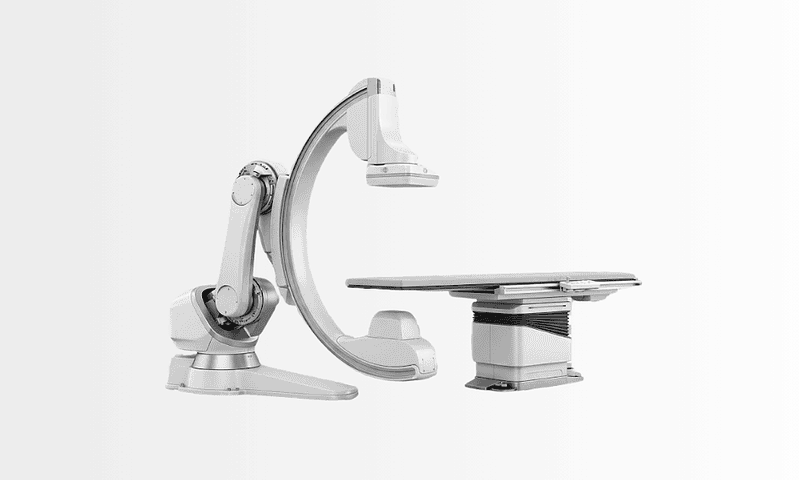
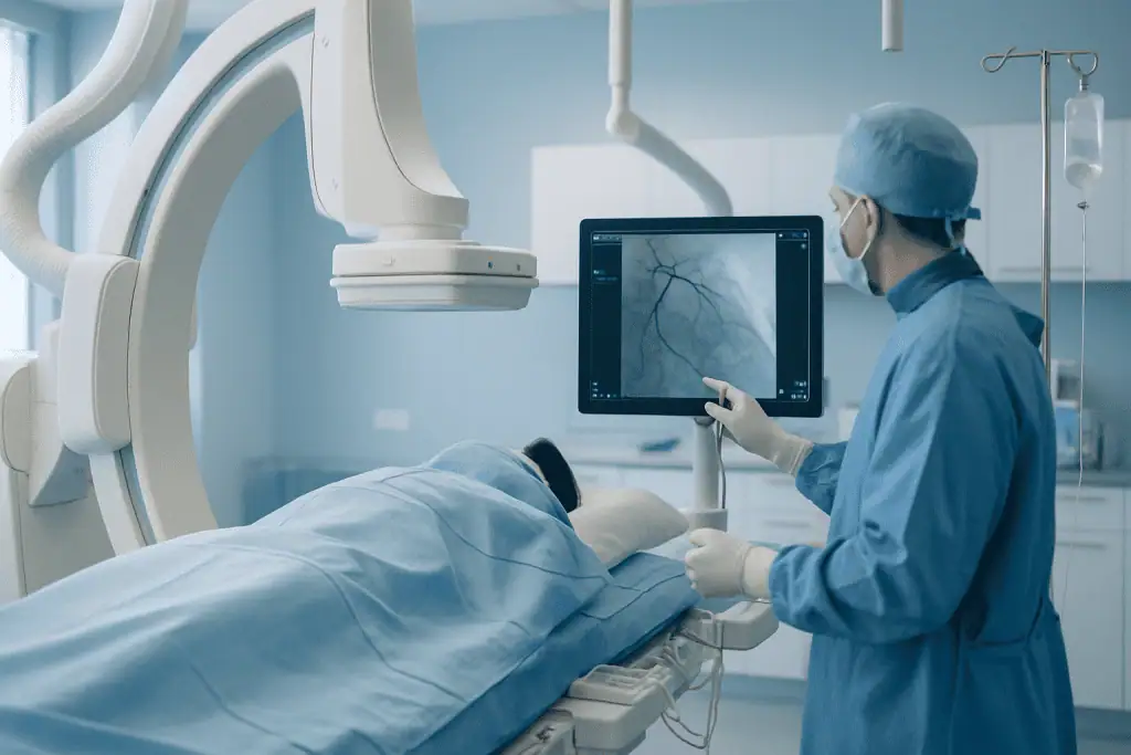
The procedure takes place in a dedicated cardiac catheterization laboratory: an environment built for advanced imaging, sterile technique, continuous monitoring and rapid treatment if an intervention becomes necessary. It is typically done under local anaesthetic with light sedation rather than general anaesthesia, although the approach varies with the patient’s condition and clinical needs. In outline, the steps are:
- Pre-procedure review of your history, medications, allergies, kidney function and recent tests, plus a short fasting period.
- Positioning on the catheterization table, with monitors tracking heart rhythm, blood pressure and oxygen level.
- Local anaesthetic at the wrist or groin, then insertion of a small sheath into the artery.
- Guiding the catheter to the openings of the coronary arteries under X-ray.
- Injecting contrast dye while images are recorded from several angles.
- Removing the catheter and sealing the access site, followed by monitored recovery.
Preparation before the procedure
Before the angiogram, the care team reviews your medical history, current medications, allergies, any previous reactions to contrast dye, kidney function, bleeding risk and recent test results. Expect questions about blood thinners, diabetes medications, kidney disease, thyroid disease, asthma, and prior stents or bypass surgery. It is worth bringing copies of recent records: ECGs, stress tests, echocardiography reports, CT scans, medication lists and discharge summaries, so the team works from a complete picture rather than repeating studies unnecessarily.
Most patients are asked not to eat or drink for several hours beforehand. Some medications are continued and others may need temporary adjustment; this is particularly relevant for anticoagulants, antiplatelet drugs and certain diabetes medications, and those decisions rest entirely with your treating cardiologist, who will give you specific instructions based on your health and the urgency of the procedure.
Blood tests commonly check kidney function, blood count, electrolytes and clotting. Kidney function matters because contrast dye is cleared through the kidneys. Where kidney impairment exists, the team takes extra precautions, careful hydration, limiting the volume of contrast, and choosing imaging strategies that avoid unnecessary dye exposure.
Are you awake during an angiogram?
Yes, in most cases you are awake, but relaxed. The procedure is usually performed under local anaesthetic at the access site, often with light sedation to ease anxiety. Staying awake is not an oversight: it is safer and more practical, because the team may ask you to take a deep breath, hold it briefly, or cough at certain moments to improve the images. You can speak to the team throughout, and if you feel uncomfortable at any point, additional sedation can usually be given. General anaesthesia is reserved for specific situations judged by the treating team.
Access through the wrist or groin
Once you are positioned on the table and connected to the monitors, the access site is cleaned and covered with sterile drapes, and local anaesthetic is injected to numb the skin. The catheter is most often introduced through the radial artery in the wrist or the femoral artery in the groin. The wrist approach usually allows earlier mobility afterwards and may reduce certain bleeding complications in suitable patients. The groin approach is preferred in some complex procedures, in patients whose wrist arteries are small or unsuitable, or when specific catheter equipment is needed. The cardiologist chooses based on safety, your anatomy and the goals of the procedure, not on habit.
Imaging the coronary arteries
With the catheter positioned at the openings of the coronary arteries, contrast dye is injected while the X-ray system captures blood flow. You may notice a brief warm sensation as the dye goes in; this is expected and passes quickly. The system records moving pictures from multiple angles so each artery segment can be evaluated clearly.
Modern catheterization laboratories use high-resolution digital imaging, dose-monitoring systems and software that optimises visualisation while managing radiation exposure. Where a narrowing looks borderline, the team may measure pressure inside the artery to determine whether it genuinely limits blood flow. In selected cases, intravascular imaging lets the cardiologist view the artery from within, useful for judging plaque structure, vessel size, stent position or complex anatomy. These tools are used when they add meaningful information, not routinely.
If a significant narrowing is found, the cardiologist may discuss whether immediate treatment is appropriate. In some cases, balloon angioplasty and stent placement, described in detail on our coronary stent applications page, proceed during the same session. In others, particularly with multiple blockages, left main disease, complex anatomy, diabetes, reduced heart function or other medical considerations, the findings go to a multidisciplinary heart team before any treatment is selected. Pausing to plan is not a delay; for complex disease it is the correct process.
Is an angiogram very painful?
Most people describe the angiogram as uncomfortable in moments rather than genuinely painful. The local anaesthetic injection stings briefly, many patients say this is the most unpleasant part. After that, you may feel pressure at the access site, and a short warm flush when the dye is injected, but the catheter moving through the blood vessels is usually not felt at all, because arteries have no pain receptors along their inner lining. Some soreness or bruising at the wrist or groin afterwards is common. Everyone’s experience differs, and the team can respond with more local anaesthetic or sedation if you are uncomfortable, so say so rather than enduring it.
How long does a coronary angiogram take?
A diagnostic angiogram often takes less than an hour, though preparation, monitoring and recovery make the overall visit considerably longer, usually most of a day. If additional measurements are taken or treatment is performed during the same session, the procedure itself takes more time. Emergency heart attack care follows a different, faster workflow focused on identifying and opening the blocked artery as quickly as is safe.
Immediately after the procedure
Once the catheter is removed, bleeding is controlled by direct pressure or a closure method. After wrist access, a compression band is usually applied and loosened gradually. After groin access, you may need to lie flat for a period while the artery seals. Nurses monitor your heart rhythm, blood pressure, the access site, your comfort and the circulation in the hand or leg. Before discharge you receive instructions covering activity, wound care, medications, hydration and follow-up arrangements.
How Serious Is Having an Angiogram?
An angiogram is an invasive procedure, so it is taken seriously, but it is also one of the most frequently performed cardiac procedures in the world, carried out in a controlled environment by teams who do it daily. For most patients the test proceeds without any significant problem, and the practical experience is closer to a long outpatient appointment than to an operation.
That said, honest information includes the risks. The most common issues are minor: bruising, soreness or a small lump at the access site, which settle over days to weeks. Less commonly, bleeding at the access site needs further treatment, or a patient reacts to the contrast dye; teams screen for allergy history beforehand and are prepared to manage reactions. Contrast can strain the kidneys, which is why kidney function is checked in advance and hydration and dye volume are managed carefully, especially in patients with existing kidney impairment. Rarely, more serious complications occur, including damage to a blood vessel, heart attack or stroke; catheterization laboratories are staffed and equipped to respond immediately if they do.
How this risk profile applies to you depends on your age, kidney function, other illnesses and the urgency of the situation, which is exactly the conversation to have with your cardiologist before consenting. The relevant comparison is not between the angiogram and doing nothing in the abstract, but between the risks of the test and the risks of leaving a possible coronary blockage undiagnosed. When the test is recommended for sound clinical reasons, that balance generally favours knowing.
Is the radiation from an angiogram harmful?
The radiation dose from a single diagnostic angiogram is kept deliberately low and, for most patients, the benefit of an accurate diagnosis clearly outweighs the exposure. Coronary angiography uses X-rays, which involve ionising radiation, and modern catheterization laboratories are designed around minimising it: dose-monitoring systems track exposure continuously, imaging angles and beam settings are adjusted to capture the necessary views efficiently, and the team avoids unnecessary imaging runs. Radiation exposure is one of the factors weighed when deciding between invasive angiography and alternatives, and it is a fair question to raise during the consent discussion. Patients who are or could be pregnant should tell the team beforehand, because that changes how, and sometimes whether, imaging is planned.
Recovery After a Coronary Angiogram
Recovery depends on three things: the access site, whether the procedure was diagnostic only or included treatment, and your overall medical condition. A purely diagnostic angiogram through the wrist involves the lightest recovery; a same-session stent, or a procedure performed during a heart attack, involves closer observation and a more structured follow-up plan.
How long are you kept in hospital after an angiogram?
Many patients who have a diagnostic angiogram go home the same day, after a few hours of monitored recovery, provided the access site is stable and there are no clinical concerns. An overnight stay is more likely if a stent was placed, if you presented with an acute coronary syndrome, if the groin was used and needs longer observation, or if other medical risks call for caution. Your team decides based on how the procedure went and how you are afterwards, not on a fixed schedule.
How do you look after the access site at home?
Looking after the access site mostly means keeping it clean and dry and giving it a short period of rest. After wrist access, patients are usually advised to avoid heavy lifting, repetitive gripping or leaning hard on that hand for a few days; after groin access, the advice typically covers avoiding strenuous activity, climbing and straining while the artery seals. Showering is generally permitted sooner than soaking, baths, pools and saunas usually wait until the site has healed, following the team’s specific instructions. A bruise, mild tenderness or a small firm lump under the skin is a normal part of healing and fades gradually. Drinking fluids in the first day, as advised, helps the kidneys clear the contrast dye. The discharge instructions you receive take priority over any general guidance, because they reflect your procedure, your access site and your medications.
| Time Period | What Patients Can Expect |
|---|---|
| Day 1 | Monitored recovery after the procedure. Mild soreness or bruising at the wrist or groin is common. Many diagnostic patients can return home the same day if clinically appropriate. |
| First Week | Most patients resume light daily activities. Heavy lifting, strenuous exercise and soaking the access site are usually avoided for a short period, following the physician’s instructions. |
| First Month | If no intervention was performed, many patients return to their normal routines. If a stent was placed or a heart attack occurred, medication planning, cardiac rehabilitation and structured follow-up become especially important. |
| Longer Term | Care focuses on controlling risk factors, taking prescribed medications, monitoring symptoms and maintaining regular cardiology follow-up to reduce future cardiac risk. |
Whether recovery is short or extended, the pattern is the same: protect the access site briefly, resume activity progressively, and treat the follow-up plan as part of the procedure rather than an optional extra. For patients who had a stent or a heart attack, a supervised cardiac rehabilitation programme is often the bridge between hospital care and confident everyday life.
Why Acting Early Matters
Coronary artery disease usually develops gradually, but its consequences can be sudden. A plaque that has narrowed an artery over many years may become unstable and trigger a clot, causing a heart attack. In other patients, progressive narrowing limits blood flow enough to cause angina, breathlessness, reduced exercise tolerance or a gradual weakening of the heart muscle.
Acting early does not mean every patient needs an invasive test immediately. It means symptoms and abnormal results deserve thoughtful evaluation without unnecessary delay. Chest pain with exertion, new shortness of breath, fainting, worsening fatigue or symptoms at rest warrant proper assessment, particularly in people who carry risk factors for coronary disease.
Delaying evaluation lets disease progress untreated. Patients restrict their activity because of symptoms, lose confidence in daily life, or experience complications that earlier clarity might have prevented. In urgent situations such as a heart attack, time matters directly, because prolonged loss of blood flow damages more heart muscle.
Early diagnosis also protects against overtreatment. Some patients learn that their arteries are not significantly narrowed, which redirects attention to other causes of their symptoms and to preventive care, rather than to procedures they do not need. For patients with meaningful blockages, early clarity supports a more precise plan. Either way, the angiogram earns its place by ending guesswork.
Benefits of Coronary Angiography
The central benefit is information: detailed, direct, decision-grade information about the blood supply to your heart, gathered in a setting where treatment can follow immediately if it is needed.
| Benefit | What It Means for You |
|---|---|
| Direct visualization of coronary arteries | Shows where arteries are narrowed or blocked and helps explain symptoms such as chest pain or shortness of breath. |
| Guides treatment planning | Helps determine whether medication, stenting, bypass surgery or continued observation is the most appropriate next step. |
| Supports urgent heart attack care | Can identify the blocked artery quickly and guide immediate treatment when time-sensitive intervention is needed. |
| Clarifies uncertain test results | Provides additional detail when stress tests, CT imaging or symptoms do not clearly answer the clinical question. |
| Allows treatment in selected cases | If appropriate, angioplasty and stent placement may be performed during the same session, avoiding a separate procedure. |
| Helps assess previous treatments | Can evaluate prior stents or bypass grafts if symptoms return or new concerns arise. |
What Influences Outcomes and a Good Result?
A good result begins with choosing the right test for the right reason. The most meaningful outcomes occur when the angiogram is one step in a broader diagnostic and treatment strategy, not an isolated event. Your symptoms, risk factors, previous test results and personal goals all shape how the findings are read and acted on.
The severity and pattern of disease matter. A short narrowing in one artery may be managed very differently from complex disease across several vessels. Disease in the left main coronary artery, blockages at vessel branches, heavily calcified plaques, long narrowed segments or reduced pumping function each call for more detailed planning. Some patients are best treated with medication, some with stenting, and some with bypass surgery, and the honest answer is often not obvious until the images exist.
Overall health also shapes the plan. Diabetes, kidney disease, anaemia, bleeding risk, previous stroke, lung disease, frailty and age influence both how the procedure is done and which treatment is chosen afterwards. Kidney function deserves particular respect because contrast dye must be used carefully where the kidneys are impaired. Medication allergies and previous contrast reactions are reviewed in advance for the same reason: outcomes improve when problems are anticipated rather than discovered.
The quality of imaging and interpretation drives decision-making. Clear views from multiple angles, appropriate contrast use, careful radiation management and the selective use of additional tools help the cardiologist judge what a narrowing really means. Pressure-based assessment can show whether a narrowing truly restricts blood flow; intravascular imaging can clarify vessel size, plaque characteristics and stent optimisation. A narrowing that looks moderate on the screen may prove insignificant under pressure testing, or the reverse, and treating the physiology rather than the picture is what separates careful practice from reflexive stenting.
Medication adherence is a major factor in the long term. Many patients with coronary disease are prescribed antiplatelet therapy, cholesterol-lowering medication, blood pressure control, diabetes management or anti-anginal drugs. After stent placement, taking antiplatelet medication exactly as prescribed matters greatly, because it protects the stent against clot formation. Any question about pausing, changing or stopping these medicines belongs with your treating cardiologist: that is a clinical decision, not a personal one.
Lifestyle and prevention carry the result forward. Stopping smoking, improving nutrition, maintaining a healthy weight, increasing physical activity as advised, treating sleep apnoea where present, and taking part in cardiac rehabilitation after a heart attack or intervention all influence future risk. The angiogram shows the anatomy on one day; ongoing prevention is what protects the heart over the years that follow.
Continuity of care is part of the outcome too. Before you leave hospital, you should understand your diagnosis, your medications, access-site care, warning symptoms, activity restrictions and the follow-up schedule. Reports and imaging should reach the physician who manages your ongoing care wherever possible, so treatment continues without a gap after discharge.
How Coronary Angiography Is Organised at Acibadem
At Acibadem, coronary angiography sits within a structured cardiovascular pathway rather than standing alone. Cardiologists, interventional cardiologists, cardiovascular surgeons, anaesthesiology teams, imaging specialists, nurses and rehabilitation professionals work within the same system, which matters most at the moments when a diagnostic test turns into a treatment decision.
When an angiogram reveals complex disease, findings can be discussed by multidisciplinary boards or specialist heart teams. This collaborative model is particularly relevant for patients with multivessel disease, left main disease, diabetes, reduced heart function, previous bypass surgery, or combined valve and coronary conditions. The question the team asks is not only what can be done during the procedure, but what should be done for the patient’s long-term health, and those are not always the same answer.
The catheterization laboratories use digital angiographic imaging to visualise blood flow in real time, with additional tools available when clinically appropriate: functional assessment of borderline narrowings, imaging from inside the artery, and support for accurate stent placement. Radiation exposure and contrast use are managed with attention to both safety and image quality. Which technology is used depends on your anatomy, kidney function, clinical urgency and treatment plan, not on a standard package.
Because an angiogram can lead onward, to further cardiac imaging, a surgical consultation, electrophysiology assessment or rehabilitation, having these services within one hospital group reduces delays and lets decisions rest on a complete clinical picture. Communication itself is treated as part of the care: before the procedure, the team explains why the angiogram is recommended, what alternatives exist, which risks deserve consideration and what would happen if a blockage is found; afterwards, patients receive a clear account of the findings and next steps, with written summaries and imaging documentation for continuing care.
Moving Forward With Clarity
Coronary angiography can feel like a significant step, but it is often the test that provides the clearest answer about blood flow to the heart. For some patients it confirms that medical therapy and risk-factor control are the right path. For others it identifies a blockage suitable for stenting, or complex disease that should be reviewed for surgery. In urgent cases it guides immediate care that can protect heart muscle.
Whatever the result, the angiogram replaces uncertainty with information, and decisions made on information tend to be better ones. Many patients find it useful to review the recommendation with their cardiologist in detail before consenting: why this test rather than a noninvasive alternative, what the likely findings are, and what each possible finding would mean for treatment. Understanding those answers is the difference between undergoing a procedure and taking part in a decision about your own heart.
Preparation
- Before coronary angiography, patients usually have blood tests, ECG, and a review of medications and allergies. Blood thinners, diabetes medicines, or kidney-related precautions may need adjustment. Fasting for several hours is typically required, and patients should arrange someone to accompany them afterward.
Aftercare
- After the procedure, the access site in the wrist or groin is monitored for bleeding or swelling. Patients are advised to rest, drink fluids if permitted, and avoid heavy lifting for a short period. Results are reviewed by the cardiology team to decide whether medication, angioplasty, stenting, or surgery is needed.
Frequently Asked Questions
What affects the cost of coronary angiography?
The final cost depends on the hospital, cardiologist, urgency, pre-procedure tests, type of angiography, contrast and medication needs, monitoring requirements, and whether additional treatment such as stenting is performed. A free consultation can help clarify what is likely to be included for your case.
How can I get a personalised quote from Acibadem?
You can share your medical reports, recent cardiac tests, medication list, and symptom history with the international patient team. A cardiology specialist reviews the information and the team prepares a personalised estimate based on the recommended care plan.
Is coronary angiography always only a diagnostic test?
It is primarily a diagnostic imaging test, but if a significant blockage is found, the cardiologist may discuss additional treatment options such as angioplasty or stenting when clinically appropriate. This can change the package scope and final cost.
What is usually included in an international patient package?
Packages may include cardiology consultation, the angiography procedure, standard hospital services, routine nursing care, basic translation support, and care coordination. The exact inclusions and exclusions should be confirmed in the written quote.
Do travel and accommodation affect the total budget?
Yes. Flights, hotel stay, companion arrangements, airport transfers, translation needs, and follow-up planning can affect the overall budget, even when they are not part of the medical fee. The international patient team can help coordinate these details.
Medically reviewed by the Acıbadem International Medical Board August 31, 2026
See our medical review board →
Update history
- PublishedJune 8, 2026
- Medical review approvedAugust 31, 2026
- Last content updateAugust 31, 2026
References2
- Coronary Angiography medlineplus.gov
- Coronary angiography nhs.uk
Trusted care for international patients
Compare options, costs and recovery
From the Acibadem Blog

Coronary Angiogram Recovery: Rest, Fluids and How Soon Normal Activity Returns
Most people go home the same day after a coronary angiogram and feel largely back to normal within about a week. Expect a few…

Angiogram: How Long It Takes and How Serious It Is
A standard coronary angiogram usually takes 30 to 60 minutes, though the whole hospital visit is often half a day because of preparation and…