

After Mole Removal: Normal Healing vs Redness, Oozing or Fever That Needs a Call

Key Takeaways
- Normal healing trends toward less each day, less redness, fluid and tenderness, while infection reverses that direction, most often within the first two weeks and up to 30 days after the procedure.
- A pencil-thin rim of pink around the wound is inflammation, which is healing; redness that widens to a thumb's width or sends streaks toward the trunk is a same-day call.
- Thin, translucent yellow fluid that dries to a honey crust is normal serous drainage; thick, opaque, yellow-green or smelly fluid is pus.
- A temperature above 100.4°F (38°C) after a minor skin procedure is never expected and should prompt same-day assessment, especially with wound changes.
- Stitch removal timing follows location, around 3 to 5 days on the face and 10 to 14 days over joints, and the scar keeps remodeling for many months afterward.
- Hydrogen peroxide, rubbing alcohol and over-the-counter antibiotic ointments do not treat an established wound infection and can damage new skin or trigger an allergic rash that mimics one.
After mole removal, a narrow rim of pink skin, mild swelling, small amounts of clear or pink fluid and itching are usually part of normal healing and ease over several days. Redness that spreads, pain that increases after day two, thick cloudy or smelly drainage, growing swelling, or a temperature above 100.4°F (38°C) can signal infection and warrant a same-day call to your care team.
Three days after a mole came off her shoulder, a reader sent us a phone photo and one question: is this pink ring normal? The dressing had slipped off in the shower. Around the small wound sat a blush of red about the width of a fingernail, and the gauze carried a faint yellow stain. Nothing hurt much. Still, she had spent the evening searching for mole removal infection signs and found a dozen pages that either shrugged or shouted.
That gap is worth closing. Most small skin wounds heal without drama, and a little redness, a little clear fluid and a firm scab are part of the script rather than a departure from it. A minority of wounds do become infected, though, and the difference is usually visible to a careful eye within a day or two.
This guide walks through what surgeons expect to see after a mole is removed, what they do not, and how to describe a change so your care team can act on it.
What actually happens when a mole is removed
A mole, medically a melanocytic nevus, is a cluster of pigment-producing cells that has settled in one patch of skin. Removing it means taking those cells out, and how deep the clinician goes depends on why the mole is coming off.
Three techniques cover most cases. A shave excision uses a fine blade to slice the raised part of the mole level with the surrounding skin; no stitches are needed, and the wound heals from the base upward like a graze. A punch excision cores out a small mole with a circular blade and is usually closed with a stitch or two. A full-thickness excision, which Mayo Clinic describes as cutting out the mole together with a margin of healthy skin, goes down to the fat layer and is closed with sutures. This is the usual choice when a mole looks unusual, because the pathologist needs the whole lesion.
Before the blade, a local anesthetic is injected around the mole. This is a medicine that temporarily blocks nerve signals so the area goes numb; the sting of that injection is often the most uncomfortable moment. The removal itself takes minutes. Bleeding is controlled with pressure or cautery, a heated tip that seals tiny vessels.
Whatever the method, the tissue is usually sent for histology, the laboratory examination of cells under a microscope. That step matters more than the wound: it tells the team whether the mole was ordinary and whether anything further is needed. Your clinician will explain the timescale for your own specimen.
You leave with a dressing, written aftercare instructions and a wound that begins closing before you reach the parking lot. The healing that follows runs in a predictable sequence, and knowing it helps you judge whether what you see is on schedule.
Who mole removal is usually for, and who is asked to wait
Two very different people sit in the same chair. One has a mole that has changed, grown, darkened, developed a ragged edge, started to itch or bleed, and the clinician wants tissue for the laboratory. The other has a mole that has never changed but catches on a razor, rubs under a strap, or simply sits where they would rather it did not. NHS guidance notes that moles are usually removed on health grounds when there is concern about them, with purely cosmetic removal handled separately, though the surgical steps are identical.

For the first group, timing is set by the mole, and clinicians rarely ask anyone to wait. For the second, a pause is sometimes sensible. Common reasons a team may suggest delaying a cosmetic removal include:
- An active skin infection near the mole, since surface bacteria can seed the fresh wound.
- Poorly controlled diabetes or another condition that slows healing, until it is better managed with the usual doctor.
- Recent sunburn or a deep tan over the area, which makes the mole harder to assess and can worsen pigment change in the scar.
- A wedding, beach trip or contact sport in the following couple of weeks, because a healing wound needs to stay clean, covered and out of pools.
- Blood-thinning medicines, which are not a bar to removal but do prompt a conversation with the prescriber about bleeding; no one should stop such a medicine on their own.
Children and pregnant patients are not excluded, but the discussion shifts: a child’s ability to hold still, or whether a mole that darkened during pregnancy, common and usually harmless, needs removal now or reassessment later. Each of these calls belongs to the treating team, who weigh the mole against the person in front of them.
Mole removal healing stages: what normal looks like, day by day
Skin heals in overlapping phases, and each has a look. Knowing them stops an ordinary Tuesday from feeling like an emergency.
Hours to day 2: clotting and inflammation. A plug of clotted blood seals the wound. White blood cells then flood in to clear debris and bacteria, and that surge is what makes the edges pink, slightly puffy and warm. A thin rim of redness hugging the wound, roughly a pencil line wide, is expected. So is a small amount of clear or faintly blood-tinged fluid on the dressing.
Days 2 to 5: the scab and the itch. Fluid dries into a crust over shave wounds; sutured wounds stay flatter. New skin cells migrate in from the edges beneath that crust. Itching often begins now and reflects regrowing nerve endings, not trouble. Redness should hold steady or start to shrink rather than march outward.
Days 5 to 14: closure. Shave wounds usually shed their scab to reveal shiny pink skin. Sutured wounds are ready for stitch removal on a schedule set by location; NHS guidance gives around 3 to 5 days for the face and 10 to 14 days over joints, with most other sites in between. Mild firmness under the line is normal as collagen is laid down.
Weeks to months: remodeling. The scar is reddest and most raised in the early weeks, then softens and fades. Pigment may sit lighter or darker than surrounding skin for a long while.
The thread through all of this is direction. Normal healing trends toward less, less redness, less fluid, less tenderness, day over day. Infection reverses that arrow, most often within the first week or two, and Johns Hopkins notes that surgical site infections can appear any time within 30 days of a procedure. That window deserves a daily glance in good light.
Mole removal infection signs: the five changes that matter most
Wound infection means bacteria, most often the staphylococcus and streptococcus species that live harmlessly on everyone’s skin, have multiplied inside the wound faster than the immune system can clear them. The body answers with a second, larger wave of inflammation, and that response is what you see. Johns Hopkins Medicine describes the classic surgical site infection as redness, delayed healing, fever, pain, tenderness, warmth or swelling, sometimes with drainage of cloudy fluid or pus.
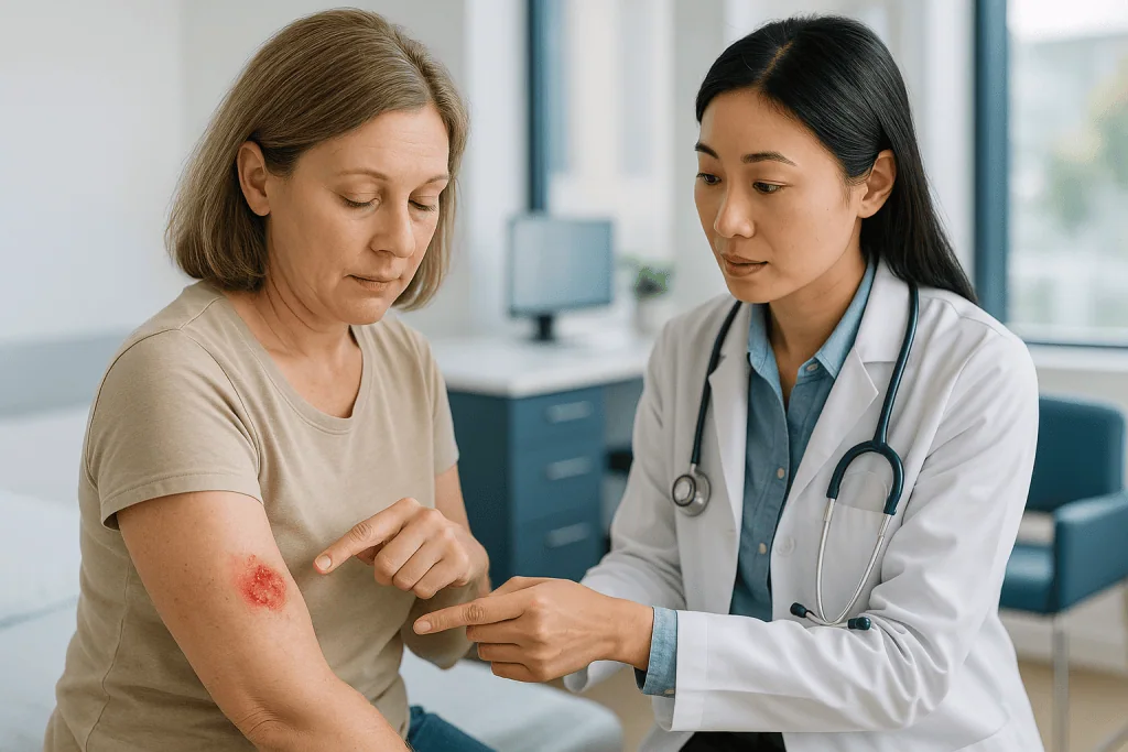
Five changes carry most of the weight:
- Spreading redness. Normal redness sits in a narrow band. Infection pushes it outward, so a pencil line becomes a thumb’s width, then a palm’s. Streaks running from the wound toward the trunk are a particular warning.
- Pain that grows instead of fading. Tenderness peaks on day one and eases. Pain that returns on day three or four, or throbs at rest, is out of sequence.
- Heat and swelling that increase. Hold your hand near the wound and near the matching spot on the other side of the body. A clear difference in warmth, or a wound that looks tighter and shinier than yesterday, matters.
- Cloudy, thick or foul-smelling drainage. Pus is a mix of dead white cells and bacteria; it is opaque, often yellow-green, and may smell. Clear or straw-colored fluid is a different thing entirely.
- Fever or feeling unwell. An infection confined to the wound rarely causes fever. When temperature climbs above 100.4°F (38°C), the threshold MedlinePlus uses for surgical wound concerns, or you feel shivery and drained, the infection may be spreading.
Any one of these alone is worth a photo and a phone call. Two together, or any of them arriving with fever, moves the conversation from keep an eye on it to be seen today. Clinicians would far rather look at a wound that turns out to be fine than hear about one that was watched for a week.
Normal redness vs mole removal infection signs: a side-by-side table
Photographs of healing wounds are easy to misread, partly because red skin looks alarming on a phone screen and partly because the same sign can be benign or serious depending on its trajectory. The table below puts the two side by side.
| What you notice | Typical of normal healing | More typical of infection | What to do |
|---|---|---|---|
| Redness | Narrow rim, stable or shrinking | Widening daily, streaks, hot raised plaque | Widening: call the same day |
| Pain | Worst on day 1, then easing | Increasing after day 2, throbbing at rest | Increasing: call |
| Fluid | Small amount, clear or pink, drying to crust | Thick, cloudy, yellow-green, smelly | Cloudy or smelly: call |
| Swelling | Mild puffiness in first 48 hours | Growing, tense, shiny skin | Growing: call |
| Warmth | Slightly warm early on | Hot compared with the opposite side | Hot plus other signs: call |
| Itch | Common from day 2 onward | Rarely the main feature | Reassuring on its own |
| Temperature | Normal | Above 100.4°F (38°C), chills | Seek care the same day |
| Stitched edges | Together, slight crusting | Gaping, wound reopening | Call; do not re-close yourself |
Two habits make this table useful. Compare against yesterday rather than against an imagined perfect wound; direction of change tells you more than any single snapshot. And photograph the wound each morning in the same light with a coin beside it for scale. A series of consistent images showing redness creeping past the coin’s edge will get you seen faster than a description of it looking a bit worse.
What the table cannot do is replace an examination. Bruising can mimic dusky redness, an allergy to dressing adhesive can mimic a spreading rash, and a small blood collection under stitches can mimic an abscess. All three are common and usually harmless, and all three are best sorted out by the team that did the procedure.
What does an infected mole removal site look like?
Ask a wound nurse to describe an infected mole removal site and the first word is usually angry. The skin around the wound loses the soft pink of healing and turns a deeper, more uniform red that feels thick under a fingertip. The edge of the redness tends to be blurred rather than crisp, and the surface may look glossy where swelling stretches it. Press gently and the brief pale spot that appears as blood is squeezed out refills more slowly than on nearby skin.
Three patterns are worth recognizing. A superficial wound infection stays within the wound itself: the crust softens and lifts, the base beneath looks yellow or grey rather than beefy pink, and cloudy fluid seeps at the margins. Stitched wounds may show small beads of pus where each suture enters, and the skin edges can begin to separate.
Cellulitis is infection that has moved from the wound into the deeper skin and the fat beneath. Mayo Clinic describes it as a swollen, hot, tender area whose redness expands, sometimes with blistering or dimpling, often with fever. On lighter skin it is red; on darker skin it may appear purple, grey or simply darker than the surrounding tissue, making warmth and swelling the more reliable clues. Cellulitis needs a clinician the same day.
An abscess is a walled-off pocket of pus. It shows as a tense, painful lump under or beside the wound that may feel fluid-filled when pressed. Abscesses seldom settle on their own; they are usually drained in clinic, a small procedure the team will explain.
What none of these look like is a dry scab with a faint pink halo and an itch. That is the ordinary picture, and the reason most people who worry about their wound end up reassured rather than treated.
Oozing after mole removal: clear, yellow or cloudy?
Fluid is the sign people find hardest to judge, because a wet dressing looks alarming whatever is on it. The color and consistency of what you find carry the information.
Clear or pale straw-colored fluid is serous drainage, the watery part of blood leaking from newly forming vessels. Small amounts are normal for the first several days, particularly from shave wounds, which fill in from below and stay moist longer than stitched wounds.
Pink or red-tinged fluid is serous fluid with a little blood, common in the first day or two and after exertion. A steadily spreading red stain soaking through the dressing is bleeding rather than oozing; it calls for firm pressure with a clean pad and, if it does not stop, a phone call. MedlinePlus wound guidance lists bleeding that will not stop with pressure among the reasons to seek care.
Thin, translucent yellow fluid that dries to a honey-colored crust is usually still serous and normal. This is the one most often mistaken for pus.
Thick, opaque yellow, green or brown fluid, especially with a smell, is purulent drainage, pus, the debris of the immune battle and a reliable marker of infection. Increasing volume over consecutive days matters as much as the color.
Two practical notes. Ointment applied under a dressing can pool and masquerade as discharge; if your team recommended one, wipe the area with a clean saline-moistened pad before judging. And any dressing worn for days smells faintly; odor counts only when it is strong or accompanies cloudy fluid.
When you call, describe the fluid in those terms, clear, pink, thick yellow, smelly, and the amount: a coin-sized spot or a soaked pad. That vocabulary lets a triage nurse decide without guessing.
Fever after mole removal: when a temperature means something
A mole removal is a small procedure, and a small procedure should not make you feverish. That single fact is the most useful thing to hold onto.
Fever is the body raising its thermostat in response to inflammatory chemicals released when infection spreads beyond a contained area. A superficial wound infection often produces no fever at all; when temperature does rise, it signals that bacteria or their signaling molecules have reached deeper tissue or the bloodstream. MedlinePlus lists a temperature above 100.4°F (38°C) among the reasons to contact a provider after surgery, and Johns Hopkins includes fever among the core signs of surgical site infection.
Timing helps. A slightly raised temperature within the first 24 hours occasionally reflects the body’s general response to any wound and settles by itself. Fever beginning on day two or later, or arriving with spreading redness, pain or pus, points toward infection and needs same-day assessment. Fever with chills, a racing heart, confusion or faintness is an emergency, because those are features of sepsisthe body’s overwhelming response to infection, and the wound, however small, may be the source.
Not every fever this week comes from the wound. A viral illness picked up at the same time is common and coincidental. The way to tell is to look: if the wound is quiet, the fever probably belongs elsewhere, though the team will still want to know.
Measure rather than guess. Use a thermometer, note the reading and the time, and repeat a few hours later. If you use an over-the-counter fever remedy, measure first, since a lowered number makes assessment harder, and mention what you took when you call. A written log of readings beside your wound photos gives the team the whole picture in one message.
How long does mole removal take to heal?
The honest answer is that a wound closes in weeks and a scar keeps changing for far longer, and the two timelines get confused.
Shave excisions heal like a deep graze: a moist wound for several days, a scab toward the end of the first week, then new skin underneath. That skin is fragile, pink and often shiny, and it lightens or darkens over subsequent months. Your surgeon’s estimate for your own site is the one to measure against.
Excisions with stitches close faster at the surface because the edges are held together. Suture removal follows location: NHS guidance gives about 3 to 5 days for the face, 7 to 10 days for the scalp and most of the body, and 10 to 14 days over joints, where tension and movement are greater. Dissolvable stitches beneath the skin break down over weeks. After stitches come out, the line still needs protection, clinicians often advise limiting stretching or heavy exertion of the area for a period they will specify, because a young scar has only a fraction of normal skin strength.
Location changes everything. Facial wounds have a rich blood supply and heal quickly. Wounds on the lower leg, especially in older adults or people with circulation problems, can take considerably longer and are more prone to infection and reopening. Wounds on the back and shoulders, where skin is thick and constantly stretched, tend to widen into broader scars.
Several factors slow every stage: smoking, which constricts the small vessels feeding the wound; poorly controlled diabetes; steroid medicines and some immune-suppressing treatments; and poor nutrition. None is a reason to avoid removal, but each is a reason for closer follow-up.
The scar is the last chapter. It is most noticeable in the early weeks and gradually flattens and pales over many months. Judging a scar at week four is like judging a painting before the varnish has dried.
What can I put on an infected mole removal?
Nothing from the bathroom cabinet, and that answer deserves an explanation rather than a scold.
A wound that is truly infected has bacteria multiplying in tissue below the surface. Creams, gels and household antiseptics act only where they touch, so they cannot reach the problem, and several make things worse. Hydrogen peroxide and rubbing alcohol destroy the new cells trying to close the wound as efficiently as they kill bacteria. Over-the-counter antibiotic ointments have not been shown to clear an established infection, and they are a frequent cause of contact allergy: a red, weeping rash around the wound that is easily mistaken for spreading infection. Kitchen remedies such as tea tree oil, table honey or toothpaste have no evidence behind them for surgical wounds and add irritation.
What an infected wound needs is assessment. A clinician will judge depth and spread, may take a swab, a sample of fluid sent to identify the bacteria, and will decide whether the wound needs cleaning, opening so pus can drain, removal of stitches, or an oral antibiotic. Antibiotics for skin infection are typically chosen from classes active against staphylococcus and streptococcus; the choice, duration and any adjustment after swab results belong entirely to the prescriber. MedlinePlus notes that surgical wound infections are managed with wound care and, when needed, antibiotics, and that a wound may be left open to heal from the inside.
Until you are seen, keep it simple. Wash your hands, cover the wound with a clean non-stick dressing, change it if it soaks through, and avoid pressing or squeezing. Do not remove stitches yourself. If your team suggested a bland ointment such as plain petroleum jelly for routine care, ask before continuing it on a wound you suspect is infected.
For the ordinary, uninfected wound the picture is much less exciting: gentle washing, a bland ointment if advised, and a dressing. The best thing to put on a healing wound is usually very little.
What are the side effects of removing a mole?
Side effect is a loose term for a skin procedure; clinicians think in terms of expected consequences and less common complications, and separating them sets fairer expectations.
A scar is not a side effect but a certainty. Every removal that goes through the full thickness of skin leaves one, and shave removals leave a flat, often paler patch. Mayo Clinic notes the scar may be more noticeable than the mole was, which is one reason cosmetic removal deserves a frank conversation beforehand. Scar quality depends on location, skin type, tension across the wound and genetics more than on technique.
Pigment change is common. New skin may be lighter (hypopigmentation) or darker (hyperpigmentation) than its surroundings, and sun exposure in the first months deepens the darker kind. People with more melanin in their skin are more prone to hyperpigmentation and to thickened scars.
Regrowth of the mole happens mainly after shave removal, because pigment cells below the shaved level remain. A regrown mole is not more dangerous, but it should be assessed, because its appearance can be irregular and confusing.
Less common complications include:
- Bleeding under the wound forming a small collection (a hematoma), which usually reabsorbs but can need draining.
- Infection, the subject of this article.
- Wound reopening (dehiscence), more likely on the back, shoulders and legs.
- Keloid or hypertrophic scars, raised, thickened scars that grow above or beyond the original wound, more frequent on the chest, shoulders and earlobes.
- Numbness or altered sensation if a small skin nerve is cut, which often improves over months but can persist.
- Reaction to dressings or tape, producing an itchy rash that outlines the adhesive.
Two things are not side effects. Removing a mole does not cause cancer and does not spread anything; a mole that turns out to be melanoma was melanoma before the blade touched it, and removal is the first step in treating it. The team will explain what your pathology means if that happens.
What people often get wrong about healing after mole removal
Wound folklore is stubborn. These are the beliefs clinicians spend the most time undoing.
“It itches, so it must be infected.” The reverse is closer to the truth. Itch accompanies regrowing nerve endings and the histamine released during normal repair. Infected wounds hurt and throb more than they itch.
“Letting it air out helps.” Wounds heal faster and scar less when kept slightly moist and covered. A dry, exposed wound forms a thick scab that new skin cells must tunnel beneath. Follow your team’s instructions on when to change the dressing and when to stop it.
“Antiseptic sting means it’s working.” Hydrogen peroxide, iodine and alcohol damage healing tissue. Clean water or saline, as directed, is what mainstream guidance recommends for routine care.
“Antibiotic cream will stop infection before it starts.” For clean, small surgical wounds the evidence does not support routine antibiotic ointment, and it causes allergic rashes in a meaningful share of users. Plain petroleum jelly, if your team suggests anything, keeps the wound moist without that risk.
“A little redness means it’s already infected.” A narrow, stable pink rim is inflammation, which is healing. Only spreading, hot, painful redness points to infection.
“Picking the scab off speeds things up.” It rips away the new skin forming underneath and reopens a door for bacteria.
“The mole came back, so cancer came back.” Regrowth after a shave removal is common and usually reflects pigment cells left below the shaved level. It warrants a look, not panic.
“If it’s going to get infected, it happens on day one.” Bacteria need time to multiply; infection typically shows several days in, and Johns Hopkins notes surgical site infections can appear up to 30 days after a procedure. Day-one redness is almost never infection.
“Sun helps it heal.” Ultraviolet light darkens new scars, sometimes permanently. Shade, clothing or sunscreen once the wound has closed is standard advice.
Why infections happen after mole removal, and what lowers the odds
Every mole removal breaches the skin, the body’s main barrier against the trillions of bacteria living on its surface. Infection follows when enough of them enter the wound, multiply and outpace the immune response. Most of the time the balance favors the patient: the wound is small, the bacteria few, and blood delivers immune cells within minutes. Knowing when the balance tips is the basis of prevention.
Bacteria arrive from three directions. Skin flora at the wound edges are the commonest source, which is why the site is cleaned with antiseptic beforehand. Hands, yours and anyone else’s, carry bacteria to dressings and wounds, and washing them before any contact remains the single most effective habit. Water from baths, pools, hot tubs and lakes carries organisms a wound was never meant to meet, so soaking is usually discouraged until closure.
Host factors shift the odds too. Diabetes impairs white cell function and small-vessel blood flow. Smoking narrows vessels and starves the wound of oxygen. Significant immunosuppression, from illness or medicines, blunts the defensive response. Lower-leg wounds heal in tissue with slower circulation and more swelling. None of these makes infection likely, but each makes it more worth watching.
Wound factors matter as well: a hematoma gives bacteria a protected pool, tension on stitches reduces blood flow at the edges, and a reopened wound exposes deeper tissue.
Prevention, then, is mostly unglamorous:
- Wash hands before touching the dressing or wound.
- Keep the dressing on for the period your team specified and change it only as directed.
- Shower rather than bathe once permitted; pat the area dry.
- Leave stitches, scabs and edges alone.
- Manage blood sugar and pause smoking if you can, ideally with support from your usual doctor.
- Rest the area for the first days if it sits over a joint or on the leg.
Routine preventive antibiotics are not standard for uncomplicated mole removal; whether any individual needs them is the surgeon’s decision, based on the wound and the person.
Questions to ask your care team before you leave
The best time to learn what your wound should look like is before you have one. Ten minutes at the end of the appointment, with these questions in hand, saves a week of second-guessing.
- Which technique did you use, shave, punch or full excision, and how does that change the way the wound heals?
- Are there stitches, are they dissolvable, and if not, when and where are they removed?
- How long should the dressing stay on, and how do I change it?
- When can I shower, and when can I swim or bathe?
- Should I apply anything to the wound, and if so, what and until when?
- How much redness, fluid and swelling do you expect in the first week for a wound in this spot?
- Which signs would you want to hear about the same day, and which can wait for office hours?
- Who do I call out of hours, and can I send a photo?
- Is the tissue going to pathology, when will results be available, and how will I hear?
- Is there anything about my health or medicines that raises my risk of slow healing or infection?
- What activities should I avoid, and for how long, given where the wound is?
- When is my follow-up, and what happens if the wound is not healing as expected by then?
Write the answers down or ask for them in print. Aftercare leaflets are generic by necessity; the surgeon’s specific expectation for your wound is what you will measure against at the bathroom mirror.
One further question is worth asking even when it feels awkward: if this were infected, what would you do? Hearing the plan, assessment, possible swab, possible antibiotics, possible drainage, takes the mystery out of the phone call you may later need to make, and makes it easier to pick up the phone early rather than late.
When to call your doctor
Mole removal wounds are small, and the threshold for calling about them should be low. No surgical team resents a phone call about a wound; they resent the infection that arrived on day eight because the patient waited.
Call the same day if you notice any of the following:
- Redness spreading beyond a narrow rim, widening day over day, or red streaks tracking away from the wound.
- Pain that increases after the second day, or throbbing at rest.
- Thick, cloudy, yellow-green or foul-smelling drainage, or drainage that increases over consecutive days.
- Swelling that grows, or a tense, painful lump under or beside the wound.
- A temperature above 100.4°F (38°C), chills, or feeling generally unwell.
- Stitches that have pulled apart or a wound that has reopened.
- Bleeding that does not stop after sustained firm pressure.
- Numbness, weakness or color change in the skin beyond the wound.
Seek emergency care if fever comes with a racing heart, breathlessness, confusion, faintness, a rapidly spreading rash, or pain far out of proportion to what you can see. These can indicate sepsis or a fast-moving soft-tissue infection, both rare after minor skin surgery and both treated as emergencies.
Mention at your next contact, without urgency: mild itching, a scab that has not yet lifted, slight pigment change, a scar that feels firm, or a painless small lump at a stitch site.
When you call, have your photos, your temperature log, the date of the procedure, and a list of your medicines and health conditions ready. Say what you see in plain terms, how wide the redness is, what color the fluid is, how the pain has changed, and let the team decide what it means. That decision, along with every choice about treatment, sits with them. Your job is to notice early and describe clearly, and you now have the vocabulary for both.
Frequently asked questions
How do I know if my mole removal is healing properly?
A properly healing wound gets quieter every day: the pink rim stays narrow or shrinks, tenderness fades after the first day, any fluid is clear or pale and decreasing, and a scab or closed stitch line forms within the first week. Itching from day two onward is a good sign. Compare each morning with the day before; a wound that is trending toward less redness, fluid and pain is on track, whatever a single photo looks like.
What are infected mole symptoms if the mole has not been removed?
An intact mole can become inflamed if a hair follicle within it is blocked or it is scratched or shaved, producing tenderness, a small pus spot, swelling and surrounding redness that settles within days. A mole that bleeds, crusts, itches or changes size, shape or color without an obvious injury is a different matter and should be examined, because those changes are the ones clinicians use to decide whether a mole needs removal and laboratory review.
Is swelling normal 24 hours after a mole removal?
Yes, mild puffiness around the wound in the first 48 hours is part of normal inflammation, and it may feel slightly warm and tight, particularly on the face or around the eyes where tissue is loose. Swelling that keeps growing after the second day, becomes tense and shiny, forms a painful lump, or arrives with spreading redness or fever is not typical and should be reported to your care team the same day.
What can I put on an infected mole removal?
Nothing you have not been told to use; an infected wound needs a clinician’s assessment rather than a product. Household antiseptics such as hydrogen peroxide damage healing tissue, and over-the-counter antibiotic ointments do not clear established infection and often cause an allergic rash. Until you are seen, wash your hands, cover the wound with a clean non-stick dressing, leave stitches alone, and let the treating team decide on cleaning, drainage or antibiotics.
What are the side effects of removing a mole?
A scar is expected after every removal, and it may be more visible than the mole was; pigment lighter or darker than surrounding skin is common and can persist for months. Regrowth of the mole happens mainly after shave removal. Less common problems include bleeding under the wound, infection, wound reopening, raised keloid or hypertrophic scars, altered sensation from a cut skin nerve, and rashes from dressing adhesive. Removal does not cause or spread cancer.
How long does mole removal take to heal?
The surface of a shave wound usually scabs within the first week and closes within a few weeks, while stitched wounds close faster at the surface and have sutures removed on a location-based schedule, about 3 to 5 days on the face and 10 to 14 days over joints, according to NHS guidance. The scar then softens and fades over many months. Lower-leg wounds, smoking and poorly controlled diabetes slow every stage.
Can I shower after mole removal?
Most teams allow a brief shower after the first day or two, keeping the dressing on or replacing it afterward and patting rather than rubbing the area dry, but the exact timing depends on the technique and location, so follow the instructions you were given. Soaking in a bath, pool, hot tub or open water is usually discouraged until the wound has fully closed, because standing water carries bacteria that can seed an open wound.
Why is my mole removal site yellow?
A thin, translucent yellow film or honey-colored crust is usually dried serous fluid, the watery part of blood that leaks from a healing wound, and is normal, particularly on shave wounds. Ointment under a dressing can also look yellow. Thick, opaque yellow or green material, especially if it smells, increases day over day, or sits on a base that looks grey rather than pink, is more consistent with pus and should be assessed by your care team.
Does a mole removal scar go away?
A scar never disappears completely, but most fade substantially: they are reddest and most raised in the early weeks, then flatten and pale over many months as collagen remodels. Location, skin type, wound tension and genetics influence the final result more than technique. Protecting the closed wound from sun helps limit darkening. If a scar becomes thick, itchy or grows beyond the original wound, ask your team about options, which they can tailor to you.
What happens if a mole removal infection is left untreated?
A superficial wound infection can progress to cellulitis, in which bacteria spread through the deeper skin and fat, producing an expanding hot red area and often fever, or to an abscess, a walled pocket of pus that usually needs draining. Rarely, infection reaches the bloodstream and causes sepsis, which is life-threatening. Delayed treatment also worsens the eventual scar. Early assessment keeps most infections small, local and simple to manage.
References
- MedlinePlus – Surgical wound infection: treatment
- NHS – Moles
- NHS – How should I care for my stitches (sutures)?
This article is for general information only and is not a substitute for professional medical advice. Please consult a qualified doctor about your individual situation.

More from the Blog
Which Injuries Need Microsurgery? Nerve Repairs, Reattachment and Free Tissue Transfer
Microsurgery treats injuries and defects too small or delicate for standard surgical tools: severed or damaged peripheral nerves, amputated fingers, hands or other body…
What an RF Microneedling Session Involves: Numbing, Needle Depth and the Heat You Feel
An RF microneedling procedure begins with a topical numbing cream left on for 30 to 60 minutes. A handpiece then pushes insulated needles a…

RF Microneedling Downtime: Redness, Grid Marks and When You Can Wear Makeup Again
RF microneedling downtime is usually short. Redness and mild swelling typically peak in the first 24 hours and fade over one to three days,…

The Best Age for a Facelift: Why Anatomy Matters More Than Birthdays
There is no single best age for a facelift. Most patients are in their 40s through 60s, but surgeons evaluate anatomy, skin laxity, jowls,…

Reconstructive Surgery in Older Adults: How Age, Heart Health and Healing Are Weighed
Reconstructive surgery in older adults is judged on overall health, not birth date. Surgical and anesthesia teams weigh heart and lung function, frailty, medicines,…
Split-Thickness vs Full-Thickness Skin Grafts: How Surgeons Choose the Graft Type
Surgeons choose between a split-thickness and a full-thickness skin graft mainly on wound size, location, and how much the graft may shrink. Split-thickness grafts…