

AI in Radiology: How Algorithms Read Mammograms and CT Scans, and Why a Radiologist Still Signs the Report
Key Takeaways
- In the Swedish MASAI randomized trial of more than 105,000 women, AI-supported screening detected about 29% more breast cancers than standard double reading while cutting radiologist reading work by roughly 44% and leaving the false-positive rate unchanged at 1.5%.
- Every AI radiology device authorized in the United States is cleared as an aid to a physician, so a radiologist reviews and signs the final report even at sites where software scores every exam.
- A screening tool with 90% sensitivity and 90% specificity would flag about 104 women in every 1,000 screened, of whom roughly five actually have cancer, which is why sensitivity alone says little about a test.
- A 2024 reader study of 140 radiologists found AI assistance improved some readers and worsened others, a pattern explained by automation bias rather than by skill level.
- US guidance now advises average-risk women to have a mammogram every two years from age 40 through 74, while the NHS invites women aged 50 to 71 every three years; AI has not changed these ages.
- Detection rates are a surrogate endpoint: no trial has yet shown that AI-supported screening lowers breast cancer deaths, and interval-cancer follow-up from MASAI is still accumulating.
AI in radiology means software trained on large image datasets that flags suspicious areas on mammograms and CT scans, sorts urgent cases to the top of a worklist, and measures structures automatically. In one large Swedish randomized trial, AI-supported mammography screening found more cancers with 44% less reading work and no rise in false positives. Regulators classify these tools as assistive, so a radiologist still reviews every image and signs the final report.
At 6:45 on a Monday morning, before the first patient has parked, a breast imaging worklist has already been reshuffled. A handful of mammograms sit at the top, each carrying a small colored marker that no human placed. The radiologist who arrives at seven did not ask for the reorder. Software did it overnight. Scenes like this explain why ai in radiology has become one of the most-searched medical technology phrases of the year.
The spark was a run of unusually strong data. A Swedish randomized trial reported in 2023 and published in full in 2025 showed that an algorithm could take over part of screening mammography reading without missing cancers, and a national German program followed with real-world numbers on hundreds of thousands of women. Social feeds compressed all of that into a punchier claim: AI now beats doctors at spotting cancer.
As of March 2026, that claim is half right and importantly wrong. What follows is the fuller picture, including the quiet reason every report you receive still carries a physician’s name.
What changed recently: why AI in radiology is trending in 2026
Three developments landed close together, and each one fed the others. The first was evidence. In August 2023, The Lancet Oncology published the interim safety analysis of the Mammography Screening with Artificial Intelligence trial, known as MASAI, which randomized 80,033 Swedish women to either standard double reading by two radiologists or an AI-supported pathway. Cancer detection rose from about 5.1 to 6.1 per 1,000 women screened, false positives stayed flat at 1.5%, and the reading workload fell by roughly 44%. The full trial results, covering just over 105,000 women, appeared in early 2025 and confirmed the pattern with a 29% relative increase in detected cancers, including more invasive and more small tumors.
The second development was regulation. By 2025 the US Food and Drug Administration’s public list of authorized AI-enabled medical devices had passed a thousand entries, and roughly three in four of those were radiology tools. None of them is authorized to issue a final diagnosis on its own; every one is labeled as an aid to a clinician. In parallel, the World Health Organization’s guidance on the ethics and governance of artificial intelligence for health, first issued in 2021 and expanded for large language models in 2024, gave health systems a shared vocabulary for transparency, accountability and human oversight.
The third was a change in who gets screened. In April 2024 the US Preventive Services Task Force finalized its recommendation that women at average risk begin mammography at 40 rather than 50, screening every two years through age 74, and the Centers for Disease Control and Prevention updated its public guidance to match. Millions more exams per year, in a country already short of breast imagers, made workload-reducing software look less like a novelty and more like infrastructure.
Put those together and you get the current moment: real trial data, a crowded market, and a rising tide of exams. The viral version of the story skipped the nuance. The rest of this piece restores it.
What does AI in radiology actually mean?
Strip away the marketing and the phrase covers four distinct jobs. Detection software points to a spot on an image that resembles something abnormal. Triage software re-sorts a queue so likely emergencies are read first. Quantification software measures things humans measure slowly and inconsistently, such as the volume of a lung nodule or the amount of calcium in a coronary artery. Report software, the newest arrival, drafts or summarizes text.
A few terms recur throughout, so here they are in plain language. An algorithm is a fixed set of computational steps that turns an input, such as an image, into an output, such as a score. Machine learning is the practice of letting a computer adjust those steps by studying many labeled examples rather than being programmed rule by rule. Deep learning is a form of machine learning that stacks many layers of simple mathematical filters, and the specific architecture that dominates imaging, the convolutional neural network, was designed to find edges, textures and shapes at increasing scales, much as the visual cortex does. A large language model is a text-prediction system trained on enormous bodies of writing; it can draft a radiology report but cannot see a pixel unless paired with an image model.
None of this is entirely new. Computer-aided detection for mammography, usually abbreviated CAD, received US clearance in 1998 and marked possible masses and calcifications with small triangles and asterisks. It was widely adopted, and large observational studies over the following fifteen years found it added recalls without reliably adding cancers. That history matters because it shows a tool can be cleared, reimbursed and popular while producing no measurable benefit for patients. The current generation differs in one fundamental way: instead of hand-coded rules about brightness and edges, it learns from hundreds of thousands of exams with known outcomes, and it produces a calibrated probability rather than a binary mark.
The honest test for any AI in radiology is therefore not whether it is clever, but whether it changes what happens to a patient.
How an AI mammogram reading works, pixel by pixel
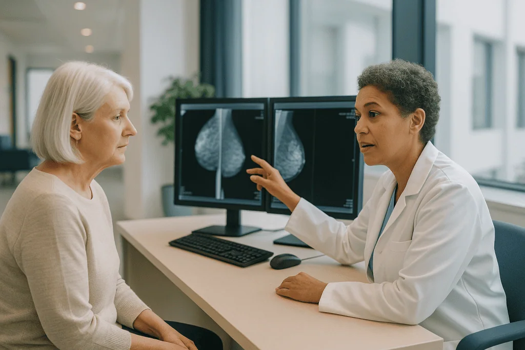
A screening mammogram is four images: two views of each breast, one from above and one at an angle. Each digital image is a grid of several million pixels, and each pixel holds a brightness value that reflects how much X-ray energy passed through the tissue at that point. Dense fibroglandular tissue appears white. Fat appears dark. Cancer, inconveniently, is also white, which is why finding it in a dense breast has been compared to spotting a snowball in a snowstorm.
The software begins by standardizing the images, correcting for differences between machine vendors and compression settings so that a picture from one manufacturer resembles a picture from another. The neural network then scans the image at multiple scales, looking for the patterns it learned during training: irregular masses with spiky margins, clusters of tiny bright calcifications arranged in certain shapes, subtle areas where the normal architecture appears pulled inward. It compares the two views of the same breast, because a true lesion should appear in both, and it may compare left with right, because asymmetry is a clue.
The output is usually two things. One is a heat map, a translucent overlay in which warmer colors mark the regions that most influenced the model’s decision. The other is a single case-level score, often on a scale of one to ten, expressing how likely the exam is to contain cancer. Screening programs set thresholds on that score. In MASAI, exams scoring in the top tenth were flagged for extra attention and read by two radiologists, while the rest went to a single reader supported by the software.
What the algorithm does not do is assign a BI-RADS category, the standardized zero-to-six rating that American radiologists use to communicate the level of suspicion and the recommended next step. That judgment belongs to the physician, who also sees the patient’s age, prior films, family history and any symptom the technologist noted. An AI mammogram score is an input to that judgment, not a substitute for it.
How algorithms read CT scans: triage, nodules and measurement
A mammogram is four pictures. A chest CT can be five hundred, each a thin cross-sectional slice of the body reconstructed from X-ray projections taken as the scanner rotates. Reading a CT means scrolling through a three-dimensional volume, and the sheer number of slices is one reason CT has become the busiest frontier for AI in radiology.
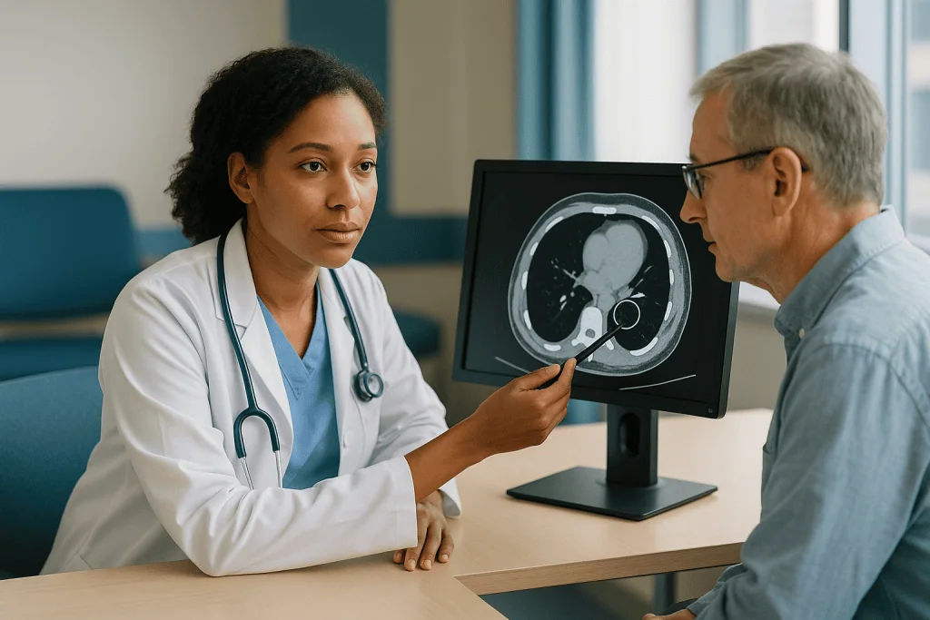
The most mature application is triage. Software running in the background examines every head CT the moment it is acquired, searching for the bright, dense signal of fresh bleeding inside the skull. If the model believes hemorrhage is present, it pushes that study to the top of the radiologist’s queue and may send an alert to the emergency team. Similar tools exist for large-vessel occlusion in stroke, where minutes of delay translate into lost brain tissue, and for pulmonary embolism, a clot in the arteries of the lung. The evidence base here consists mainly of retrospective accuracy studies and before-and-after time-to-notification comparisons, which consistently show faster flagging; whether faster flagging improves neurological outcomes is far less well demonstrated.
The second application is nodule detection on lung CT. Screening for lung cancer with low-dose CT is recommended in the United States for adults aged 50 to 80 with a 20 pack-year smoking history who currently smoke or quit within the past 15 years, according to CDC guidance. Those scans contain many small nodules, most benign. AI marks candidate nodules and measures their volume, and volumetric change over time is a more reproducible signal of malignancy than a diameter measured by hand with a digital caliper.
The third is opportunistic measurement. Every abdominal or chest CT contains a spine and often the heart. Algorithms can now estimate bone density from vertebrae, count coronary artery calcium, and quantify liver fat on scans ordered for entirely different reasons. These findings can be clinically useful, but they also raise a question radiology has not fully answered: who is responsible for acting on an incidental result that no one ordered?
What the evidence actually says, graded by strength
Medical evidence comes in tiers, and it is worth being explicit about which tier each claim rests on. Randomized controlled trials, in which participants are assigned by chance to one approach or another, sit at the top because they balance out hidden differences between groups. Prospective observational studies follow real patients forward in time without randomization. Reader studies ask radiologists to interpret a curated set of cases with and without software. Retrospective studies apply an algorithm to old images and compare its output with what eventually happened.
For AI-supported breast screening, there is now one completed randomized trial, MASAI, with more than 105,000 participants. Its core finding is robust: within a European double-reading program, using AI to decide which exams need two readers and which need one preserved cancer detection, modestly increased it, and cut reading work by about 44% without increasing false positives. That is high-quality evidence for that specific use in that specific setting.
It comes with two honest caveats. Sweden and most of Europe read every screening mammogram twice; the United States reads once, so the workload math does not transfer directly. And detection is a surrogate endpoint. The outcomes patients actually care about, fewer cancers appearing between screens and fewer deaths, require years of follow-up that the field is only beginning to accumulate.
Below the trial sits a large prospective observational study from Germany, published in 2025, in which more than 460,000 women were screened with or without AI support and the AI-supported group had a roughly 18% higher detection rate with no increase in recalls. Observational data cannot rule out that screening sites choosing AI differed in other ways, but the direction and size agree with the trial.
Below that are dozens of retrospective studies, which regularly report that a standalone algorithm matches or exceeds an average radiologist. These are the studies that generate headlines. They are also the ones most vulnerable to a problem called dataset shift, in which a model trained on one population, one scanner brand or one era of imaging performs worse when moved elsewhere. For CT applications, most of the evidence is still at this retrospective and workflow-timing level.
Grading it all: strong for AI as a workload-reducing second reader in double-reading mammography; moderate for detection gains; weak to absent for mortality benefit; and mostly preliminary for CT beyond speed of alerting.
AI radiology tools at a glance: what they do and how strong the evidence is
The phrase AI in radiology covers tools with very different track records. A single summary helps keep them straight. The middle column reflects the best available evidence as of early 2026; the final column is the same for every row, which is the point.
| Type of tool | Typical task | Strength of evidence | Who signs the report |
|---|---|---|---|
| Mammography second reader or triage | Scores each screening exam; routes high-scoring cases to two radiologists | One large randomized trial plus large prospective observational data; detection maintained or improved, workload reduced | Radiologist |
| Emergency CT triage | Flags suspected brain bleed, stroke or lung clot and moves it up the queue | Retrospective accuracy and time-to-alert studies; faster flagging shown, patient-outcome benefit not yet proven | Radiologist |
| Lung nodule detection and volumetry | Marks candidate nodules on CT and measures growth between scans | Reader studies and retrospective validation; improves reproducibility of measurement | Radiologist |
| Opportunistic quantification | Estimates bone density, coronary calcium or liver fat on scans ordered for other reasons | Retrospective and correlational; clinical pathways for acting on results still developing | Radiologist |
| Report drafting with language models | Turns dictated findings into structured text or a patient-friendly summary | Early usability studies; accuracy of generated text requires physician verification | Radiologist |
Two patterns stand out. The strongest evidence exists where the task is narrow, the outcome is countable and the setting is a population screening program with decades of quality-control habits. The weakest evidence sits where the tool is newest and the output is words rather than a location on an image.
The other pattern is that regulatory status and evidence quality are not the same thing. Every tool in the table can be legally marketed somewhere. Only the first row has been tested in a randomized trial. When a health system chooses software, the difference between those two facts should drive the decision, and when a patient hears that AI is used at a screening site, it is fair to ask which row they are talking about.
AI cancer detection: what the headlines about beating radiologists leave out
A claim like the algorithm found 90% of cancers sounds decisive until you ask what else it found. Two numbers describe any test. Sensitivity is the share of true cancers the tool flags. Specificity is the share of healthy exams it correctly leaves alone. In screening, where most people are well, specificity does most of the work.
Run the arithmetic on a typical screening population, where roughly five to six cancers exist in every 1,000 women screened. A tool with 90% sensitivity and 90% specificity would catch about five of those cancers. It would also flag about 99 of the 994 women without cancer. Of roughly 104 women called back, five have the disease. That ratio, a positive predictive value near 5%, is not a failure; it is close to what human screening achieves. But it shows why a headline about sensitivity alone tells you almost nothing about the experience of the women in the waiting room.
Retrospective comparisons carry a second, quieter distortion. When researchers report that a model outperformed radiologists on a set of old mammograms, the radiologists in that comparison usually read the images blind, without prior films, clinical history or the option to call the patient back for extra views. Real radiologists have all three. The comparison measures the algorithm against a diminished version of the human it claims to beat.
A third issue is what kind of cancer gets found. Screening can detect ductal carcinoma in situ, abnormal cells confined to the milk ducts that may never progress, and very slow-growing invasive tumors that would not have harmed the person in her lifetime. Finding more of those raises detection statistics while adding treatment without adding survival, a phenomenon called overdiagnosis. MASAI’s full results were reassuring on this point, with gains concentrated in invasive cancers, but it will take years of interval-cancer and mortality data to settle the question.
The responsible way to read any AI cancer detection claim is to ask three things: sensitivity and specificity together, compared against what, and cancers of which kind.
Why a radiologist still signs the report
Start with the legal reality. Every AI radiology device authorized in the United States is cleared as a tool to assist a qualified physician. The labeling says so in plain terms, and the professional societies, the malpractice framework and the payment rules all assume a licensed radiologist made the final interpretation. In Europe, a small number of products carry a mark allowing them to classify certain chest X-rays as normal without human review, but even there the pathway is narrow, audited, and applied to a minority of studies. Nowhere is an algorithm the physician of record.
The clinical reasons run deeper than paperwork. An image is one piece of a puzzle whose other pieces are text: the reason the scan was ordered, the medications the patient takes, the biopsy done three years ago, the incidental finding on a different modality last spring. Breast imaging software examines the breast; it does not notice that the same woman’s lymph nodes looked different in a recent chest CT. A radiologist reading the study inside an electronic record can.
The report itself is a communication, not a classification. It tells a referring physician what was seen, what it probably means, what should happen next and how urgently. It may recommend a short-interval follow-up rather than a biopsy, or suggest a different imaging test entirely. Those recommendations weigh a patient’s age, comorbidities and preferences. No cleared algorithm attempts that step.
There is also the matter of what happens when the software is wrong. A model trained overwhelmingly on one manufacturer’s images may misjudge another’s. It may be confused by a pacemaker, a surgical clip, motion blur or an unusual body habitus. It has no mechanism for saying I am not sure, call the patient back for another view. The radiologist supplies exactly that kind of judgment, along with a name to which a question can be addressed.
The Swedish trial, so often cited as evidence that AI can do the job, illustrates the point precisely. It did not remove the radiologist. It removed one of two radiologists for most exams, and the remaining physician signed every report.
Does AI help or hurt a radiologist's performance? The evidence is mixed
The intuitive model of AI assistance is additive: a good reader plus a good tool equals a better reader. The research says human-machine teams are more complicated than that.
In a 2024 study published in Nature Medicine, researchers asked 140 radiologists to interpret hundreds of chest X-rays with and without support from a well-validated algorithm. On average, accuracy improved slightly. Underneath the average, the effect ran in both directions. Some radiologists became meaningfully better with the tool. Others became worse, and the pattern was hard to predict from experience level alone. The study is a reader experiment rather than a clinical trial, so it grades as moderate evidence, but it matched what human-factors researchers have described for decades in aviation and other fields.
The mechanism has a name: automation bias. When a trusted system says nothing is there, a person tends to look less carefully. When it marks a spot, attention gravitates toward the mark and away from the rest of the image. Neither tendency is laziness; both are how human attention economizes under time pressure. A radiologist reading a hundred screening mammograms in a session is precisely the kind of expert, working at precisely the kind of pace, that automation bias affects most.
A related concern is deskilling. A 2025 observational study of colonoscopy in Poland, a different specialty but a similar dynamic, found that endoscopists who had worked with AI polyp detection for several months found fewer polyps when the software was switched off than they had before its introduction. Whether the same erosion occurs in imaging is unknown, and the study design cannot prove causation, but it is the first real-world signal of a risk that had been largely theoretical.
None of this argues against the technology. It argues for how the technology is deployed. Software that shows its confidence honestly, that can be toggled off for periodic unassisted reading, and whose performance is audited against biopsy results at each site behaves as an instrument. Software presented as an oracle invites over-reliance. The difference is a design and governance decision, and it belongs to health systems rather than to the algorithm.
Is AI going to replace radiology? Are radiologists safe from AI?
In 2016 a prominent computer scientist suggested that medical schools should stop training radiologists because software would soon outperform them. A decade later, the United States has a documented shortage of radiologists, imaging volumes continue to rise faster than the workforce, and residency positions in the specialty fill every year. The prediction was not irrational. It simply misjudged what the job is.
Reading images is the visible part of radiology. The rest includes deciding which scan to order and how to perform it, tailoring radiation dose and contrast to a patient with kidney disease, performing image-guided biopsies and drainages, sitting in tumor boards, explaining a finding to a frightened patient, and catching the one-in-a-thousand study where the question the referrer asked is the wrong question. Algorithms today do none of these.
What the evidence does support is that certain narrow reading tasks can be partly automated. Screening mammography second reads are the clearest example. Excluding clearly normal chest X-rays is another; a Danish retrospective analysis found that a commercial tool could confidently label a substantial share of chest radiographs as normal with very high accuracy, and a few European sites now allow such studies to bypass a human read under audit. Expect the list of automatable subtasks to lengthen.
Expect, too, that the freed time will be absorbed. The 2024 change in US screening age, the aging of the population and the expansion of lung cancer screening all add exams. Radiologists who work with well-governed AI are likely to read more studies per hour and to spend a larger share of their day on the tasks the software cannot touch. That is a change in the shape of the job, not its disappearance.
Honesty requires acknowledging uncertainty. No one can say what image interpretation will look like in 2046. But the credible near-term forecast, grounded in what has actually been demonstrated, is a specialty transformed rather than replaced, with a physician’s signature on the report for the foreseeable future.
Which AI is best for radiology? The question behind the question
People searching this phrase usually want a ranking. There is no honest one, for the same reason there is no single best medication: the right choice depends on the task, the setting and the population. What can be offered is a set of criteria that separates a tool worth trusting from one that merely looks impressive in a demonstration.
Start with the specific indication. A device cleared to flag intracranial hemorrhage on non-contrast head CT has been evaluated for exactly that, not for detecting a subtle stroke or a tumor. Using it outside that scope is an experiment, not a clinical service. The same applies to mammography software cleared on two-dimensional images and then pointed at three-dimensional tomosynthesis studies.
Ask where the validation happened. A model that performs beautifully on images from the hospital that built it may stumble on scanners from another manufacturer or on a population with a different age structure, breast density distribution or disease prevalence. External validation, meaning testing on data from institutions that had no part in development, is the minimum. Prospective evaluation in a setting resembling the one where the tool will be deployed is better. A randomized trial, still rare, is best.
Look for calibration, not just accuracy. A tool that says 80% likely should be right about 80% of the time it says so. Overconfident software fuels exactly the automation bias described earlier.
Insist on monitoring. Scanners get upgraded, protocols drift, populations shift. Performance measured at installation is not performance two years later. Good deployments compare algorithm output against biopsy and follow-up results continuously and have a plan for when the numbers slide.
Finally, weigh how the output is shown. A heat map that explains where the model looked supports a radiologist’s judgment; a bare score encourages deference. For a patient, the practical version of all this is simpler: it is reasonable to ask a screening center whether AI is used, for which purpose, and whether a radiologist reviews every study. The answer to the last question should always be yes.
How to become an AI radiologist, and what the title really means
There is no board certificate that reads AI radiologist. The phrase describes a radiologist who understands machine learning well enough to evaluate, deploy and study it, or occasionally to build it. The path runs through the same door as every other radiologist: an undergraduate degree, four years of medical school, a clinical internship year, a four-year diagnostic radiology residency, and usually a one- or two-year fellowship in a subspecialty such as breast, neuroradiology or thoracic imaging. Board certification follows. That is roughly thirteen to fourteen years after high school before independent practice.
The AI layer is added alongside or after that training. Some residents pursue a master’s degree in biomedical informatics or data science. Others complete a clinical informatics fellowship, a two-year accredited program recognized by the American Board of Preventive Medicine, which qualifies a physician for a second board certification in clinical informatics. Many teach themselves the basics of statistics, programming and model evaluation through online coursework and by joining research projects during residency, when protected academic time exists.
The skills that matter most are not the ones people expect. Writing neural-network code is a small part of the work and is often done by engineers. The radiologist’s distinctive contribution is defining the clinical problem precisely, judging whether a labeled dataset actually reflects the patients who will be scanned, designing a validation study that would convince a skeptic, spotting the failure modes an engineer would never anticipate, and translating a performance metric into a decision about a patient. Fluency in study design and epidemiology, meaning the science of how disease is distributed and measured in populations, is more valuable than fluency in any particular software library.
The field also has room for people who are not physicians. Medical physicists, imaging scientists, data engineers and regulatory specialists all work on AI in radiology, and hybrid teams are the norm. For a student weighing the choice, the safest generalization is that the specialty is recruiting people who can hold two kinds of skepticism at once: about the limits of algorithms and about the limits of unaided human vision.
Common myths about AI in radiology, corrected
Viral posts thrive on compression. Here are the claims circulating most widely and what the record shows.
Sweden has replaced radiologists with AI. It has not. The MASAI trial replaced one of two human reads for most screening exams. A radiologist reviewed and signed every report, and the highest-scoring tenth of studies received more human attention than before, not less.
AI finds every cancer. No screening method does. Even in the AI-supported arm of MASAI, detection was about six cancers per thousand women, and some cancers still surface between screening rounds. Sensitivity near 100% is achievable only by recalling nearly everyone, which is not a screening program.
If AI is used, no human looks at my scan. In the United States, a radiologist is legally responsible for every interpretation. A very small number of European sites allow certain chest X-rays that software classifies as normal to skip a human read under strict audit; that is the exception, disclosed and monitored, not the rule.
Algorithms are objective, so they cannot be biased. Models learn from the data they are given. If a training set underrepresents certain ages, ethnic groups, breast densities or scanner types, performance in those groups can be worse, and the tool will not announce it. Bias in AI is a measurable engineering property, which is exactly why external validation and ongoing monitoring matter.
A high AI score means I have cancer. A score is a probability estimate for the image, not a diagnosis of the person. Most women recalled after screening, whether flagged by software or by a radiologist, turn out not to have cancer. Diagnosis requires further imaging and, when indicated, a biopsy.
You can upload your own mammogram to a chatbot and get an answer. General-purpose chatbots are not cleared medical devices, are not validated on medical images, and can produce confident, fluent and wrong statements. Results from your imaging should be discussed with the clinician who ordered the test or with the radiology department that performed it.
AI makes radiology faster, so results arrive faster. Sometimes. Triage tools speed up urgent cases, but routine reporting turnaround depends on staffing, not on whether an algorithm ran in the background.
What AI means for you at your next mammogram or CT
For most patients, the arrival of AI in radiology changes nothing about the visit itself. The compression is the same, the scanner is the same, the technologist is the same. What may differ is the notice. Some screening programs now inform patients that software assists the reading, and a few ask for consent to include images in research databases. Reading the paperwork is worth thirty seconds.
Screening recommendations have not changed because of AI, and they remain the foundation of any decision. In the United States, CDC guidance based on the 2024 US Preventive Services Task Force recommendation advises women at average risk to have a mammogram every two years from age 40 through 74. Women at higher-than-average risk, for example those with a strong family history or a known genetic variant, may be advised to start earlier or to add other imaging; that plan is made with a clinician. In England, the NHS Breast Screening Programme invites women aged 50 to 71 every three years, with a first invitation arriving within three years of the 50th birthday. Lung cancer screening in the US applies to adults 50 to 80 with a 20 pack-year smoking history who still smoke or quit within the past 15 years. Screening outside those age ranges is a conversation, not a default.
Since September 2024, US mammography reports must also tell you whether your breast tissue is dense. Density lowers the sensitivity of mammography for humans and algorithms alike and modestly raises breast cancer risk. Whether that warrants supplemental imaging depends on your overall risk profile and is, again, a decision to make with your clinician.
If you are recalled after a screening exam, remember what the arithmetic earlier in this piece showed: the large majority of recalls, whether prompted by a physician’s eye or by a heat map, end without a cancer diagnosis. Recall means the picture needs clarifying, not that something has been found.
Reasonable questions to ask any imaging center include whether AI is used in interpretation, for what purpose, and whether a radiologist reviews every study. You are entitled to clear answers, and to a copy of your report with a physician’s name on it.
When to see a doctor
No algorithm, however well validated, replaces the basic rule of breast and lung health: a new change in your body is a reason to see a clinician, regardless of when your last scan was or what it showed. Screening looks for disease in people without symptoms. A normal mammogram or a low AI score last month does not rule out a problem that has become noticeable this month.
Per NHS and Mayo Clinic guidance, the changes that should prompt a prompt appointment rather than a wait for the next screening date include a new lump or area of thickening in the breast or underarm, a change in the size, shape or skin of one breast such as dimpling or puckering, a nipple that has newly turned inward or is leaking fluid, redness or a rash around the nipple, and persistent pain in one spot. Most of these turn out to have benign causes, but each deserves examination and, usually, diagnostic imaging targeted to the area rather than a routine screening exam.
For CT, the relevant moments are different. If you received a letter or a message about an incidental finding, meaning something noticed on a scan ordered for another reason, contact the clinician who ordered the study to ask what follow-up is recommended and when. Do not assume that no news means no finding; ask for your report. If you have persistent cough, coughing up blood, unexplained weight loss or shortness of breath, see a doctor, whether or not you qualify for screening.
A few situations call for urgent care rather than an appointment. Sudden severe headache, new weakness or numbness on one side of the body, trouble speaking, or sudden chest pain with shortness of breath are emergencies. The triage software described in this article exists precisely because those conditions are time-critical, and the first step is a call to emergency services, not a scan you arrange yourself.
Finally, never adjust a follow-up interval, skip a recommended biopsy or delay a scan because a consumer app or a chatbot offered a reassuring reading of your images. Those tools are not cleared for that purpose. The decision about what happens next belongs to you and the clinician who knows your history, informed by a report a radiologist has reviewed and signed.
Frequently asked questions
Is AI going to replace radiology?
Not on current evidence, and not in the foreseeable future. Algorithms have proven able to take over narrow reading tasks, most convincingly the second read in double-reading mammography programs. Radiologists also choose and tailor scans, perform image-guided procedures, integrate findings with a patient’s history and sign the legal report. Every authorized AI device is labeled as an aid to a physician. The realistic forecast is a specialty whose daily work shifts, not one that disappears.
How accurate is an AI mammogram reading compared with a radiologist?
In the strongest study to date, a randomized trial of over 105,000 Swedish women, an AI-supported pathway detected about 6.4 cancers per 1,000 screens versus 5.0 with two radiologists, with identical false-positive rates. Retrospective studies often show standalone algorithms matching average radiologists, but those comparisons strip the humans of prior films and clinical history. Accuracy also varies by scanner, population and breast density, which is why site-level monitoring matters.
Does AI cancer detection mean fewer cancers are missed?
It can mean more cancers are found at screening, which the Swedish trial and a large German observational study both showed. Whether that translates into fewer cancers appearing between screening rounds, and ultimately fewer deaths, is not yet proven; those outcomes take years of follow-up. Some additional detections may also be slow-growing tumors that would never have caused harm. The early data on tumor type are encouraging but not conclusive.
Are radiologists safe from AI, or will fewer be trained?
The United States currently has a shortage of radiologists, imaging volumes are rising, and residency programs fill every year. A 2016 prediction that training should stop has not held up. Certain tasks will be partly automated, and radiologists who work with well-designed software will likely read more studies per hour. The job is changing in shape, with more time on procedures, consultation and oversight, rather than shrinking in demand.
Which AI is best for radiology?
There is no single best system, because tools are cleared for specific tasks on specific image types. What separates a trustworthy tool is external validation on data from institutions that did not build it, prospective testing in a setting like the one where it will be used, honest calibration of its confidence scores, and ongoing monitoring against biopsy results after installation. Only AI-supported screening mammography has randomized trial evidence so far.
How do I become an AI radiologist?
Complete the standard route first: medical school, an internship year, a four-year diagnostic radiology residency and usually a fellowship, then board certification. Add the AI layer through a clinical informatics fellowship, which leads to a second board certification, a master’s degree in biomedical informatics or data science, or research training during residency. The most valuable skills are study design, epidemiology and the judgment to spot how a model can fail on real patients.
If AI flags my mammogram, does that mean I have cancer?
No. A flag or a high score is a probability estimate for the image, not a diagnosis. Most women recalled after screening, whether by a radiologist or by software, do not have cancer; the recall means the picture needs clarifying with additional views or ultrasound. A biopsy, if recommended, is what establishes a diagnosis. The radiologist who reviewed your images decides on recall, and your clinician explains what comes next.
Can I upload my own scan to a chatbot to get a second opinion?
It is not advisable. General-purpose chatbots are not cleared medical devices, have not been validated on medical images, and can produce confident, fluent statements that are wrong. They also cannot see prior studies or your history. For a second opinion, ask your clinician for a referral to another radiologist, who can review the full images with the appropriate software and context and take responsibility for the interpretation.
Does AI make radiology reports come back faster?
Sometimes, but not universally. Triage software does speed up urgent cases such as suspected brain bleeds by moving them to the front of the reading queue, and before-and-after studies show shorter times to alert. For routine outpatient studies, turnaround depends mostly on how many radiologists are available and how the department is staffed. Whether an algorithm ran in the background rarely changes when a screening result reaches you.
At what age should I start breast or lung cancer screening, and does AI change that?
AI does not change screening ages. In the United States, CDC guidance based on the 2024 US Preventive Services Task Force recommendation advises average-risk women to have a mammogram every two years from 40 through 74. The NHS invites women aged 50 to 71 every three years. US lung screening applies to adults 50 to 80 with a 20 pack-year smoking history who still smoke or quit within 15 years. Higher-risk plans are set with a clinician.
References
- Screening for Breast Cancer: CDC
- Screening for Lung Cancer: CDC
- Breast screening (mammogram): NHS
- Ethics and governance of artificial intelligence for health: WHO
This article is for general information only and is not a substitute for professional medical advice. Please consult a qualified doctor about your individual situation.

More from the Blog
AI in Healthcare: What It Already Does in Hospitals, and Where It Falls Short
AI in healthcare currently helps hospitals read medical images, flag patients at risk of deterioration such as sepsis, draft clinical notes from recorded visits,…
Home Blood Pressure Monitors: Wrist vs Arm and How to Take a Reading That Counts
For most adults, the best home blood pressure monitor is an automatic upper-arm cuff that has passed independent validation testing and fits your arm…
Telehealth: What Can Be Diagnosed on a Video Call and What Cannot
Telehealth can reliably assess conditions a clinician diagnoses mostly by history and by looking: many rashes, mental health concerns, medication follow-ups, chronic disease check-ins…

Recovering from Radiotherapy: How Long Until You Feel Better
Most people start to feel noticeably better within a few weeks of their last radiotherapy session, although side effects such as fatigue and skin…
Patient Portals: How to Read Your Own Lab Results Without Panicking
A patient portal is the secure website or app your health system uses to show you appointments, messages, and test results, and since 2021…

Over-the-Counter CGMs: What Glucose Data Means for People Without Diabetes
Over-the-counter continuous glucose monitors such as Stelo and Lingo show adults without diabetes how blood sugar rises and falls through the day. For most…