

Allograft Bone Grafting Risks: Infection, Graft Non-Union and Signs to Report Early

Key Takeaways
- Processed allograft has its donor cells removed, so the failures patients call 'rejection' are almost always infection, exposure or non-union rather than an immune reaction.
- The CDC counts a surgical site infection as one appearing within 30 days of surgery, or within 90 days when an implant such as a bone graft is left in place.
- MedlinePlus gives 2 weeks to 3 months as the typical recovery window for bone grafting, with incorporation and remodeling continuing well beyond that.
- Small particulate grafts are replaced by living bone relatively quickly, while large structural allografts may retain a core of non-living bone indefinitely and can fracture years later.
- Bacteria on a graft can form a biofilm that antibiotics cannot penetrate, which is why an established graft infection often requires removing the material rather than medication alone.
- The single most useful warning sign is a change in direction: pain, swelling or drainage that was improving and then worsens deserves a same-day call to the surgical team.
Allograft bone grafts use processed donor bone to fill or rebuild a defect, and their main complications are surgical site infection, graft non-union or resorption (the graft fails to knit with living bone or is absorbed), wound breakdown, and rarely disease transmission from donor tissue. Increasing pain, fever, persistent drainage, loose implants or exposed graft material should be reported promptly to the treating team.
The consent form is three pages long, and somewhere on page two a line reads “donor bone.” That is usually the moment a patient looks up from the paper. Whose bone? Could it carry something? What if my body refuses it? A surgeon can answer those questions in a minute, but the worry tends to travel home and sit at the kitchen table for a week.
Allograft bone graft complications are real, they are mostly predictable, and most of them announce themselves early if you know what to watch for. That is the useful part. Infection and non-union, the two outcomes that matter most, rarely appear out of nowhere; they follow a pattern of pain that stops improving, swelling that returns, or a wound that will not settle.
This explainer walks through how donor bone actually becomes your bone, where the process can stall, and which signs deserve a phone call rather than another day of waiting.
What is an allograft bone graft and where does the bone come from?
An allograft is bone taken from one person and placed in another. The donor is a deceased human being whose family consented to tissue donation, and the bone passes through a licensed tissue bank before it ever reaches an operating room. That distinguishes it from an autograft, which is bone moved from one part of your own body (often the hip crest or the chin) to another, and from synthetic or animal-derived materials that mimic bone mineral.
Tissue banks do far more than store the material. Donors are screened by medical history and laboratory testing, and the bone itself is cleaned, stripped of cells and marrow, and usually sterilized or treated with chemicals, irradiation or freeze-drying. What remains is mostly the mineral scaffold and collagen framework, sometimes with the natural growth proteins partly preserved. Cleveland Clinic describes this processed donor bone as a common choice for dental grafting precisely because it spares the patient a second surgical site.
Allograft arrives in several physical forms, and the form matters for how complications unfold:
- Particulate or granular bone, packed into a socket or sinus floor in dentistry.
- Blocks or wedges, used to restore height in a jaw ridge or to fill a defect around a joint.
- Structural segments, such as a shaft of long bone, used in spine fusion cages, revision joint replacement, or tumor reconstruction.
- Demineralized bone matrix, a putty or paste in which the mineral has been removed to expose growth proteins.
Small particles are quickly surrounded by new blood vessels and are replaced relatively fast. A large structural piece, by contrast, may only ever be remodeled at its edges, which is why orthopedic literature on structural allograft focuses so heavily on late fracture and slow incorporation. The same word, allograft, covers a sliver of jawbone filler and a load-bearing femoral segment, and their risk profiles are not interchangeable.
How allograft bone graft complications actually develop
Donor bone does not stay donor bone. Because the cells have been removed, the graft is essentially a scaffold, and your body treats it as a construction site rather than a foreign invader. Over weeks and months, blood vessels grow into the scaffold, bone-dissolving cells called osteoclasts chew away the old mineral, and bone-forming cells called osteoblasts lay down new living bone in its place. Surgeons call this creeping substitution. When it works, the graft is gradually replaced, and what remains a year later is largely your own tissue on the donor’s blueprint.
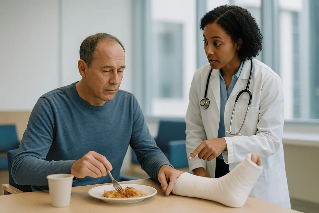
Every major complication is a failure at one step of that sequence. Infection happens when bacteria colonize the scaffold before your cells do; a graft has no blood supply of its own, so it cannot deliver immune cells or antibiotics to its interior, and bacteria that settle there are hard to clear. Non-union happens when the blood vessels never fully arrive, often because the graft is moving, the gap is too wide, or the surrounding bone is poorly perfused. Resorption, the graft shrinking or vanishing on imaging, happens when the dissolving phase outpaces the rebuilding phase, which is more likely in smokers, in people with uncontrolled diabetes, and when the graft is not held still.
Timing follows the biology. MedlinePlus notes that recovery from bone grafting typically spans 2 weeks to 3 months depending on graft size and location, and that the graft continues to remodel well beyond that. Problems in the first two weeks are almost always wound or infection issues. Problems that surface at three to six months are more often about incorporation: the graft simply did not become bone. Understanding which phase you are in helps you interpret a symptom instead of fearing it.
Is allograft bone safe? Screening, processing and disease transmission
The question people are often embarrassed to ask out loud is whether donor bone can give them a disease. The honest answer is that transmission has been documented in the medical literature, that it is rare, and that the modern screening and processing system exists because of those early cases.
Donor safety rests on layers rather than a single test. Tissue banks review the donor’s medical and social history, test blood for HIV, hepatitis B and C, syphilis and other agents, and reject donors with cancer that could involve bone, systemic infection, or unexplained neurological disease. The bone is then processed to remove marrow and blood, which is where most infectious material lives, and many products are terminally sterilized. Cleveland Clinic and Mayo Clinic both describe processed allograft as a standard, widely used material in dental and orthopedic reconstruction.
The CDC has published investigations of bacterial infections linked to contaminated musculoskeletal tissue in the past, and those reports drove tighter regulation of how tissue is recovered and processed. Reading them is oddly reassuring: they show a surveillance system that detects clusters and traces them to a source. What they do not show is a steady background of infections from routine allograft use.
Two caveats are worth stating plainly. Sterilization methods that kill every microorganism can also weaken the graft’s natural growth proteins, so tissue banks balance safety against biological activity, and surgeons choose products with that trade-off in mind. Also, no screening system reduces risk to zero, which is why the consent form mentions transmission at all. If this concern weighs on you, ask your surgeon which tissue bank supplies their graft and how it is processed. That is a reasonable question, not a paranoid one, and a good team will answer it directly.
How does infection happen after a bone graft?
A bone graft is, in surgical terms, an implant: a piece of material with no blood supply placed inside living tissue. Bacteria love such surfaces. They can form a biofilm, a slimy protective layer that shields them from immune cells and from antibiotics circulating in the blood. Once a biofilm establishes itself on graft particles or a block, the infection often cannot be cleared without removing the material.
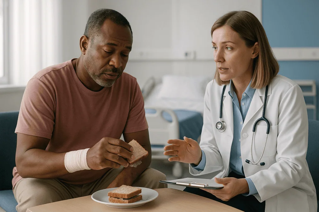
Bacteria reach the graft by three main routes. They can enter during surgery from the skin or, in dental work, from the mouth, which is why sterile technique and careful wound closure matter so much. They can arrive later through a wound that opens or a gum flap that pulls back and exposes the graft. They can also travel through the bloodstream from an infection elsewhere, such as a urinary or dental infection, and settle on the poorly perfused scaffold.
The CDC defines a surgical site infection as one that occurs within 30 days of an operation, or within 90 days when an implant is left in place. That longer window applies to bone grafts. Most infections declare themselves in the first few weeks, but a low-grade infection around a structural allograft or a jaw graft can smolder for months and present as unexplained aching, a small draining sinus, or hardware loosening on an X-ray.
Certain factors raise the odds and are worth discussing before surgery rather than after:
- Smoking or vaping nicotine, which narrows small blood vessels and delays healing.
- Poorly controlled diabetes, which impairs white blood cell function.
- Medicines that suppress immunity, including long-term steroids.
- Active gum disease or untreated tooth infection near a dental graft site.
- Prior radiation to the jaw or the surgical region.
None of these forbid grafting. They change how closely you are watched and what preparation your team may ask of you.
Bone graft infection symptoms versus ordinary healing
Some discomfort after grafting is not a warning sign; it is the price of admission. Swelling, bruising and soreness peak around the second or third day and then recede. Small bone granules working their way out of a dental socket in the first week are common and usually harmless. What separates ordinary healing from trouble is direction of travel. Healing gets better, unevenly but steadily. Infection gets better and then worse, or never gets better at all.
| Sign | Usually part of normal healing | Suggests a developing problem |
|---|---|---|
| Pain | Improves after day 3, controlled with prescribed relief | Increases after initial improvement, throbbing, wakes you at night |
| Swelling | Peaks by day 2–3, then softens | Returns after settling, feels hot or tense |
| Drainage | Slight blood-tinged ooze in the first 1–2 days | Thick, cloudy, foul-smelling, or any drainage after the first week |
| Temperature | Mild warmth at the site | Fever, chills, feeling generally unwell |
| Graft material | A few granules shed early (dental) | Grey or exposed block, membrane visible, large loss of granules |
| Function | Gradual return of chewing, walking or range of motion | Function plateaus or regresses; implant or hardware feels loose |
Two patterns deserve special mention. A wound that looked closed and then gapes open, sometimes with a visible edge of graft, is called dehiscence, and exposed allograft is at high risk of infection because it has no living cover. Second, a fever combined with a red, hot surgical site is a same-day call, not a wait-and-see. Photographing the site daily in good light can help you and your team judge whether the trajectory is right; a picture from Tuesday makes Friday’s swelling much easier to interpret.
What graft non-union and resorption mean, and why they happen
Non-union is the term surgeons use when bone that should have joined has not. In the context of allograft, it means the boundary between the graft and your native bone has remained a gap or a layer of scar rather than becoming continuous bone. Resorption is a related outcome: the graft has been dissolved by your osteoclasts faster than it could be rebuilt, so the volume you gained on the day of surgery has shrunk on later imaging.
Neither is an immune rejection. Processed allograft has had its cells removed, so there is little for the immune system to target in the way it would target a transplanted kidney. The failure is mechanical and biological: too little blood supply, too much movement, too wide a gap, or a scaffold that was consumed before it could be replaced.
The known drivers cluster into a few groups:
- Blood supply. Bone heals from its edges inward. Scarred, irradiated or heavily smoked-on tissue delivers fewer vessels, so incorporation stalls.
- Stability. A graft that shifts under chewing, walking or spinal loading keeps breaking the fragile early bridges of new bone. Plates, screws, membranes and activity restrictions exist to hold it still.
- Graft size. Small particles incorporate quickly. A large structural block may never be fully replaced and can fracture years later as it slowly weakens.
- Host factors. Uncontrolled diabetes, vitamin D deficiency, some anti-inflammatory and anti-resorptive medicines, and advanced age all slow bone turnover.
Detection is usually radiographic. At follow-up your team looks for bridging bone across the junction, loss of the sharp line between graft and host, and preserved height or width. A persistent lucent line at several months, hardware loosening, or a graft that has visibly shrunk are the objective signs. Symptoms may be subtle: aching under load, a dental implant that fails to tighten, or a fusion that still moves. Sometimes the only clue is that recovery has quietly stopped progressing.
Signs of bone graft failure after a dental bone graft
People searching for signs that a dental bone graft has failed are usually somewhere between week two and month four, staring at a socket that does not feel right. A few descriptions come up again and again in that situation, and each has a plausible explanation.
The gum over the site pulling back to expose grey or white granules is the most common early concern. A handful of loose particles in the first days is normal. A membrane or block that is visible and stays visible is not; exposed graft dries out, collects bacteria, and is often lost. Persistent bad taste or odor from the site, especially with a little pus at the margin, points the same direction.
Later signs are quieter. The ridge that felt firm may feel soft or spongy, or the gum may look sunken where height was supposed to be gained. At the implant stage, the surgeon may find that the drill meets soft, non-bony material, or that the implant does not achieve stability. On X-rays, the graft may appear to have melted away rather than blended into the surrounding jaw.
The question of what percentage of dental bone grafts fail does not have a single honest number. Published series vary widely depending on the site (a small socket preservation graft behaves very differently from a large sinus lift), the material, the patient’s smoking status and health, and how failure is defined. Cleveland Clinic and Mayo Clinic both describe dental grafting as a routine, well-established step before implants, while noting that healing takes several months and that smoking, poor oral hygiene and certain health conditions reduce the chances of success. Rather than anchoring on a percentage from a website, ask your surgeon how often grafts in your specific situation need to be redone in their practice, and what they do differently the second time.
Warning signs with orthopedic and spinal allografts
Outside the mouth, allograft carries load. A wedge in a spinal fusion, a strut in a revision hip replacement, or a segment replacing bone removed for a tumor is expected to bear weight while it is slowly incorporated, and the complications that follow are shaped by that demand.
Deep infection presents much as it does anywhere in orthopedics: increasing pain at rest, warmth and redness over the incision, drainage that persists beyond the first days, and fever. Because these grafts sit deep, the skin may look calm while the problem develops underneath; new or worsening pain that does not match the expected recovery curve is often the earliest clue. Around the spine, infection or non-union can also produce new nerve symptoms if a cage shifts or collapses.
Non-union in a fusion means the vertebrae still move at the operated level. Patients describe a return of the original back or neck pain after an initial honeymoon, sometimes with a sense of instability. The surgeon looks for a lucent halo around screws, broken rods or screws, and absence of bridging bone on CT. In a long-bone or joint reconstruction, non-union shows as pain with weight bearing, and a structural allograft that never fully incorporates can fracture months or years later, because the portion that remains dead bone gradually loses strength.
Hardware loosening deserves its own line. Screws and plates are designed to hold bone still while it heals, not to carry load forever. If the graft does not unite, the metal eventually fatigues. A new click, grinding sensation, or sudden increase in pain at a site that had been improving is worth a call and an X-ray, not a wait until the next scheduled visit.
The pattern across all these settings is consistent: the direction of recovery changes. Watch the trend rather than any single bad day.
Who is usually offered an allograft, and who is asked to wait
Allograft tends to be chosen when the surgeon wants to avoid harvesting the patient’s own bone, when the defect is larger than an autograft could comfortably fill, or when the patient is older or frail enough that a second surgical site is a real burden. In dentistry that covers socket preservation after extraction, ridge augmentation before implants, and sinus floor lifts. In orthopedics it covers spinal fusion, filling defects after tumor removal or trauma, and revision joint replacement where the original bone has been lost.
Candidates generally share a few features: a defect that is stable or can be stabilized, reasonably healthy surrounding tissue, and control of conditions that would sabotage incorporation. Surgeons are also weighing how much biological help the graft will need. A small gap in a well-perfused jaw can rely on allograft alone. A large structural defect may need a mix of allograft for volume and autograft for living cells.
Some people are asked to wait, and the reasons are about improving the odds rather than closing a door:
- Active infection at or near the site, including untreated gum disease or a draining tooth abscess.
- Current smoking or nicotine use; many teams ask for a period of abstinence before and after surgery because nicotine reduces blood flow to healing bone.
- Poorly controlled diabetes, since high glucose impairs both immune function and bone formation.
- Recent or planned radiation to the region.
- Certain medicines that alter bone turnover, where timing may need coordination with the prescribing clinician; never adjust these on your own.
A few situations shift the choice away from allograft altogether, for example where the surgeon needs living cells and growth factors that only autograft provides, or where a patient declines donor tissue for personal or religious reasons. Alternatives include autograft, synthetic ceramics and animal-derived mineral, and the decision about which fits your anatomy and health sits with the treating team.
Allograft bone graft complications compared with other graft types
No graft material is free of downsides. Choosing between them is a trade of one set of risks for another, and seeing them side by side clarifies why a surgeon might pick allograft in one case and your own bone in the next.
| Graft type | Source | Main strengths | Characteristic complications |
|---|---|---|---|
| Autograft | Your own bone (hip, chin, ramus, rib) | Living cells and growth factors; no transmission risk; fastest incorporation | Pain, bleeding, nerve injury or infection at the donor site; limited quantity; longer surgery |
| Allograft | Processed donor human bone | Unlimited supply; no second surgical site; available as particles, blocks, structural segments | Slower incorporation; higher resorption in some settings; rare disease transmission; late fracture of large structural pieces |
| Xenograft | Animal-derived mineral, usually bovine | Very slow resorption, holds volume well in the jaw | Little biological activity; residual particles may persist for years; unacceptable to some patients |
| Synthetic | Ceramics such as calcium phosphate, bioactive glass | No biological transmission risk; predictable supply | No living cells or proteins; some dissolve too fast or too slowly; variable strength |
The pattern in the table is a sliding scale between biology and convenience. Autograft is the most biologically active and the most expensive in terms of patient morbidity, because it costs you a second wound. Allograft removes that wound but relies entirely on your body to supply the cells, so it is more vulnerable to the factors that starve a graft of blood and stability. Synthetics and xenografts push further along the same line.
Surgeons frequently combine materials, layering allograft particles over a core of autograft to get volume and biology together, or mixing allograft with a synthetic to slow its resorption. If your consent form lists more than one material, that is usually why. Ask what each component is meant to do; the answer tells you a lot about which complication your team is most concerned to prevent in your case.
What the days and weeks after an allograft usually look like
The first 48 hours are about bleeding and swelling. The site is protected, activity is restricted, and the graft is at its most vulnerable because nothing yet holds it except sutures, a membrane, or hardware. Bruising and a dull ache are expected. For dental grafts, that means soft food, no straws, no vigorous rinsing, and no smoking; for orthopedic grafts it means the weight-bearing or brace instructions your surgeon gave you, followed exactly.
Days three through fourteen are the wound phase. Swelling should be receding and pain easing day by day. Sutures may dissolve or be removed. This is the window in which most infections and wound breakdowns appear, and it is when a daily look at the site pays off. Any reversal of the improving trend in this period is worth a call.
Weeks two through twelve are the quiet phase. From the outside nothing seems to happen, but this is when blood vessels invade the scaffold and the first living bone forms at the junction. MedlinePlus describes overall recovery from bone grafting as taking 2 weeks to 3 months depending on graft size and site. Activity restrictions often continue through this period precisely because early bone bridges are fragile.
Months three to six and beyond are the incorporation phase. Your team will typically image the site to look for bridging bone and preserved volume. For dental grafts, Mayo Clinic notes that healing before implant placement can take several months, and the overall implant journey commonly stretches across many months. For spinal fusion or structural reconstruction, radiographic confirmation of union can take longer still, and structural allograft continues to remodel for years.
These are typical ranges from published patient information, not promises. Your own timeline depends on the graft’s size, your health, and how the site behaves, and your surgeon will adjust the plan as the imaging comes in.
How long does a bone graft last, and what is its average lifespan?
The phrase “lifespan of a bone graft” contains a hidden assumption: that the graft is a part with a service life, like a hip prosthesis. For most allografts that is the wrong mental model. A successfully incorporated graft is not a component that wears out; it has been replaced by your own living bone, which remodels for the rest of your life like any other bone. In that sense the graft does not have a lifespan at all. It becomes you.
Where the question does make sense is in two situations. The first is large structural allograft, such as a long-bone segment or a big strut in a revision hip. These pieces are too large to be fully replaced, so a core of non-living donor bone often persists indefinitely. Dead bone slowly accumulates microdamage without the repair that living bone performs, and the orthopedic literature on structural allograft describes late fracture of the graft as a recognized long-term complication, sometimes years after surgery. Surgeons monitor these grafts with periodic imaging for exactly that reason.
The second situation is the jaw. A dental graft that healed well can still shrink over the years if the tooth it was meant to support is not placed, because bone that is not loaded tends to resorb. That is why dentists often sequence implant placement within a defined window after grafting, and why an unused grafted ridge may need touching up before an implant years later.
What keeps a graft healthy long term is unglamorous: not smoking, keeping diabetes and gum disease controlled, loading the bone appropriately through normal chewing or weight bearing, and attending follow-up imaging. If you are told your graft has incorporated, the more useful question than “how long will it last” is “what would make it weaken, and how will we know?”
What people often get wrong about bone graft rejection and failure
Misconceptions about allograft are remarkably consistent, and correcting them removes a good deal of unnecessary dread.
“My body might reject the donor bone.” True rejection, the immune attack seen in organ transplants, does not occur in the way people imagine with processed allograft. The donor’s cells, which carry the markers the immune system reacts to, have been removed. What people call rejection is almost always infection, exposure, or non-union: a biological or mechanical failure, not an immune one. No anti-rejection medicines are used with bone allograft.
“If a few granules come out, the graft has failed.” Shedding some particles from a dental socket in the first week is common. Loss of a large amount, or a visible membrane or block, is the concern.
“Donor bone is a frequent source of infection.” Most graft infections come from the patient’s own skin or mouth bacteria, not from the graft. Contamination of processed tissue has occurred and has been investigated by public health agencies, but it is uncommon.
“Antibiotics will fix any graft infection.” Antibiotics reach infected bone poorly and cannot penetrate a mature biofilm on a scaffold with no blood supply. Established graft infection often requires removing the graft and regrafting later. Early reporting matters because a superficial wound problem caught in days is far easier to manage than a deep one found in weeks.
“Once the X-ray looks good, I can do anything.” Radiographic bridging is a milestone, not a finish line. Remodeling continues for many months, and structural grafts are watched for years.
“Failure means I can never have the procedure again.” A failed graft is usually removed, the site is allowed to heal, and regrafting is often possible, sometimes with a different material or added autograft. The decision rests with the treating team and depends on why the first attempt failed.
Questions to ask your care team before and after grafting
Good questions do two things: they surface information that changes your behavior, and they signal to the team what you are worried about so they can address it directly. The following have earned their place in that conversation.
Before surgery:
- Why allograft rather than my own bone or a synthetic in my case, and what is each material in the plan meant to do?
- Which tissue bank supplies the graft, and how is it processed and sterilized?
- What in my health or habits raises my risk of infection or non-union, and is there anything you want me to change beforehand?
- Should any of my regular medicines be timed differently around surgery? (Any change should come from the prescribing clinician, never self-directed.)
- How will the graft be held still, and what activity restrictions follow from that?
After surgery:
- What does a normal day three, day seven and day fourteen look like for this specific graft, so I can judge whether I am on track?
- Which signs should prompt a same-day call, and which can wait for the next visit?
- When will you image the site, and what are you looking for on that scan?
- How often do grafts like mine need to be redone in your experience, and what usually causes that?
- If the graft does not incorporate, what are the options, and when would you decide?
Bring a written list; post-operative appointments are short and anesthesia dulls memory. Ask permission to photograph the site so you have a baseline. If English is not your first language or medical vocabulary is unfamiliar, ask the team to define terms; “dehiscence,” “lucency” and “incorporation” are jargon, and you are entitled to plain versions. None of these questions second-guesses your surgeon. They give the team the chance to tailor advice to you rather than to the average patient, and they make you a better observer of your own recovery, which is the single most useful thing a patient can be in the weeks after a graft.
When to call your doctor: red-flag signs after an allograft bone graft
The surgical team would rather take ten unnecessary calls than miss one early infection. That is the spirit in which the following list is offered. It is not a checklist for diagnosing yourself; it is a prompt to pick up the phone and let the people who placed the graft decide what it means.
Call the same day if you notice any of these:
- Pain that was improving and is now worsening, or pain that is severe, throbbing and not controlled by the relief you were prescribed.
- Fever, chills, or feeling generally unwell in the weeks after surgery.
- Swelling that returns after it had settled, or a site that is hot, red and spreading.
- Any drainage after the first few days, particularly if it is cloudy, thick or foul-smelling, or a persistent bad taste at a dental site.
- The wound opening, or graft material, a membrane or a block becoming visible.
- A dental implant, plate, screw or rod that feels loose, clicks, or shifts.
- New numbness, tingling, weakness or a change in bladder or bowel control after a spinal graft.
- Bleeding that does not stop with firm pressure for the time you were told.
Seek emergency care immediately if you develop difficulty breathing or swallowing, rapidly spreading facial or neck swelling, a high fever with confusion, or sudden severe weakness in the legs. These are rare but cannot wait for a scheduled appointment.
Beyond the acute period, contact your team if recovery seems to have stalled: a fusion site where the original pain has returned, a grafted ridge that feels soft or looks sunken, or a limb that hurts more with weight bearing month after month. Those are the quiet presentations of non-union, and earlier imaging gives more options.
Keep the after-hours number somewhere obvious. In every one of these situations, the decision about what to do next belongs to the treating team, and the most valuable thing you can contribute is a clear, early description of what has changed.
Frequently asked questions
What are the signs that a dental bone graft has failed?
The clearest signs are graft material or a membrane that becomes and stays exposed, persistent bad taste or pus at the site, pain or swelling that returns after settling, and a ridge that later feels soft or looks sunken. On imaging, a failed graft appears to have shrunk rather than blended into the jaw, and at implant placement the surgeon may find soft material instead of bone. Report these to your dental surgeon promptly.
Is allograft bone safe?
Processed allograft is widely used and regarded as a standard graft material in dental and orthopedic surgery. Donors are screened by history and blood testing, and the bone is cleaned, stripped of cells and marrow, and usually sterilized. Transmission of infection from donor tissue has been documented in the past and is rare; public health investigations of such cases led to tighter tissue-bank regulation. No screening reduces risk to zero, which is why consent forms mention it.
What is the average lifespan of a bone graft?
A successfully incorporated graft does not have a lifespan in the usual sense, because it is gradually replaced by your own living bone, which remodels for life. The exception is large structural allograft, where a core of non-living donor bone may persist indefinitely and can weaken or fracture years later, so surgeons monitor it with periodic imaging. A grafted jaw ridge left without an implant may also slowly resorb over time.
What percentage of dental bone grafts fail?
There is no single honest percentage. Published series vary widely depending on the site, the material, how failure is defined, and patient factors such as smoking and diabetes. Patient information from Cleveland Clinic and Mayo Clinic describes dental grafting as a routine, well-established step before implants while noting that healing takes several months and that smoking and poor oral hygiene lower the chance of success. Ask your surgeon about outcomes in situations like yours.
What are the most common bone graft infection symptoms?
Infection usually shows as pain that worsens after initially improving, swelling that returns or feels hot and tense, drainage that is cloudy, thick or foul-smelling after the first few days, and sometimes fever or chills. At a dental site a persistent bad taste or odor is common. Deep infections around spinal or orthopedic grafts may produce few skin changes at first, so unexplained increasing pain is often the earliest clue.
Can your body reject donor bone in a bone graft rejection?
Not in the way organs are rejected. Processed allograft has had the donor’s cells removed, and those cells carry the markers the immune system reacts against. No anti-rejection medicines are used with bone grafts. When a graft is lost, the cause is nearly always infection, exposure of the material, movement, or poor blood supply preventing incorporation, which surgeons call non-union or resorption rather than rejection.
How long does it take for allograft bone to integrate?
Integration is gradual. MedlinePlus describes overall recovery from bone grafting as typically 2 weeks to 3 months depending on graft size and site, and Mayo Clinic notes that healing after a dental graft before implant placement can take several months. Small particulate grafts incorporate faster than large blocks, and structural allograft may remodel for years. Your surgeon confirms integration with imaging rather than by the calendar.
Why did my bone graft dissolve or shrink?
Resorption happens when the cells that dissolve old bone work faster than the cells that build new bone, so the scaffold is consumed before it is replaced. Common contributors are smoking, uncontrolled diabetes, movement of the graft, a wide gap, poor blood supply in scarred or irradiated tissue, and, in the jaw, lack of loading when no implant is placed. Your team can usually identify the likely cause from your history and imaging.
Can a failed allograft bone graft be redone?
Often, yes. A failed or infected graft is typically removed, the site is allowed to heal and any infection is treated, and regrafting is considered later. The surgeon may choose a different material, add some of your own bone for living cells, or change how the graft is stabilized, depending on why the first attempt failed. Whether and when to regraft is a decision for the treating team based on your specific situation.
Does smoking really affect bone graft healing?
Yes. Nicotine narrows small blood vessels and reduces oxygen delivery to healing tissue, and smoking impairs the cells that form new bone and fight infection. Mayo Clinic and Cleveland Clinic both list smoking among the factors that lower the chance of a dental graft or implant succeeding. Many surgical teams ask patients to stop nicotine in all forms before and after grafting; discuss timing and support with your care team.
References
This article is for general information only and is not a substitute for professional medical advice. Please consult a qualified doctor about your individual situation.

More from the Blog

NHS Knee Replacement Waiting Times: Your Realistic Options in 2026
In England, the NHS standard is that treatment should start within 18 weeks of referral, but that target has not been met nationally since…

What Bunion Pain Feels Like, What Bunions Look Like, and Whether They Are Hereditary
Bunion pain usually feels like a deep, throbbing ache or burning soreness at the base of the big toe, worse in snug shoes and…
Hip Replacement Abroad: Costs, Safety Checks and Aftercare Planning
A total hip replacement abroad typically costs a fraction of UK or US private prices: our guide range is EUR 9,100–16,900, compared with roughly…

How Is Pain Managed After Shoulder Replacement? Nerve Blocks and the First Nights Explained
Pain after shoulder replacement is usually managed in layers: a regional nerve block placed before surgery numbs the shoulder for roughly the first day,…

Robotic Knee Replacement Cost: Is the Premium Worth Paying?
Robotic assistance typically adds a premium of roughly EUR 2,000-6,000 on top of a conventional total knee replacement price, with similar surcharges in the…

How Recovery Differs After Complex Joint Reconstruction: Slower Milestones, Explained
Recovery after complex joint reconstruction, such as revision replacement, repair of a fracture around a joint or rebuilding lost bone and ligament, is usually…