

Allograft vs Autograft Bone Grafting: Why Your Surgeon May Choose Donor Bone
Key Takeaways
- Autograft supplies living bone-forming cells, intact growth signals and a scaffold, while processed allograft keeps the scaffold, loses the cells and retains variable signaling depending on how it was treated.
- The most common reasons a surgeon chooses donor bone are that more bone is needed than your body can spare, and that harvesting your own bone creates a second surgical site with its own pain and complication risk.
- Comparative studies of routine single-level spinal fusion generally find similar fusion with allograft plus local bone versus iliac crest autograft, which is why pelvic harvest is now reserved for harder cases.
- Several systematic reviews report higher ACL graft failure with allograft in young, high-demand athletes, while the gap narrows in older recreational patients, so age and sport drive the ACL graft decision.
- Processed bone allograft does not trigger organ-style rejection because the donor cells have been removed, and modern screening has made disease transmission an extremely rare event.
- MedlinePlus puts overall bone graft recovery anywhere from about two weeks to three months depending on the site, and the Mayo Clinic notes spinal fusion takes several months to become solid regardless of graft type.
An autograft is bone taken from your own body; an allograft is sterilized, screened bone from a deceased donor. Autograft contains living cells and is often considered the reference standard, but harvesting it adds a second surgical site with its own pain and risks. Surgeons choose allograft when they need more bone than you can spare, want to avoid donor-site problems, or judge the extra biology unnecessary for that particular repair.
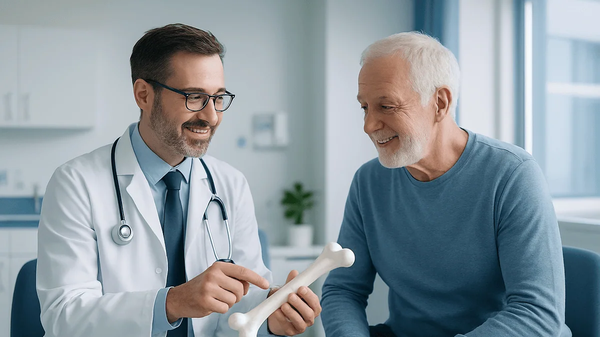
The consent form lists two options, and a patient who came in worried about a wobbly knee or a collapsing vertebra now has a new question they never expected: whose bone will be inside them? Their own, taken from the hip or shin during the same operation, or a piece from someone who died and left their tissue to strangers?
It is a reasonable thing to pause over. Most people have a gut instinct that their own bone must be better, and for some repairs that instinct is right. For others, it is simply not the whole story. The allograft vs autograft bone graft decision is one of the quieter judgment calls in orthopedics, dentistry and spine surgery, shaped by the size of the defect, the age and health of the patient, the job the graft has to do, and the surgeon’s own experience.
What follows is the version of that conversation you rarely get in a fifteen-minute clinic slot: how each graft behaves once it is in place, where donor bone earns its keep, where your own bone still wins, and what the evidence actually shows.
What is an allograft bone graft, and what is an autograft?
A bone graft is a piece of bone, or a bone-like material, placed where the body needs help rebuilding: a gap after a tumor is removed, a fracture that refuses to knit, a spine that must be fused, a jaw too thin to hold an implant, or a torn ligament that needs a new anchor. The graft acts as scaffolding. Your own bone-forming cells then move in, lay down new bone and gradually replace the graft with living tissue, a process surgeons call incorporation.
An autograft is bone harvested from your own body during the same operation. Common sources are the iliac crest, the ridge of pelvic bone you can feel just below your waistline, along with the shin, the fibula, a rib, or, in knee ligament surgery, a strip of tendon with small bone blocks attached at each end.
An allograft is bone donated by another person, almost always a deceased donor whose family consented to tissue donation. The bone is recovered under sterile conditions, tested, processed and stored by an accredited tissue bank until a surgeon requests it. It arrives as chips, blocks, wedges, struts, or as whole tendons with bone attached, depending on the job.
Two neighbors in this family are worth naming so they are not confused with the above. A xenograft comes from an animal source, usually bovine, and is processed until only the mineral framework remains; it is common in dental work. A synthetic graft is a laboratory-made ceramic or polymer designed to mimic bone mineral. MedlinePlus lists all of these as accepted options, and many operations combine more than one, for example a small amount of your own bone mixed with donor chips to stretch a limited supply.
Allograft vs autograft bone graft: how each one actually works in the body
Bone healing needs three ingredients, and this is where the allograft vs autograft bone graft comparison becomes concrete rather than emotional. Surgeons describe them as osteoconduction, osteoinduction and osteogenesis.
Osteoconduction means providing a structure that new bone can grow along, like trellis for a climbing plant. Osteoinduction means releasing signaling proteins that recruit nearby stem cells and instruct them to become bone-building cells. Osteogenesis means the graft itself contains living cells that start making bone on day one.
Fresh autograft brings all three. It is your own tissue, so there is no immune reaction, its proteins are intact, and some of its cells survive the transfer. That is why textbooks still call it the reference standard for biology.
Allograft is a different proposition. Processing removes the donor’s cells to lower the risk of immune reaction and disease transmission, so osteogenesis is essentially gone. What remains is a sound mineral scaffold, strong on osteoconduction, with varying amounts of osteoinductive protein depending on how it was treated. Freeze-dried allograft keeps structure but loses more signaling; demineralized bone matrix, an allograft that has had its mineral removed, keeps more protein but has no structural strength and is used as a putty or paste.
Incorporation follows the same steps for both: bleeding and clot around the graft, new blood vessels growing in, old bone resorbed and new bone deposited. The Cleveland Clinic describes this as a gradual replacement rather than a sudden switch. In practice, allograft incorporates more slowly and, in large structural pieces, may never be fully replaced, leaving a core of dead but mechanically useful bone. For many repairs that is perfectly acceptable. For others, it is the reason your own bone is preferred.
Where does donor bone come from, and how is it screened?
The word donor makes some patients picture a living person giving up a piece of themselves. That is rare for bone. Almost all allograft comes from deceased donors, recovered in the hours after death alongside organs, corneas, skin and heart valves, with consent from the donor or their next of kin. A smaller share comes from living donors having a hip replaced, whose removed femoral head would otherwise be discarded.
Screening happens in layers. The donor’s medical and social history is reviewed to exclude cancers, infections, autoimmune disease and high-risk behaviors. Blood is tested for HIV, hepatitis B and C, syphilis and other transmissible infections. The recovered tissue is cultured for bacteria. Tissue banks in the United States operate under federal oversight and, in most cases, voluntary accreditation standards that specify how tissue must be recovered, tested, processed, labeled and tracked.
Processing is where the trade-offs are made. Bone may be washed, treated with solvents to strip fat and cells, freeze-dried, frozen, or irradiated. Each step lowers the risk of transmitting anything from the donor and reduces the chance of an immune response. Each step also strips away some of the biological signals that help bone incorporate. A tissue bank is constantly balancing safety against biological activity, and different products sit at different points on that line.
Every graft carries a unique identifier so it can be traced back to the donor and forward to the recipient. If a problem were ever detected in one graft from a donor, every other graft from that donor could be located. Modern testing and processing have made disease transmission from processed bone allograft an exceptionally rare event, though no biological product carries a zero risk and your surgeon should say so plainly.
Why your surgeon may choose donor bone
The single most common reason is arithmetic. Your body has a limited supply of bone that can be safely borrowed. The iliac crest yields a modest amount; a rib or a piece of fibula yields more but at a cost. A long-segment spinal fusion, a large defect left after removing a bone tumor, or a hip revision where bone has been eaten away around a loose implant can need far more than any single donor site can provide. Donor bone comes in whatever quantity and shape the operation requires.
The second reason is the donor site itself. Harvesting bone means a second incision, more time under anesthesia, more blood loss and a new place that can hurt, bleed, become infected or, uncommonly, fracture. Iliac crest pain is the classic complaint and in some patients it outlasts the pain from the main operation. If a surgeon believes the repair will heal well with donor bone, they may reasonably decide the second wound is not worth inflicting.
Structure is the third. A large machined allograft strut or wedge can bear load in a way that morselized autograft chips cannot. When the job is to hold a space open, for instance between two vertebral bodies or under a collapsed joint surface, mechanical strength may matter more than biological activity.
Patient factors round out the list. Children have less spare bone and growth plates to protect. Older adults may have thin, osteoporotic bone that yields a poor-quality graft. People with significant heart or lung disease benefit from shorter operations. Someone who has already donated from one hip for an earlier surgery may have nothing convenient left to give.
None of these reasons means donor bone is better. They mean the surgeon has judged that, for this repair, its advantages outweigh what is lost.
The case for your own bone: what autograft still does best
If donor bone solved every problem, autograft would have faded away. It has not, and the reasons are biological rather than sentimental.
Living cells matter most where healing is hardest. A fracture that has failed to unite after months, a bone weakened by previous radiation therapy, a smoker’s spine, a diabetic foot: these are environments where the body’s own repair signals are muted. Autograft brings its own workforce and its own chemical instructions, and surgeons often reserve it for exactly these situations.
Speed of incorporation is the second advantage. Because autograft is recognized as self, revascularization begins promptly and new bone forms along the scaffold sooner. In a young athlete’s knee, faster maturation of the graft is part of why many sports surgeons still favor the patient’s own tendon, a point taken up in the ACL section below.
Infection risk is lower with respect to the graft itself. There is no possibility of transmitting a donor infection, and living tissue with its own blood supply resists bacteria better than a dead scaffold. This is one reason autograft is often preferred when a previous infection has been treated at the same site.
Vascularized autograft is a special case. A segment of fibula can be moved with its own artery and vein and reconnected under a microscope to vessels near the defect. The transplanted bone remains alive throughout and can even grow in a child. It is technically demanding and used for the largest, most compromised defects, but nothing in the allograft world can match a living, perfused bone.
Against all this sit the donor-site problems already described. The honest summary is that autograft buys better biology at the price of a second wound, and the surgeon’s job is to decide whether that purchase is worth making for you.
Allograft bone graft disadvantages, told honestly
Patients searching for allograft bone graft disadvantages usually find either alarm or reassurance. The evidence supports neither extreme, so here is the list as clinicians actually weigh it.
Slower and less complete incorporation is the central drawback. Without living cells and with reduced signaling proteins, donor bone relies entirely on the recipient’s biology. In healthy, well-vascularized tissue this is usually adequate. In compromised sites it can mean delayed union, or a graft that is resorbed faster than new bone replaces it, leaving a gap.
Graft failure and nonunion are more likely in large structural allografts than in small chips. A long strut may crack under repeated loading before host bone has grown across it. Surgeons plan for this with plates, cages or rods that share the load while incorporation proceeds.
Disease transmission is the fear most people voice. Historical cases involving hepatitis and HIV drove the modern screening system, and with current testing and processing the risk from processed bone is considered extremely low. Bacterial contamination is also rare but not zero, which is why grafts are cultured before release.
Immune reaction is uncommon with processed bone because the cells that carry the strongest immune signals have been removed. Low-grade reactions can still slow incorporation, and surgeons consider this when choosing between fresh-frozen and more heavily processed products.
Ethical and personal factors count too. Some patients have religious or cultural objections to receiving tissue from another person, and a few decline for reasons they do not need to justify. A good surgeon asks before assuming.
What allograft does not do is transmit the donor’s personality, appearance, blood type or DNA in any way that affects you. Processed bone contains no living cells, and the mineral scaffold is eventually replaced by tissue your own body has made.
Autograft vs allograft ACL: the sports medicine debate
The anterior cruciate ligament, the rope-like band in the center of the knee that stops the shin sliding forward, is reconstructed rather than repaired when it tears. A new ligament is built from a tendon graft, and the autograft vs allograft ACL choice is one of the most argued questions in sports medicine.
Autograft options include a strip of the patellar tendon with a small bone block at each end, a section of hamstring tendon, or part of the quadriceps tendon. Each leaves the donor site weaker for a time: patellar tendon grafts are associated with pain at the front of the knee and kneeling discomfort; hamstring grafts can leave a measurable deficit in hamstring strength that most people never notice but elite sprinters might.
Allograft tendon avoids all of that. The operation is shorter, early pain is often less, and there is no second scar. The Mayo Clinic notes that both approaches are in routine use.
The catch lies in who is receiving it. Several systematic reviews and large registry analyses have reported higher graft failure rates with allograft in young, high-demand athletes, particularly teenagers returning to cutting and pivoting sports. The proposed reasons are slower incorporation and the effect of irradiation and chemical processing on tendon strength. In older recreational patients, the difference narrows and many studies find comparable outcomes.
This is why the same surgeon may recommend an autograft for a seventeen-year-old soccer player and an allograft for a fifty-year-old who wants to hike without instability, and both recommendations can be evidence-based. Return to pivoting sport after ACL reconstruction commonly takes nine months or longer regardless of graft type, according to the Mayo Clinic, because the new ligament must mature before it can take repeated shearing loads.
Spinal fusion: comparing fusion outcomes with allograft versus autograft
Spinal fusion joins two or more vertebrae so they heal into one solid bone, removing painful motion or stabilizing a deformity or fracture. Bone graft is the essential ingredient; the metal screws and rods that patients focus on are only temporary scaffolding while the graft does its work. The Mayo Clinic describes fusion as taking several months to become solid, with bone continuing to mature well beyond that.
Historically, surgeons harvested iliac crest bone for nearly every fusion. Over the past two decades that has shifted. Local bone, the shavings removed from the spine itself while preparing the surface, is now routinely collected and reused, effectively a free autograft with no separate donor site. When more volume is needed, surgeons often add allograft chips or demineralized bone matrix rather than opening the pelvis.
Structural allograft has a distinct role in the front of the spine. Machined wedges of donor bone placed between vertebral bodies restore height and hold the space open while fusion proceeds. Many surgeons alternatively use synthetic cages packed with graft, and the evidence does not clearly favor one over the other for most patients.
What the comparative studies show, taken together, is that for single-level fusions in otherwise healthy adults, fusion rates with allograft plus local bone are broadly similar to those with iliac crest autograft, while donor-site complications are avoided. For multi-level fusions, revisions of failed fusions, smokers, and people with conditions that impair healing, many surgeons still reach for iliac crest bone because the biology is more forgiving.
Pediatric spine surgery has its own literature, largely because children heal briskly and have limited donor bone. Comparative studies of posterior cervical fusion in children have reported acceptable fusion with allograft in many series, but decisions in this group are highly individualized and belong with the treating team.
Dental and jaw bone grafts: why the choice looks different here
The jaw is the one place many otherwise healthy adults meet bone grafting. After a tooth is lost, the bone that held it shrinks, and a dental implant, a titanium post that acts as an artificial root, needs enough bone to sit in securely. Grafting rebuilds that volume, either at the time of extraction to preserve the socket or later to widen a thin ridge or lift the sinus floor.
Small volumes change the calculation. Autograft is available from inside the mouth, from the chin or the back of the lower jaw, but even a small intraoral harvest means a second surgical site in a sensitive area. Because the quantities needed are modest and the mouth heals well, many dentists and oral surgeons use allograft, xenograft or synthetic material as the default, reserving autograft for larger reconstructions or repeated failures.
Patients often ask which hurts more, the graft or the implant. Discomfort is individual, but a common pattern is that a graft using donor or synthetic material and an implant placement each produce a few days of soreness and swelling similar to an extraction, while an autograft adds soreness at the harvest site. The Cleveland Clinic describes bone graft pain as usually manageable with the measures your surgeon prescribes; your dentist will discuss specifics with you.
Why dental insurance frequently does not cover bone grafts is a policy question rather than a medical one. Many dental plans classify grafting and implants as elective or restorative procedures outside their basic benefit design, while medical plans may consider the jaw a dental matter. Coverage varies by plan, and the answer for your situation lies with your insurer and your dental office, not with any general rule.
Allograft vs autograft bone graft at a glance
The table below distills the comparison into the features surgeons actually weigh. No single row decides the question; the choice comes from how the rows add up for a particular repair and a particular patient.
| Feature | Autograft (your own bone) | Allograft (donor bone) |
|---|---|---|
| Living bone-forming cells | Present at transfer | Removed during processing |
| Growth-signaling proteins | Fully intact | Reduced; varies with processing method |
| Scaffold for new bone | Yes | Yes |
| Available quantity | Limited by donor site | Effectively unlimited; any shape or size |
| Second surgical site | Required | None |
| Operating time and blood loss | Longer, more | Shorter, less |
| Speed of incorporation | Faster | Slower; large pieces may never fully remodel |
| Immune reaction | None | Rare with processed bone |
| Infection transmission | Not possible | Extremely rare with modern screening |
| Structural strength in large pieces | Limited unless vascularized fibula | Good in machined struts and wedges |
| Typical uses | Nonunion, compromised healing, young athletes’ ACL, small volumes | Large defects, revisions, dental ridges, older patients’ ACL, spine supplements |
Two caveats. First, the rows for incorporation and failure describe tendencies across populations, not certainties for you; plenty of allografts incorporate beautifully and some autografts fail. Second, the categories blur in practice. A surgeon may mix local autograft with allograft chips, or pack an allograft cage with your own bone shavings, capturing some of the strengths of each. When you read your operative report and see both words, that is usually why.
Who is usually offered which graft, and who is asked to wait
Grafting decisions start long before the operating room, with a conversation about the patient rather than the material.
Autograft tends to be favored for people whose healing is expected to be sluggish and for repairs where speed of incorporation matters most. That includes established nonunion of a fracture, bone previously exposed to radiation, revision of a fusion that failed the first time, and young athletes having ACL reconstruction. It is also the default when the amount needed is small enough that the donor site adds little.
Allograft tends to be favored when volume or structure dominates: large tumor resections, hip and knee revisions with bone loss, multi-level spinal fusions, and most routine dental ridge work. Older adults with osteoporotic bone, children with limited donor supply, and patients with significant medical conditions who benefit from shorter anesthesia often fall into this group as well.
Some people are asked to wait, and the reasons are about optimizing the environment the graft will land in. Active infection at or near the site is treated first, because no graft incorporates well in infected tissue. Uncontrolled diabetes raises the risk of infection and slows healing, so surgeons often want blood sugar improved before elective grafting. Smoking is the great enemy of bone healing; nicotine narrows the small vessels that must grow into the graft, and many spine and dental surgeons ask patients to stop for a period before and after surgery. Certain medicines that affect bone turnover or the immune system may prompt a conversation with the prescriber about timing, never a unilateral change by the patient.
In a growing child, the graft must not interfere with growth plates, and vascularized autograft or carefully sized allograft may be planned around that. All of this is the treating team’s territory; the useful thing a patient can do is answer the pre-operative questions completely and honestly.
Bone graft recovery time: what the first days and weeks usually look like
Bone graft recovery time depends far more on where the graft went and what it must do than on whether it was donor or your own bone. MedlinePlus gives a broad range, from around two weeks for small grafts to about three months for larger ones, and cautions that the graft itself keeps maturing long after you feel recovered.
The first few days are about swelling, bruising and protecting the site. With autograft there are two sites to protect, and many patients report that the hip or shin donor site is the sorer of the two in the first week. Expect a dressing over each incision and instructions on when it can get wet.
Weight-bearing rules vary enormously. A dental graft may need nothing more than soft food and gentle cleaning for a week or two. A grafted ankle or hip may require crutches or a boot for weeks so the graft is not crushed before new bone has grown across it. A spinal fusion often comes with movement restrictions and sometimes a brace. The Mayo Clinic notes that spinal fusion takes several months to become solid, and that physical therapy is typically part of the plan.
Follow-up imaging is how your team watches incorporation. Plain X-rays at intervals show whether the graft edges are blurring into surrounding bone. CT may be used if union is uncertain. Allograft can look unchanged on early films because dead bone keeps its density, so a slow-looking allograft is not automatically a failing one.
ACL reconstruction has the longest visible timeline. Walking without crutches comes within days to weeks, straight-line running months later, and return to pivoting sport commonly at nine months or beyond per the Mayo Clinic, because a tendon graft must first be replaced cell by cell with ligament-like tissue.
What people often get wrong about donor bone
Myths gather around anything that involves tissue from another human being. Here are the ones that surface most, corrected with what the evidence shows.
Donor bone is rejected like a transplanted kidney. It is not. Rejection in organ transplants is driven by living donor cells displaying foreign markers. Processed bone allograft has had those cells removed, so recipients do not take anti-rejection medicines and organized rejection is not a recognized complication. Slow incorporation is a separate phenomenon.
You could catch a disease from it. Historically this happened in isolated cases and it reshaped tissue banking. With current donor screening, blood testing, tissue culture and processing, transmission from processed bone is considered extremely rare. The risk is not zero, and a surgeon who says otherwise is overstating.
Your own bone always works better. In biology yes, in outcomes not always. For many routine repairs the comparative studies find similar union rates, and the donor-site complications of autograft are real, not theoretical.
Donor bone is a permanent foreign object. Over months to years, most morselized allograft is resorbed and replaced by bone your body makes. Large structural pieces may retain a dead core indefinitely, but they function as bone and are not treated as implants.
The graft will make you set off metal detectors. Bone graft contains no metal. Screws, plates, rods and cages placed alongside it may.
Bone grafts are only for the elderly or for accidents. A large share of grafting is elective: dental ridge augmentation in middle age, ACL reconstruction in the young, spinal fusion for degenerative disease.
A graft is a guarantee. No graft, donor or own, promises union. Smoking, infection, poor blood supply, uncontrolled diabetes and mechanical instability can defeat either type, which is why surgeons spend as much effort preparing the recipient as choosing the material.
Questions to ask your care team
The most useful consultation is the one where you understand why the plan is the plan. These questions help surface the reasoning, and none of them should provoke defensiveness from a surgeon who has thought the decision through.
- For my particular repair, what job does the graft have to do: fill a gap, bear load, encourage a stubborn fracture to heal, or replace a ligament?
- Which graft are you recommending, and what is the main reason it fits my situation better than the alternative?
- If you plan an autograft, where will you take it from, what will that site feel like afterward, and what complications can it cause?
- If you plan an allograft, how has it been processed, and what does that processing do to its strength and its ability to incorporate?
- Are you combining materials, and if so, why?
- How will you know the graft is incorporating, and when will you check?
- What signs would suggest the graft is not taking, and what would the next step be?
- Which of my medical conditions or medicines could affect graft healing, and should anything be reviewed with my prescriber before surgery?
- What restrictions on weight-bearing, driving, lifting, sport or chewing should I expect, and for roughly how long?
- Is there any reason, medical or personal, that would make you reconsider the graft type for me?
Bring someone with you if you can, and write the answers down. Patients who leave understanding the trade-offs tend to follow restrictions more faithfully, and those restrictions are part of the treatment. If you have religious or personal reservations about receiving donated tissue, say so early; the team can plan around it far more easily before the operation than during it.
When to call your doctor
Most recovery after bone grafting is uneventful: soreness that eases week by week, bruising that fades, a stiffness that loosens with the exercises you were given. Some symptoms, though, need a same-day call to the surgical team, and a few need emergency care.
Contact your surgeon promptly for any of the following at either the graft site or a donor site: fever or chills; redness spreading beyond the incision edges; warmth, increasing swelling or new pain after a period of improvement; pus, cloudy fluid or a foul smell from the wound; a wound that opens or bleeds persistently; numbness, tingling or weakness that is new or worsening; and, in the case of dental grafts, loose graft particles, a bad taste, or swelling that keeps growing after the first few days. Pain that steadily climbs rather than settles is worth reporting even without a fever.
Go to an emergency department or call emergency services for chest pain, sudden shortness of breath, coughing up blood, or a calf that becomes swollen, hot and tender, particularly after spine or lower-limb surgery, because these can signal a blood clot. Sudden loss of bladder or bowel control, or rapidly progressive leg weakness after spinal fusion, also needs immediate assessment.
Later in recovery, tell your team if a site that had gone quiet becomes painful again, if you feel or hear grinding or instability, or if a limb that was bearing weight comfortably suddenly cannot. These can indicate a graft that has shifted, cracked or failed to unite, and imaging can usually sort out what is happening.
Your surgical team will give written instructions specific to your operation, and those override anything general written here. When you are unsure whether something is normal, the safest assumption is that a phone call costs nothing and a delay might.
Frequently asked questions
What is an allograft bone graft made of?
An allograft bone graft is human bone from a deceased donor that has been screened, processed and sterilized by a tissue bank. Processing removes the donor’s living cells and fat, leaving the mineral framework and some of the natural proteins. It is supplied as chips, putty, blocks, wedges or struts, and once implanted it acts as a scaffold that your own cells gradually replace with new bone.
What are the downsides of an allograft?
The main downsides are slower and sometimes incomplete incorporation, because the graft carries no living cells; a slightly higher risk of failure in large structural pieces under load; and a very small residual risk of bacterial contamination or disease transmission despite rigorous screening. Some people also have personal or religious objections to donated tissue. Against these sit the advantages of unlimited supply and no donor-site wound.
What is the success rate of allograft bone grafts?
There is no single success rate, because outcomes depend on the site, the size of the graft, the patient’s health and smoking status, and how the graft is stabilized. For routine single-level spinal fusion in healthy adults, comparative studies generally show fusion rates similar to autograft. In young athletes’ ACL reconstruction, reviews have found higher failure with allograft. Your surgeon can describe expected outcomes for your specific repair.
Autograft vs allograft ACL: which should a young athlete choose?
Many sports surgeons recommend autograft for teenagers and young adults returning to cutting and pivoting sports, because several systematic reviews have reported higher graft failure with allograft in that group. For older or recreational patients, outcomes are often comparable and allograft avoids donor-site pain. The choice should be made with your surgeon, weighing age, sport, activity goals and your own preferences.
Which is more painful, a bone graft or an implant?
Pain is individual, but in dental care a graft using donor or synthetic material and an implant placement usually each cause a few days of soreness and swelling similar to a tooth extraction. An autograft harvested from the chin or jaw adds a second sore area, so it tends to hurt more in the first week. Larger orthopedic grafts, especially from the iliac crest, can be more uncomfortable than the main surgical site.
Why doesn't dental insurance cover bone grafts?
Many dental plans classify bone grafting and implants as elective or restorative procedures that fall outside their basic benefit design, while medical plans often treat the jaw as a dental matter. That leaves grafting in a coverage gap for many patients. Policies vary widely, so the accurate answer for your situation comes from your insurer and your dental office, not from any general rule about insurance.
What is the typical bone graft recovery time?
MedlinePlus gives a range from about two weeks for small grafts to around three months for larger ones, with the graft continuing to mature afterward. Spinal fusion takes several months to become solid according to the Mayo Clinic, and return to pivoting sport after ACL reconstruction commonly takes nine months or longer. Your surgeon’s instructions for weight-bearing and activity will be specific to your operation.
Can my body reject donor bone?
Processed bone allograft does not cause the kind of rejection seen with organ transplants, because the donor cells that carry strong immune markers have been removed and recipients take no anti-rejection medicines. Low-grade immune responses can occur and may slow incorporation, and a graft can fail to unite for mechanical or healing reasons, but organized rejection is not a recognized complication of processed bone.
Is autograft always better than allograft?
Not always. Autograft has better biology because it brings living cells and intact growth signals, and it is preferred where healing is expected to be difficult. For many routine repairs, however, comparative studies find similar union rates with allograft, and allograft avoids the pain, blood loss and complications of a second surgical site. The better choice depends on the job the graft must do and on your health.
Do surgeons ever mix autograft and allograft?
Yes, frequently. In spinal fusion, bone shavings removed while preparing the vertebrae are often combined with allograft chips or demineralized bone matrix to extend the volume. In hip and knee revisions, a small amount of your own bone may be mixed into donor bone. Mixing captures some of autograft’s biology while using allograft for bulk, so seeing both words in an operative report is common.
References
This article is for general information only and is not a substitute for professional medical advice. Please consult a qualified doctor about your individual situation.

More from the Blog
Which Tests Come Before Upper Limb Surgery? Nerve Studies, Vascular Checks and Imaging
Before upper limb surgery, most people have a physical examination with orthopedic special tests, and then targeted investigations chosen for their problem: nerve conduction…
Slipped, Bulging, Herniated: The Disc Terms Explained
A bulged disc and a slipped disc are not quite the same thing, though the terms overlap in everyday speech. 'Slipped disc' is an…
Joint Replacement in Your Seventies and Eighties: How Surgeons Assess Fitness for Surgery
Age alone rarely rules out joint replacement in older adults. Surgeons assess fitness by reviewing heart and lung function, frailty and muscle strength, nutrition,…
Robotic vs Conventional Hip Replacement: Where the Two Approaches Differ for the Patient
Robotic and conventional hip replacement use the same implants and the same basic operation; the difference lies in planning and execution. Robotic systems build…
Cheilectomy vs Big Toe Fusion for Hallux Rigidus: How Surgeons Choose the Procedure
Surgeons usually offer a cheilectomy, which trims the bone spur off the top of the big toe joint and keeps the joint moving, when…
Can You Walk, Run and Wear Heels After Big Toe Fusion? Life After Hallux Rigidus Surgery
Most people can walk in a stiff-soled surgical shoe within days of big toe fusion, usually heel-weighted at first, and return to ordinary flat…