

Bladder Scanner vs Catheter Measurement: Two Ways to Do a Post Void Residual Test Compared

Key Takeaways
- Post void residual is the urine left in the bladder immediately after voiding, and it should be measured within roughly ten to fifteen minutes because the kidneys keep producing urine the whole time.
- A residual under about 50 milliliters is generally normal, 50 to 100 is often accepted especially in older adults, and readings above about 200 milliliters usually prompt further evaluation.
- Portable bladder scanners typically agree with catheter drainage to within a few tens of milliliters, with the least precision at very small and very large volumes.
- Ovarian cysts, pregnancy, ascites, scar tissue, blood clots and an off-center probe are the main reasons a scanner can report a misleading volume.
- Catheter measurement is the reference standard and also drains the bladder and yields an uncontaminated urine sample, but each insertion carries a small risk of infection and urethral irritation.
- Trends across repeated readings matter far more than any single number, so a borderline result is usually repeated rather than acted on immediately.
A bladder scan and a catheter measurement both check post void residual, the urine left in the bladder right after urinating. The scanner estimates volume from outside the body with ultrasound and involves no tube, while catheter measurement drains the bladder through a thin tube and measures the urine directly. Catheter measurement is the traditional reference method; the scanner is usually accurate enough for screening and trend-tracking, with the choice depending on the clinical situation.
The nurse has already handed over the plastic jug. You have done what was asked, and now she is explaining that the next part is finding out how much your bladder kept back. “We can check with the scanner,” she says, “or, if that’s unclear, with a small catheter.” The word catheter lands harder than she intends.
That small moment plays out on hospital wards, in urology clinics, and in recovery rooms every day. The bladder scan vs catheter residual question is rarely about which test is “better” in the abstract. It is about which one gives a trustworthy number for a particular person, on a particular day, with the least discomfort and risk.
Both methods answer the same question. They arrive at it very differently, and understanding that difference makes the conversation with your care team far easier to follow.
What a post void residual test actually measures
Post void residual, usually shortened to PVR, is the volume of urine still sitting in the bladder immediately after you have finished urinating. A healthy bladder empties almost completely; a bladder that is weak, blocked, or poorly coordinated with the nerves that control it leaves some urine behind.
The test itself is simple in concept. You are asked to urinate as you normally would, without straining and without holding on longer than usual. Then, as soon as practical afterward, someone measures what remains. Timing matters more than people expect. Your kidneys never stop producing urine, so the longer the gap between finishing and measuring, the more the number drifts upward for reasons that have nothing to do with how well your bladder emptied. Clinical guidance describes measuring within roughly ten to fifteen minutes of voiding as the standard approach (StatPearls, NIH).
Why do clinicians care? Urine that lingers in the bladder can encourage infection, because bacteria have a warm, still pool to multiply in. Over months and years, chronic retention can stretch the bladder wall, weaken the muscle further, and in more severe cases push pressure back toward the kidneys (NIDDK). A PVR reading is one of the fastest ways to tell whether a person’s symptoms, such as frequent trips to the bathroom, a weak stream, or a sense of never quite finishing, are linked to incomplete emptying.
Two tools do this job. One listens with sound waves from outside the body. The other goes in and drains. Everything else in this article flows from that single distinction.
Bladder scan vs catheter residual: the core difference in plain language
A bladder scanner is a handheld ultrasound device. It sends harmless sound waves through the lower abdomen, records the echoes that bounce back from the fluid-filled bladder, and calculates an estimated volume. Nothing enters the body. The reading appears on a small screen within seconds (Mayo Clinic describes the general principle of ultrasound imaging).
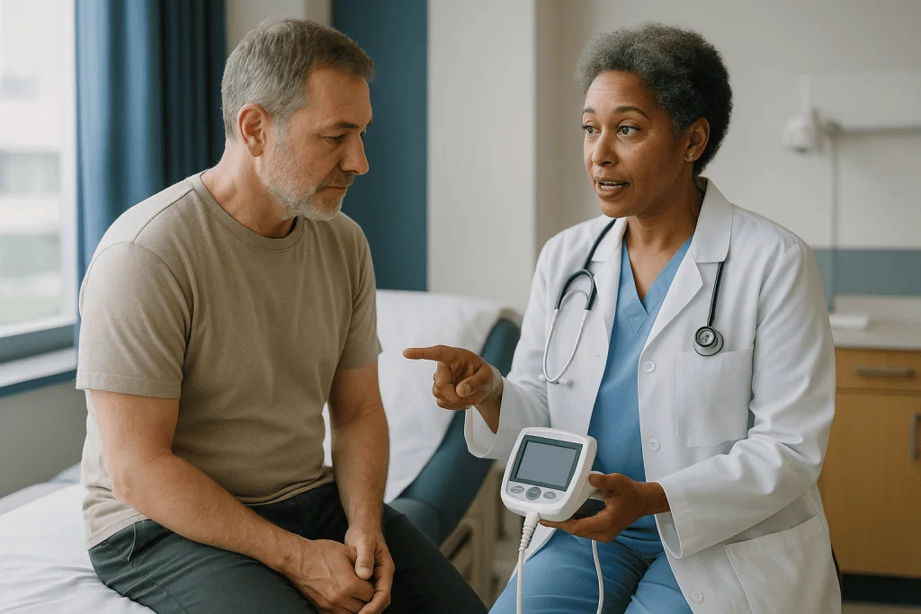
Catheter measurement takes the opposite approach. A clinician passes a thin, flexible tube, the catheter, through the urethra, the channel that carries urine out of the body, and into the bladder. Urine drains into a graduated container, the amount is read off directly, and the tube is removed. This is often called a straight catheter, or an in-and-out catheter, to distinguish it from one that stays in place (MedlinePlus).
Here is the honest framing. The catheter gives a direct measurement of what was actually there. The scanner gives a mathematical estimate based on the shape and size of the echo. That is why catheter drainage has long been the reference standard against which scanners are tested (StatPearls, NIH).
The scanner, though, has an enormous practical advantage: it carries almost no risk. No urethral irritation, no chance of introducing bacteria, no need for a sterile setup. For most routine situations, guidance now favors the scanner first and reserves catheterization for cases where the scan result is doubtful, where the bladder needs draining anyway, or where a clean urine sample is required.
So the bladder scan vs catheter residual choice is less a rivalry than a sequence. Scan first, catheterize when there is a good reason. Your care team’s job is to know when that reason exists.
How a portable bladder scanner works, step by step
The procedure takes a couple of minutes and usually happens with you lying flat or propped up slightly. A small amount of water-based gel goes on the skin just above the pubic bone. The gel feels cool, occasionally startlingly so, and its only job is to remove the tiny layer of air between the probe and your skin, because sound waves travel poorly through air.
The clinician presses the probe gently downward and angles it toward the bladder. Many devices display a targeting image, often a crosshair over an outline, so the operator can tell whether the bladder is centered. Some models ask the operator to select a setting for whether the person has a uterus, because the uterus sits directly behind the bladder and can be mistaken for it (StatPearls, NIH).
Inside the device, the software measures the bladder across several planes and applies a geometric formula to estimate volume. Older scanners assumed the bladder is roughly egg-shaped; newer ones capture more slices for a three-dimensional estimate. Either way, the number on the screen is a calculation, not a direct count of milliliters.
Good technique matters. Repeating the reading two or three times and using the highest or most consistent value is common practice, because a single off-center aim can undercount. The clinician wipes off the gel, and you are done.
There is no radiation involved. Ultrasound uses sound, not X-rays, and has been used for decades in pregnancy precisely because of its safety profile (Mayo Clinic).
What catheter measurement involves and how long it takes
Catheter measurement is a short clinical procedure rather than a bedside reading. The clinician washes their hands, puts on gloves, and cleans the opening of the urethra with an antiseptic wipe. A sterile, single-use catheter is coated with lubricating gel, which often contains a mild numbing agent, though whether one is used depends on local practice and your own history.

The tube is guided into the urethra and advanced until urine begins to flow, which signals it has reached the bladder. Urine drains into a measuring container. When the flow stops, the tube is slid out. From preparation to removal, the whole thing usually takes only a few minutes (NHS).
Anatomy shapes the experience. In women the urethra is short, and the catheter reaches the bladder quickly. In men it is longer and curves around the prostate, a gland that sits below the bladder and can enlarge with age. An enlarged prostate can make insertion slower and more uncomfortable, and clinicians may choose a slightly different catheter design in that situation (MedlinePlus).
Catheter measurement has one feature the scanner cannot match: it empties the bladder. If a person is in acute retention, unable to pass urine and in pain, the catheter is both the test and the treatment. The volume drained is recorded, and the immediate problem is relieved at the same time (NIDDK).
Another practical bonus is the sample. Urine collected this way has not touched the skin, so it can be sent for culture without the contamination that often muddies a cup sample.
Post void residual normal range: how much residual is normal?
Ask three clinicians for the exact cutoff and you may get three slightly different answers, and that is not carelessness. The normal range depends on age, symptoms, and what else is going on.
The commonly cited framework runs like this. A residual under about 50 milliliters is generally considered normal. Between roughly 50 and 100 milliliters is often accepted, particularly in older adults whose bladders naturally empty a little less completely. Readings above about 200 milliliters are widely regarded as showing inadequate emptying and usually prompt further evaluation (StatPearls, NIH).
To picture those numbers: 50 milliliters is roughly a shot glass. Two hundred milliliters is a small coffee cup. A bladder that comfortably holds around 400 to 600 milliliters when full is leaving nearly half its capacity behind at that level (Cleveland Clinic).
The context matters as much as the figure. A person with no symptoms and a single reading of 120 milliliters is a very different case from someone with recurrent infections and readings that keep climbing. Clinicians look at trends, at whether the number is stable or worsening, and at how it sits alongside a physical exam and, where needed, tests of kidney function.
One reading rarely decides anything on its own. If the number is borderline, the usual response is not alarm but repetition, sometimes on a different day, sometimes with the other method. Your treating team will interpret the figure in light of your whole picture, which is exactly how it should be.
How accurate is a bladder scan? What the evidence really shows
People searching this question often want a yes or no. The truthful answer is: accurate enough for most purposes, but not exact, and the gap is predictable.
Studies comparing portable scanners against catheter drainage generally find good agreement, with the scanner reading typically differing from the drained volume by a few tens of milliliters in either direction (StatPearls, NIH). That is small when the question is “is this person retaining more than 200 milliliters?” It is large when the question is “is this 40 milliliters or 90?”
Accuracy also varies with volume. Scanners tend to be least reliable at very small volumes, where the bladder is nearly collapsed and hard to outline, and can lose precision at very large volumes, where the bladder extends beyond the probe’s field. In the middle range, where most clinical decisions are made, performance is at its best.
Operator skill is part of the equation. A probe aimed slightly off center, insufficient gel, or a reading taken with the person tensing their abdominal muscles can all skew the number. Training and repeat readings narrow that variability considerably.
What this means in practice is that the scanner excels as a screening and monitoring tool. It can tell a ward team that a patient’s retention is easing, or that a threshold has been crossed, without a single catheter. When a precise figure will change management, or when the reading simply does not match how the patient looks and feels, clinicians reach for the catheter to confirm. The two methods are strongest used together, each covering the other’s blind spot.
Situations where the scanner can be fooled
Because the scanner detects fluid and shape rather than urine specifically, anything that looks like a fluid-filled sac in the pelvis can confuse it. Knowing these situations helps explain why a clinician might say the scan “doesn’t look right” and ask to confirm another way.
Common culprits include:
- Ovarian cysts or a fluid-filled uterus, which sit close to the bladder and can be counted in the volume.
- Pregnancy, where the uterus dominates the pelvis and displaces the bladder.
- Ascites, which is free fluid in the abdominal cavity, often linked to liver disease.
- Significant abdominal wall thickness, which weakens the echo and blurs the outline.
- Scar tissue or surgical staples from previous pelvic or abdominal operations.
- Blood clots inside the bladder, which reflect sound differently from urine.
- Wrong device setting for pelvic anatomy, or the probe aimed off center (StatPearls, NIH).
The catheter has fewer ways to mislead, but it is not immune. If the tube drains only part of the bladder because it sits in a pocket or is withdrawn too early, the volume is undercounted. Sediment or clots can block the eyelets of the tube. And, as with the scanner, a long delay after voiding inflates the number regardless of method.
None of this makes the scanner unreliable in general. It means a thoughtful clinician treats a surprising reading as a prompt to look again rather than as a final answer. When a scan shows 500 milliliters in someone who feels comfortable and just passed a good stream, the mismatch itself is the clue.
Who usually gets a scan, who gets a catheter, and who is asked to wait
The scanner is the default for most routine checks. That includes people recovering from surgery, where anesthesia and pain medicines can temporarily slow bladder emptying; older adults being assessed for urinary symptoms; people with neurological conditions such as multiple sclerosis, Parkinson’s disease, or spinal cord injury that affect bladder nerves; and anyone being monitored over days or weeks where repeated catheterization would carry cumulative infection risk (NIDDK).
Catheter measurement tends to be chosen when the scan result is doubtful because of one of the confounders described earlier, when the bladder needs to be drained anyway because the person is in painful retention, when a clean urine sample is needed for culture, or when a scanner simply is not available. Some specialist assessments, such as urodynamic testing that measures bladder pressure and flow, use catheters as part of the procedure itself.
Who is asked to wait? Sometimes the answer is to wait a few minutes. If the last void was over an hour ago, or if you have been drinking freely since, a clinician may ask you to urinate again and repeat the measurement to get a meaningful number. Catheterization is generally deferred, unless a specialist advises otherwise, in people who have recently had surgery on the urethra or prostate, or who have a known urethral injury or stricture, which is a narrowing from scar tissue, because passing a tube could cause harm (NHS).
The decision is rarely made in isolation. It sits with the treating team, weighing the accuracy they need against the comfort and safety of the person in front of them.
Bladder scan vs catheter residual at a glance: comparison table
Side by side, the trade-offs become easy to see. The table below summarizes what mainstream clinical guidance says about each method.
| Feature | Portable bladder scanner | Straight catheter measurement |
|---|---|---|
| What it does | Estimates bladder volume from outside using ultrasound | Drains the bladder and measures urine directly |
| Invasiveness | None; gel and probe on the lower abdomen | Tube passed through the urethra |
| Accuracy | Good agreement; typically differs by tens of milliliters; less precise at very small or very large volumes | Reference standard; direct measurement |
| Infection risk | Essentially none | Small risk with each insertion |
| Discomfort | Cool gel, light pressure | Brief stinging or pressure, stronger urge to void |
| Time | About one to two minutes | A few minutes including preparation |
| Empties the bladder | No | Yes |
| Provides urine sample | No | Yes, uncontaminated |
| Can be confused by | Cysts, pregnancy, ascites, scarring, positioning | Incomplete drainage, blocked tube |
| Typical use | Screening, post-surgery checks, trend monitoring | Confirmation, acute retention, sample collection |
Sources for the figures and characteristics: StatPearls via NIH, MedlinePlus, NHS.
The pattern is consistent. Where risk and comfort matter most, the scanner wins. Where precision, drainage, or a sterile sample matter most, the catheter wins. A well-run service uses both and knows which question it is trying to answer before choosing.
Does a bladder scan hurt? Comfort compared honestly
A bladder scan does not hurt. The gel is cool, the probe presses lightly, and the entire contact lasts under a minute. Some people notice mild pressure if the bladder still holds a lot of urine, because pushing on a full bladder intensifies the urge to go. That is the extent of it. Children and people who are anxious about medical procedures often find it reassuring to watch the screen.
Catheter measurement is a different experience, and it deserves a straight answer rather than reassurance. Most people describe a brief stinging or burning as the tube passes, followed by a strong urge to urinate as it reaches the bladder. The sensation typically lasts seconds to a minute or two. Lubricating gel, sometimes containing a local anesthetic, reduces the sting. Slow, steady breathing and relaxing the pelvic floor, the sling of muscles at the base of the pelvis, genuinely help, because tensing narrows the urethra (NHS).
Men often report more discomfort than women because of the longer urethra and the passage past the prostate. People with an enlarged prostate, previous urethral surgery, or a history of difficult catheterization should say so beforehand; clinicians can adjust technique or catheter type accordingly.
After removal, a mild burning during the next one or two urinations is common and usually settles within a day (MedlinePlus). Persistent pain beyond that is not expected and should be reported.
There is no shame in asking for the scan first. Most clinicians will already be planning that, and the catheter is reserved for when the number truly needs confirming.
Risks of catheter measurement and why scanners reduce them
Every time a catheter enters the bladder, it can carry skin bacteria with it. The risk from a single, brief in-and-out catheter is small, but it is not zero, and it accumulates with repetition. Urinary tract infection is the most common complication linked to catheter use in health care settings, which is a major reason guidance encourages non-invasive measurement wherever it is adequate (NHS; NIDDK).
Physical injury is the second concern. The urethra is delicate. Forcing a tube past resistance can cause bleeding, create a false passage where the tube tunnels into tissue rather than following the natural channel, or, over repeated episodes, contribute to stricture formation. Men with prostate enlargement are at higher risk because the tube must navigate a narrowed, curved path. Skilled, unhurried technique keeps these events rare.
Some people experience bladder spasm, a cramping sensation, for a short period afterward, and a little pink-tinged urine is occasionally seen after a difficult insertion. Both usually resolve quickly (MedlinePlus).
The scanner’s risk profile is close to blank. Ultrasound at diagnostic intensities has no known harmful effects, there is no skin break, and no bacteria are introduced. The only real risk is a misleading number, which is why understanding its confounders matters.
This asymmetry shapes modern practice. If a scan can safely answer the question, the small but real harms of catheterization are avoided entirely. If it cannot, the catheter’s benefits, direct measurement, drainage, and a clean sample, justify accepting those harms. Your treating team weighs this balance each time, and you are entitled to hear their reasoning.
What the following days and weeks usually look like
After a bladder scan, there is nothing to recover from. You wipe off any remaining gel and carry on. If you are on a hospital ward, the scan may be repeated every few hours or daily to follow a trend, for example while a temporary post-surgical slowdown in bladder emptying resolves (NIDDK).
After catheter measurement, the first day is the one to notice. Mild burning with the first couple of urinations is common and expected. Drinking normally, rather than restricting fluids to avoid the sting, helps flush the urethra. Faintly pink urine on one occasion is not unusual after a difficult insertion, though it should clear quickly (MedlinePlus).
What happens next depends on the number. A normal or low residual usually closes the question, or redirects attention to other explanations for your symptoms. A borderline reading typically leads to a repeat measurement, sometimes on another day and sometimes by the other method, before any conclusions are drawn.
A clearly raised residual opens a conversation rather than an alarm. Common next steps include a review of medicines, since some drug classes such as anticholinergics, certain antihistamines, and opioids can relax the bladder muscle or tighten the outlet and raise residual volume; assessment for an enlarged prostate or pelvic floor problem; blood tests of kidney function; and referral to a urologist. Some people are taught intermittent self-catheterization, a technique for draining the bladder at home several times a day, which many find far easier than they feared once shown properly (Cleveland Clinic).
Any change to a prescribed medicine sits with the prescribing clinician. The test simply gives them better information to work with.
What people often get wrong about residual urine tests
Misunderstandings cluster around a few themes, and clearing them up saves a lot of unnecessary worry.
“A high residual means my kidneys are failing.” Not usually. Most raised residuals are caused by problems below the kidneys, such as a weak bladder muscle or an obstructed outlet. Kidney strain is a possible consequence of severe, prolonged retention, not a given, and it is checked separately with blood tests (NIDDK).
“The scanner is exact.” It is an estimate. The reading can differ from the true volume by tens of milliliters, which is why borderline results are repeated (StatPearls, NIH).
“The catheter is always more accurate, so I should ask for it.” It is more direct, but it carries small risks that the scanner does not. For a screening question, the scanner is the appropriate first tool.
“The test tells me why my bladder isn’t emptying.” It measures how much is left. The cause requires other assessments.
“One reading settles it.” Residual volume varies with hydration, timing, position, and anxiety. Trends across several readings carry far more weight than any single figure.
“I should drink a lot beforehand so there’s something to measure.” The point is to measure what remains after a normal void. Overfilling the bladder distorts the result.
“Bladder scans use radiation.” They use sound waves, the same technology used throughout pregnancy (Mayo Clinic).
“Needing a catheter means something is seriously wrong.” Often it simply means the scan was unclear, or the team wanted a clean urine sample. The method chosen says less about severity than people assume.
Questions to ask your care team before the test
A short conversation before the measurement helps you understand what the result will mean and keeps the decision transparent. The following questions are ones clinicians are used to answering and generally welcome.
- Will you use the scanner first, and what would make you decide a catheter is needed?
- How soon after I urinate will the measurement be taken, and should I let you know the moment I finish?
- What residual volume would you consider normal for someone my age, and what number would change the plan?
- If the reading is borderline, will it be repeated, and when?
- Is there anything about my history, such as pelvic surgery, cysts, or pregnancy, that could affect the scan’s accuracy?
- If a catheter is used, will a numbing gel be applied, and what should I expect to feel?
- I have had a difficult catheterization before, or I have an enlarged prostate or a known stricture. Does that change your approach?
- Could any of my current medicines be contributing to incomplete emptying? (Any changes remain the prescriber’s decision.)
- What symptoms after the test would you want to hear about straight away?
- If the residual is high, what are the likely next steps, and who will coordinate them?
Writing the answers down, or bringing someone to listen alongside you, is worth doing. Test results delivered quickly on a busy ward are easy to misremember, and a residual figure without its context can sound more frightening than it is.
Your care team is the right place for every one of these decisions. Their job is to choose the method that answers the question safely; yours is to make sure you understand what they found.
When to call your doctor: red-flag signs after either test
Most people have no problems after a bladder scan, and only minor, short-lived discomfort after a catheter measurement. Certain symptoms, however, need prompt attention rather than waiting for a routine appointment.
Seek urgent care the same day, or go to an emergency department, if you:
- Cannot pass urine at all and have a painful, swollen lower abdomen, which suggests acute retention (NIDDK).
- Develop fever, chills, or shaking after a catheter procedure, which can signal a urinary infection spreading beyond the bladder.
- Pass heavy or clotted blood in your urine, rather than a faint pink tinge that clears after one or two voids.
- Have new pain in your back or side at kidney level, especially with fever.
- Notice sudden confusion or drowsiness in an older adult, which can be the first sign of infection.
Contact your doctor or care team within a day or two if burning on urination continues beyond about 48 hours after catheterization, if you are urinating far more often than usual or feel unable to hold urine, if you notice leaking without sensation, or if your urine becomes cloudy or foul-smelling (NHS; MedlinePlus).
Tell the team about anything that feels different from what you were told to expect. A quick call that turns out to be nothing is far preferable to a delay that allows an infection or retention to progress.
The residual test is a measurement, not a verdict. Whatever the number, the interpretation, the follow-up, and any change to your treatment belong to the clinicians who know your full history, and they will want to hear from you if something is not right.
Frequently asked questions
What is the difference between a bladder scan and a post-void residual urinary test?
A post-void residual test is the measurement itself, the amount of urine left after urinating, and a bladder scan is one of two ways to perform it. The scan estimates volume with ultrasound from outside the body. The other method drains the bladder with a thin catheter and measures the urine directly. Both answer the same question; the scan is non-invasive, the catheter is more direct.
How much bladder residual is normal?
Under about 50 milliliters is generally considered normal, and 50 to 100 milliliters is often accepted, particularly in older adults. Readings above roughly 200 milliliters are widely regarded as incomplete emptying and usually lead to further assessment. The exact threshold varies with age, symptoms and the reason for testing, and clinicians place more weight on repeated readings than on a single value.
How reliable are bladder scans compared with a catheter?
Reliable enough for screening and monitoring in most people. Studies comparing portable scanners with catheter drainage generally show good agreement, with differences typically in the range of tens of milliliters. Accuracy falls at very small or very large volumes and when cysts, pregnancy, abdominal fluid or scarring confuse the image. When a precise figure will change the plan, clinicians confirm with a catheter.
How painful is a bladder scan?
A bladder scan is not painful. You feel cool gel and light pressure from the probe on your lower abdomen for under a minute. If your bladder still holds a lot of urine, that pressure can heighten the urge to go, but there is no sting and no tube. Catheter measurement is different, usually causing a brief burning sensation and a strong urge to urinate for a minute or two.
What is the post void residual normal range for older adults?
Older adults often retain slightly more urine after voiding, so many clinicians accept residuals up to about 100 milliliters in this group when there are no troubling symptoms. Volumes above roughly 200 milliliters are still regarded as significant at any age. Because age, medicines and prostate or pelvic floor changes all influence the number, your care team interprets it alongside your symptoms rather than in isolation.
Does a bladder scan hurt if you have just had surgery?
The scan itself does not hurt, but gentle pressure on a tender abdomen after pelvic or abdominal surgery can be uncomfortable. Clinicians usually press lightly and avoid incision sites. Post-surgical scans are common because anesthesia and pain medicines can temporarily slow bladder emptying, and the scanner allows repeated checks without the infection risk of repeated catheterization.
How accurate is a bladder scan in pregnancy or with ovarian cysts?
Less accurate, because the scanner detects fluid-filled shapes rather than urine specifically. A pregnant uterus or a cyst lying next to the bladder can be counted in the volume, producing a falsely high reading. In these situations clinicians interpret the scan cautiously and may confirm with a catheter measurement if the result would change management.
Why did the nurse repeat the bladder scan several times?
Repeating the reading is standard technique, not a sign of a problem. A probe aimed slightly off center or a moment of abdominal tensing can undercount the volume, so operators commonly take two or three readings and use the highest or most consistent one. The repetition improves accuracy and reduces the chance of an unnecessary catheter.
What happens if my post void residual test is high?
A high reading opens a conversation rather than triggering immediate treatment. Typical next steps include repeating the measurement, reviewing medicines that can affect bladder emptying, examining for prostate enlargement or pelvic floor problems, checking kidney function with blood tests, and possibly referral to a urologist. Some people are later taught to drain the bladder themselves at home. All decisions rest with your treating team.
Can a bladder scan tell why my bladder is not emptying?
No. It measures how much urine remains, not the cause. Incomplete emptying can result from a weak bladder muscle, an obstructed outlet such as an enlarged prostate, nerve conditions, or certain medicines. Identifying the reason requires a history, physical examination and sometimes additional tests such as flow studies or urodynamics, which your care team will arrange if needed.
References
- Bladder Post Void Residual Volume – StatPearls (NIH National Library of Medicine)
- Urinary Retention – NIH National Institute of Diabetes and Digestive and Kidney Diseases
- Urinary catheters – MedlinePlus Medical Encyclopedia
- Urinary catheter – NHS
This article is for general information only and is not a substitute for professional medical advice. Please consult a qualified doctor about your individual situation.

More from the Blog

How Uremic Syndrome Is Treated: Stabilization, Dialysis When Needed and Longer-Term Care
Uremic syndrome treatment has three layers. First, clinicians stabilize the patient by correcting dangerous potassium, acid, fluid and heart-rhythm problems and treating any reversible…

Leaks, Urgency or Repeat Infections: How Female Bladder Care Is Planned Step by Step
Female bladder problems treatment starts by identifying which problem is present: stress leaks, urgency, repeat infections, or a mix. Care then moves in steps,…

How Do You Read a Urodynamic Report? Capacity, Pressure-Flow Curves and Leak Points Explained
A urodynamic report is read in order: the header and patient details, then the flow curve and the volume left after voiding, then the…

When Can You Lift, Drive and Exercise Again After Robotic Prostate Surgery?
Most people walk the day of robotic prostate surgery, drive again once the catheter is out and they are no longer taking opioid painkillers,…
How Peritoneal Dialysis Exchanges Work: Fill, Dwell and Drain Explained in Plain Words
A peritoneal dialysis exchange uses the lining of your own abdomen as a filter. Cleansing fluid flows through a soft catheter into the belly…

Reading a Bladder Disease Treatment Plan: Questions to Ask Your Urologist Before You Decide
Before agreeing to a bladder disease treatment plan, ask your urologist which specific condition is being treated, what test results support that diagnosis, why…