

Bladder Stones vs Kidney Stones: Why They Form Differently and Why Treatment Differs
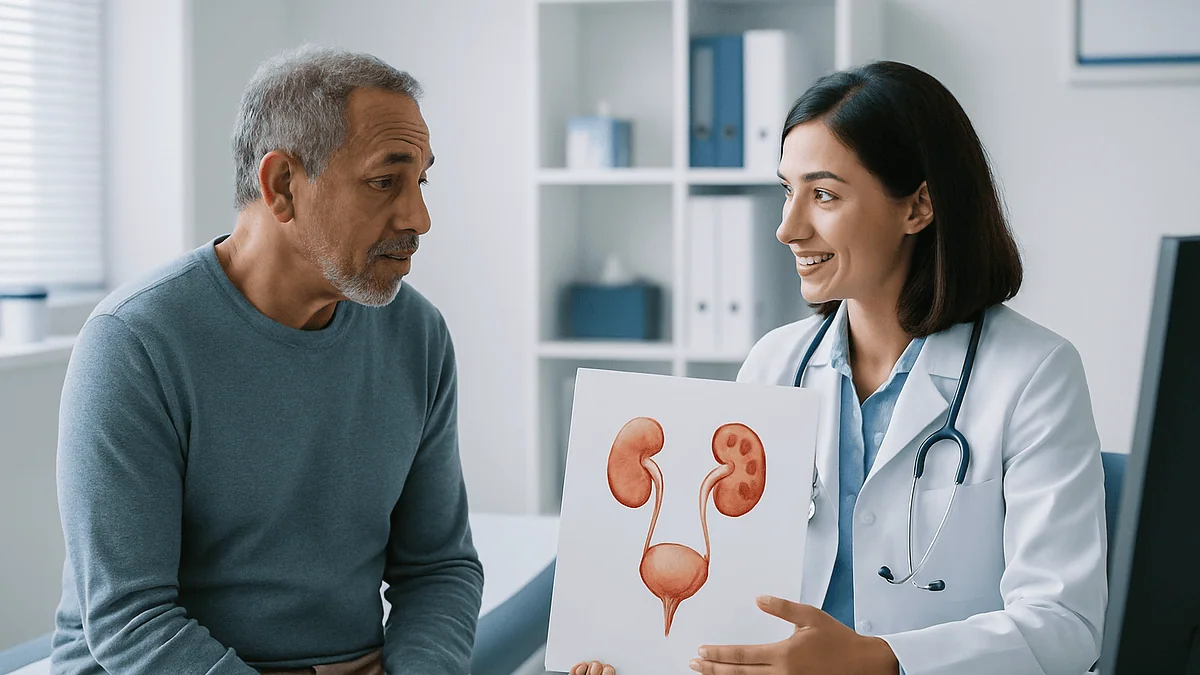
Key Takeaways
- Kidney stones form when urine becomes supersaturated with minerals inside the kidney, while bladder stones usually form because the bladder fails to empty completely, most often behind an enlarged prostate.
- The NIDDK estimates about 11 percent of men and 6 percent of women in the United States have a kidney stone at some point, whereas bladder stones are far less common and concentrated in men over 50.
- Kidney stone pain comes from a ureter blocked by a moving stone and arrives in waves in the flank and groin; bladder stones more often cause a dull low-abdominal ache and a stream that starts and stops.
- Calcium oxalate dominates kidney stones, but bladder stones are frequently uric acid or infection-related struvite, which is why laboratory analysis of a removed stone points back to its cause.
- Small kidney stones often pass on their own over days to weeks, but bladder stones rarely pass in men because the same obstruction that let them form blocks their exit, so a scope-based procedure is usual.
- Both conditions recur unless the cause is addressed: kidney stone follow-up centers on fluids and urine chemistry, while bladder stone follow-up centers on fixing the drainage problem.
Kidney stones form when minerals crystallize in concentrated urine inside the kidney and typically cause severe, wave-like flank pain as they travel down the ureter. Bladder stones usually form because the bladder does not empty completely, most often in older men with an enlarged prostate, and cause lower-abdominal discomfort and a disrupted urine stream. Small kidney stones often pass on their own; bladder stones more commonly need a scope-based procedure.
He had been up three times in the night again, standing at the toilet while the stream started, stopped, and started once more. His daughter, who had passed a kidney stone the previous spring, told him it sounded exactly like hers. It did not. She had spent an afternoon on the bathroom floor with pain that came in waves; he had a dull ache low in his belly and a stream that behaved like a faulty tap.
That gap between two stories is the whole point of understanding bladder stones vs kidney stones. Both are hard lumps of mineral that form in urine. Both can bleed, both can hurt, and both show up on the same scan. Yet they begin in different places for different reasons, and the reason a stone formed usually decides how a urologist will remove it and what happens afterward.
This explainer walks through the mechanics, the pain, the imaging, and the procedures, so the conversation with your care team starts from evidence rather than a relative’s memory.
What is the difference between bladder stones and kidney stones?
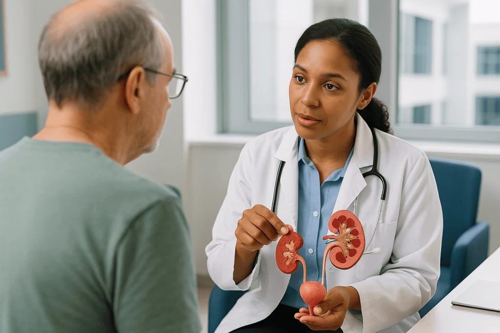
The urinary tract is a plumbing system with two reservoirs. The kidneys filter blood and make urine continuously, sending it down two narrow tubes called ureters into the bladder, which stores it until you choose to empty it through the urethra. A stone is simply a crystal that grew large enough to matter, and where it grows tells you a great deal about why.
Kidney stones form in the collecting system of the kidney itself. The mechanism is chemical: urine becomes so concentrated with calcium, oxalate, uric acid or other salts that they can no longer stay dissolved, so they precipitate out much as sugar crystallizes in an over-sweetened cup of tea left overnight. Most people never notice a kidney stone until it leaves the kidney and lodges in a ureter, and that blockage is what produces the famous pain.
Bladder stones form in the bladder, and the mechanism is usually mechanical rather than purely chemical. The Mayo Clinic describes them as developing when the bladder does not empty completely, leaving urine to sit, concentrate and crystallize. Something is blocking the exit, or the bladder muscle is not contracting well, or a foreign object such as a long-term catheter gives crystals a surface to grow on. In most cases the stone is a symptom of a drainage problem, not a problem in its own right.
That distinction drives almost everything that follows. A kidney stone is typically a supersaturation problem, so the long-term work is about urine chemistry and fluids. A bladder stone is typically an emptying problem, so removing the stone without addressing why urine was pooling tends to invite a repeat. Two stones, two mechanisms, two treatment logics.
Why do kidney stones form in the first place?
Kidney stones are common. The National Institute of Diabetes and Digestive and Kidney Diseases (NIDDK) estimates that about 11 percent of men and 6 percent of women in the United States have a kidney stone at some point in their lives. Most are calcium oxalate stones, followed by calcium phosphate, uric acid, struvite (infection-related) and, rarely, cystine stones linked to an inherited condition.
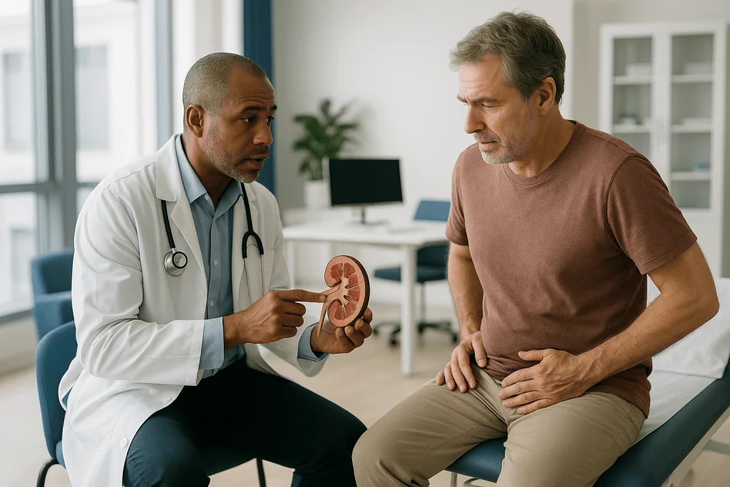
Formation depends on a balance. Urine normally contains substances that discourage crystals, chiefly citrate and simple volume, and substances that encourage them, mainly calcium, oxalate, sodium and uric acid. When the promoters outweigh the inhibitors, microscopic crystals seed and grow. Not drinking enough is the single most consistent contributor, because concentrated urine leaves less room for salts to stay in solution. The Mayo Clinic also lists diets high in sodium, animal protein and sugar; obesity; certain digestive diseases and surgeries that change absorption; hyperparathyroidism; and a family or personal history of stones.
Urine acidity matters too. Uric acid stones favor acidic urine, which is one reason they are seen more often alongside gout and metabolic syndrome. Struvite stones grow in alkaline urine produced by certain bacteria during urinary tract infections and can enlarge quickly into branching shapes that fill the kidney.
The stone itself is usually painless while it sits in the kidney. Trouble begins when a fragment enters the ureter, a tube only a few millimeters wide. The ureter spasms against the obstruction, urine backs up, and the kidney capsule stretches. That stretch, not the stone scraping tissue, is the source of renal colic, the medical term for the intense cramping pain that comes in waves. Understanding this explains why pain can ease abruptly the moment a stone drops into the bladder, even before it has left the body.
Why do bladder stones form differently?
A healthy bladder empties almost completely each time you urinate, flushing away any early crystals before they can grow. Bladder stones appear when that flush fails. According to the NHS, they are much more common in men over 50, and the Mayo Clinic explains why: the enlarging prostate that many men develop with age squeezes the urethra where it leaves the bladder, so urine is retained after each void. The leftover pool concentrates, and stones begin.
Nerve damage is the second major route. Conditions such as spinal cord injury, stroke, diabetes and multiple sclerosis can disrupt the signals that tell the bladder muscle to contract, a situation clinicians call neurogenic bladder. Urine stagnates, infections become more frequent, and stones follow. People who rely on indwelling catheters face a related risk, because bacteria and mineral deposits accumulate on the catheter surface and the balloon can serve as a nucleus for crystal growth.
Chronic inflammation from repeated infections or prior radiation to the pelvis can also make the bladder lining more hospitable to stone formation. Less commonly, a stone forms around a foreign object, such as a fragment of a surgical stitch or a device that migrated into the bladder.
Stone chemistry differs as a result. Bladder stones in men with prostatic enlargement are frequently uric acid stones, while those associated with infection tend to be struvite. Calcium oxalate stones, dominant in the kidney, are a smaller share of bladder stones. The lesson urologists draw from this is practical: a bladder stone is a signpost pointing at a cause. Removing it relieves symptoms, but the MedlinePlus entry on bladder stones is explicit that stones can return if the underlying cause is not addressed. The prostate, the nerves, or the catheter usually needs attention too.
Do bladder stones start out as kidney stones?
Sometimes, but less often than people assume. A kidney stone that travels down the ureter and reaches the bladder normally continues on its way and is passed in urine within hours or days, because the urethra is wider than the ureter it has just navigated. The Mayo Clinic notes that kidney stones can become bladder stones only when they fail to leave the bladder, which usually happens for the same reason primary bladder stones form: something is preventing complete emptying.
Picture the bladder as a sink. A small pebble washed into a sink with an open drain disappears. The same pebble in a sink whose drain is half blocked sits in the standing water and, if that water is rich in minerals, grows. So a migrated kidney stone can act as the seed for a bladder stone, but the enlargement that follows is the bladder’s own work.
Most bladder stones, however, form entirely in the bladder. Their composition often gives this away. If a stone retrieved from the bladder is uric acid in a man with an enlarged prostate and no kidney stone history, it almost certainly grew where it was found. A stone with a calcium oxalate core and a different outer layer may tell the opposite story, and laboratory analysis of removed stones is routine for this reason.
The distinction is not academic. If the bladder stone began as a kidney stone, the person carries two risks, one in the kidney and one in the bladder, and follow-up imaging of the kidneys becomes part of the plan. If it formed locally, the priority shifts to the drainage problem. Either way, the decision about which investigations to pursue rests with the treating urologist, who will weigh stone analysis, imaging and the person’s history together.
How do you tell if you have a bladder or kidney stone?
Honestly, you often cannot tell from home, and no reputable source suggests trying. What clinicians look for is a pattern, and the two conditions do tend to produce different patterns.
Kidney stones announce themselves when they move. The Mayo Clinic describes severe, sharp pain in the side and back below the ribs that radiates toward the lower abdomen and groin, arriving in waves and shifting location as the stone travels. Nausea and vomiting are common companions, driven by the shared nerve supply between the kidney and the gut. Blood may tint the urine pink or brown, and if infection is present, fever and chills appear. People with renal colic characteristically cannot find a comfortable position; they pace.
Bladder stones speak a different language. The NHS and Mayo Clinic list lower abdominal pain, pain or burning during urination, a need to go more often, a stream that starts and stops, difficulty starting the stream, dark or cloudy urine, and blood in the urine. Men sometimes report pain at the tip of the penis. Many bladder stones are found by chance on a scan done for another reason, because a stone sitting quietly in a roomy bladder may cause nothing at all until it rolls against the outlet.
Two caveats matter. First, urinary tract infection can mimic either condition, which is why urine testing is part of every workup. Second, symptoms overlap: a large kidney stone lodged at the very bottom of the ureter, near the bladder, can produce urgency and frequency that feel like a bladder problem. Imaging settles the question, and a clinician who suspects a stone will usually order it rather than guess. The right response to any of these patterns is an appointment, not a self-diagnosis.
Which is more painful, kidney stones or bladder stones?
Kidney stones, as a rule, and the reason is anatomical. Pain from a kidney stone is caused by obstruction: a stone wedged in a ureter blocks the flow of urine, pressure builds inside the kidney, and the capsule surrounding it stretches. The Mayo Clinic describes this as pain so severe that many people seek emergency care, and it is frequently compared to labor, though comparisons of that kind vary enormously from person to person.
Bladder stones rarely produce that kind of obstruction because the bladder is a large, distensible sac. A stone can roll around in it for months with only intermittent discomfort. The pain people do report tends to be a dull, persistent ache low in the abdomen, a burning at the end of urination, or a sharp catch when the stone momentarily blocks the bladder outlet and the stream stops mid-flow. Uncomfortable and wearing, but seldom the pacing, vomiting agony of renal colic.
There are exceptions in both directions. A small bladder stone that lodges in the urethra can cause sudden severe pain and an inability to pass urine, a genuine emergency. Conversely, a kidney stone that stays put in the kidney may cause a vague backache or nothing at all for years.
Pain intensity is also a poor guide to danger. A kidney stone blocking one ureter while the other kidney works normally can be excruciating yet not immediately threatening, whereas a person with a neurogenic bladder may have a large infected bladder stone and feel little because sensation is impaired. Clinicians assess fever, urine output, kidney function on blood tests and imaging findings, not pain scores alone, when deciding how urgently to act. If pain is severe or accompanied by fever, the answer is the same for either stone: seek care that day.
Bladder stones vs kidney stones at a glance
Placing the two side by side clarifies why a urologist approaches them so differently. The table below summarizes the mainstream picture drawn from the NHS, Mayo Clinic and NIDDK descriptions; individual cases can and do depart from it.
| Feature | Kidney stones | Bladder stones |
|---|---|---|
| Where they form | Inside the kidney’s collecting system | Inside the bladder |
| Main mechanism | Urine supersaturated with minerals | Incomplete bladder emptying, foreign body or infection |
| Who is most affected | Adults of both sexes; more men than women (NIDDK) | Predominantly men over 50 (NHS) |
| Typical composition | Calcium oxalate most common | Uric acid and struvite common |
| Classic pain | Severe wave-like flank pain radiating to groin | Dull lower-abdominal ache, burning, interrupted stream |
| Often silent? | Yes, while still in the kidney | Yes, frequently found incidentally |
| Chance of passing naturally | Good for small stones | Possible for small stones; less likely when outlet is obstructed |
| Usual procedures | Shock wave lithotripsy, ureteroscopy, percutaneous nephrolithotomy | Cystolitholapaxy (scope-based fragmentation); open surgery for very large stones |
| Key to preventing recurrence | Fluids, diet, urine chemistry | Treating the drainage problem |
Two rows deserve emphasis. The composition row explains why stone analysis after removal is standard: it points backward to the cause. The recurrence row explains why treatment plans look so different even when the stones themselves look alike on a scan. A kidney stone patient leaves with advice about water and salt; a bladder stone patient often leaves with a plan for the prostate or the bladder’s nerve supply.
How are bladder and kidney stones diagnosed?
Diagnosis rests on three pillars: a conversation, a urine sample, and pictures.
The conversation covers where the pain is, how it behaves, how the stream behaves, previous stones, prostate symptoms, catheter use, neurological conditions, medications and family history. A clinician listening to a man describe an intermittent stream and a low ache is already leaning toward the bladder; a woman describing flank pain that made her vomit points toward the ureter.
A urine test looks for blood, crystals, signs of infection and acidity. Blood tests check kidney function and, in recurrent kidney stone formers, calcium and uric acid levels. Neither identifies where a stone sits, but both shape urgency and treatment.
Imaging gives the answer. The Mayo Clinic notes that a non-contrast CT scan is highly sensitive for kidney stones and shows size and location precisely, which matters because size largely predicts whether a stone will pass. Ultrasound avoids radiation and is preferred in pregnancy and often in children; it is good at showing a swollen kidney and reasonably good at spotting bladder stones, which appear as bright objects casting a shadow. A plain X-ray can show many stones but misses uric acid stones, which are radiolucent, a limitation worth knowing because uric acid is a common bladder stone type.
For the bladder specifically, urologists sometimes use cystoscopy, in which a thin lighted tube is passed through the urethra to look directly inside. MedlinePlus lists it among standard tests for bladder stones. It is diagnostic and, frequently, the first step of treatment, since the same instrument used to see the stone can fragment it. A bladder scan measuring how much urine remains after voiding may be added, because that residual volume is the mechanical heart of the problem.
Can you pass bladder stones the way you pass kidney stones?
Small ones sometimes, but the odds are stacked differently, and it helps to understand why.
Kidney stones have a reasonable track record of passing on their own because the bottleneck they face is the ureter, and once a stone has squeezed through that, the rest of the journey is easier. The NHS and Mayo Clinic both note that most kidney stones are small enough to pass in urine, typically with pain relief, fluids and time, and sometimes with a medication class that relaxes the ureter to ease passage. Size is the key predictor: the Mayo Clinic describes small stones as often passing without intervention, while larger stones increasingly need help. The NHS describes natural passage as taking anywhere from days to a few weeks depending on size and position, and clinicians commonly arrange follow-up imaging to confirm the stone is gone rather than assuming so.
Bladder stones face a different bottleneck. The Mayo Clinic acknowledges that small bladder stones may pass without treatment, and this is more common in women, whose shorter, wider urethra offers less resistance. In men, the very obstruction that allowed the stone to form, usually an enlarged prostate, also blocks its exit. A stone that grew because urine could not get out is unlikely to get out itself. Bladder stones also tend to be larger by the time they are found, because a roomy bladder tolerates growth silently in a way a narrow ureter never could.
Drinking more water, the standard advice for kidney stones, does not unblock a prostate. That is the practical reason urologists move to a procedure sooner for bladder stones. A person with a small bladder stone and no outlet obstruction may reasonably be offered a period of watching; the choice depends on stone size, symptoms, infection and the cause, and it belongs to the treating team.
How are bladder stones removed compared with kidney stones?
The stone’s address determines the route in. For bladder stones, the standard procedure is cystolitholapaxy, a long word for a simple idea: a thin telescope called a cystoscope is passed through the urethra into the bladder, the stone is located, and an energy source, most often a laser or ultrasound, or a mechanical crushing device, breaks it into fragments small enough to flush or suction out. The NHS describes it as usually performed under general or spinal anesthesia, and the Mayo Clinic notes that very large or very hard stones may instead require a small incision in the lower abdomen to remove the stone whole. Because bladder stones are a symptom of poor emptying, the same operation is sometimes combined with treatment of the enlarged prostate.
Kidney stones offer three main routes, chosen by size, location, hardness and anatomy. Shock wave lithotripsy delivers focused sound waves from outside the body that fracture the stone so the pieces can pass; it suits smaller stones in accessible positions. Ureteroscopy passes a fine scope up the urethra, through the bladder and into the ureter or kidney, where a laser fragments the stone and a basket retrieves pieces; a temporary stent is often left to keep the ureter open while it heals. Percutaneous nephrolithotomy reaches large or complex stones through a small incision in the back directly into the kidney.
The contrast is instructive. Bladder stone surgery is almost entirely about access to a single large stone in a spacious organ; kidney stone surgery is about reaching a small target through narrow tubes without damaging them. Risks differ accordingly. The Mayo Clinic lists urinary tract infection and bladder wall injury for cystolitholapaxy; kidney procedures add ureteral injury, bleeding and stent discomfort. Which option fits a given person is a judgment for the urologist, weighing imaging against health and preferences.
Who is usually offered a procedure, and who is asked to wait?
Guideline-level practice sorts people into three broad groups, and the sorting is different for each stone.
For kidney stones, watching is common. The Mayo Clinic and NHS both describe conservative management, meaning pain relief, hydration and time, for small stones causing manageable symptoms in a person with a normal second kidney and no infection. Follow-up imaging checks progress. A procedure is offered when a stone is too large to pass, has failed to move over weeks, is causing intractable pain or repeated vomiting, is blocking a solitary kidney, or is accompanied by infection or declining kidney function. Infection behind an obstructing stone is treated as an emergency, because trapped, infected urine can rapidly become sepsis; the priority is drainage first and definitive stone treatment later.
For bladder stones, the threshold for intervention is lower because waiting rarely works. The NHS notes that small stones may pass, but most bladder stones need removal, and MedlinePlus advises that stones causing symptoms, infections or bleeding should be treated. People asked to wait are typically those with a very small stone, no outlet obstruction, and mild symptoms, often women, and the wait is short and monitored.
Some people are asked to wait for a different reason: they are not yet ready for anesthesia. A recent heart event, an active infection needing treatment first, blood-thinning medication that must be managed around surgery, or uncontrolled diabetes may all delay a scheduled procedure. Pregnancy changes the picture too; ultrasound replaces CT, shock wave lithotripsy is avoided, and temporary drainage may be preferred until after delivery.
None of this is a checklist a person can apply to themselves. The same stone can warrant urgent surgery in one person and patience in another, and the treating team weighs the whole picture.
What the days and weeks after treatment usually look like
Recovery timelines differ because the procedures differ, and everything below is a typical range from the cited sources, not a promise.
After cystolitholapaxy for a bladder stone, the NHS says most people go home the same day or after a short hospital stay of a day or two. A catheter may drain the bladder for a brief period while the lining settles. Seeing blood in the urine for several days is expected, as is some burning and a need to urinate more often. The NHS advises rest for a few days and avoiding heavy exertion for a period the surgeon will specify. If the prostate was treated at the same time, that operation’s own recovery guidance applies and is usually longer.
After shock wave lithotripsy for a kidney stone, fragments pass over the following days to weeks, and the Mayo Clinic notes this can itself cause discomfort and blood in the urine; a follow-up scan confirms clearance. After ureteroscopy, the stent is the dominant experience: it commonly causes urgency, frequency and flank twinges until it is removed, which the Mayo Clinic describes as typically within days to a few weeks according to the surgeon’s plan. Percutaneous nephrolithotomy involves a hospital stay and a longer return to full activity because it goes through the kidney itself.
The weeks that follow are where the two conditions diverge most sharply. The kidney stone patient usually receives stone analysis results and, for recurrent formers, a metabolic evaluation of urine chemistry that leads to fluid and dietary advice and sometimes preventive medication, decided by the clinician. The bladder stone patient’s follow-up centers on the cause: how well the bladder now empties, whether the prostate needs further treatment, whether catheter care should change. Fixing the plumbing, not the stone, is the point.
What people often get wrong about bladder and kidney stones
Misconceptions cluster around a few themes, and each one can lead someone to delay care or worry about the wrong thing.
The first is that stones are stones. In fact the two conditions have different causes, populations and treatments, as the earlier sections show. Advice that works for one, such as drinking a great deal of water to flush a kidney stone, does little for a bladder stone sitting behind an enlarged prostate.
The second is that cutting out calcium prevents kidney stones. The NIDDK and Mayo Clinic point out that dietary calcium binds oxalate in the gut so less reaches the urine, and very low calcium diets can raise stone risk. Excess sodium and animal protein are more consistent dietary culprits. Calcium supplements are a separate question best raised with a clinician.
The third is that a stone that stopped hurting has gone. Renal colic often eases when a stone drops into the bladder or when the kidney adapts to partial blockage, yet the stone may still be present. Follow-up imaging exists because relief and resolution are not the same thing.
The fourth is that bladder stones are a men’s problem only. They are far more common in men because of the prostate, but women with neurogenic bladder, long-term catheters or pelvic surgery can form them too.
The fifth is that lemon juice, herbal blends or so-called stone-dissolving supplements reliably break stones down. Evidence for these is weak or absent for most stone types; the NIDDK notes that only certain uric acid stones can be reduced by altering urine acidity under medical supervision, and no such approach applies to calcium stones or to bladder stones behind an obstruction.
The last is that passing one stone means immunity. Recurrence is common for both conditions unless the cause is addressed, which is exactly why the follow-up conversation matters more than the procedure.
Questions to ask your care team
A stone consultation moves quickly, and people often leave with the scan explained but the plan fuzzy. Writing questions down beforehand changes that. The following are the ones urologists say they are glad to be asked.
- Where exactly is the stone, how large is it, and is there more than one?
- Based on its size and position, is watching a reasonable option for me, and for how long before we reconsider?
- If I am waiting, what signs should make me call you or go to the emergency department without waiting for the next appointment?
- Which procedure are you recommending, why that one over the alternatives, and what are the specific risks in my case?
- Will I have a catheter or stent afterward, roughly how long, and how will it be removed?
- Will the stone be sent for analysis, and how will the result change my follow-up?
- For a bladder stone: what is causing my bladder not to empty, and are we treating that at the same time or separately?
- For a kidney stone: do I need a metabolic evaluation, and what dietary changes does the evidence actually support for my stone type?
- Which of my current medicines, if any, might affect stone risk or need to be managed around the procedure? I understand any change is your decision, not mine.
- When will imaging confirm the stone is gone, and how often should my kidneys be checked afterward?
One further question is worth asking of yourself before the appointment: what matters most to you? For some people it is avoiding a stent at all costs; for others it is the fastest return to work, or the lowest chance of a repeat procedure. Saying so helps the team weigh options against your priorities rather than against an average patient who does not exist.
When to call your doctor
Most stones are uncomfortable rather than dangerous, but a small number of situations need same-day or emergency assessment, and they apply whether the stone is thought to be in the kidney or the bladder.
Seek emergency care if pain is severe enough that you cannot sit still or find any position of relief, if pain is accompanied by fever or shaking chills, if you are vomiting and cannot keep fluids down, or if you are unable to pass urine at all. The combination of a suspected stone and fever is the most urgent of these, because the Mayo Clinic and NHS both warn that infection above a blockage can progress rapidly to a bloodstream infection. A person with a single kidney, a transplanted kidney, or known kidney disease should treat any suspected obstructing stone as urgent.
Call your doctor promptly, within the day, if urine is visibly bloody rather than faintly tinged, if you notice a sudden interruption of the stream with pain, if burning on urination is worsening, or if a stone you were told to watch has produced symptoms that are changing rather than settling. After a procedure, contact the team about heavy bleeding, clots blocking the catheter or stream, fever, pain that escalates instead of easing, or a stent that seems to have shifted.
Between those extremes sits the routine call: a follow-up scan that is due, a stone analysis result you have not heard about, or emptying symptoms that have crept back months after a bladder stone was removed. That last one matters because it may signal the underlying cause was never fully addressed.
When in doubt, call. Every decision about investigation, timing and treatment rests with the clinicians who can see your imaging and know your history, and a phone call that turns out to be unnecessary costs far less than a delay that does not.
Frequently asked questions
Which is more painful, kidney stones or bladder stones?
Kidney stones are usually far more painful, because a stone lodged in the narrow ureter blocks urine flow and stretches the kidney, producing intense wave-like pain the Mayo Clinic notes often drives people to emergency care. Bladder stones sit in a roomy organ and more commonly cause a dull lower-abdominal ache, burning and an interrupted stream. A bladder stone that lodges in the urethra is the exception and can cause sudden severe pain with inability to urinate.
How do you tell if you have a bladder or kidney stone?
You generally cannot tell with certainty at home; imaging is needed. Clinicians look for patterns: flank pain radiating to the groin with nausea suggests a kidney stone moving through the ureter, while lower-abdominal discomfort, burning and a stream that stops and starts point toward the bladder. Urine tests rule out infection, and a CT scan or ultrasound shows where the stone actually is. Any of these symptoms deserves a medical appointment rather than self-diagnosis.
Do bladder stones start as kidney stones?
Occasionally, but most bladder stones form in the bladder itself. A kidney stone that reaches the bladder usually passes in urine within hours or days, because the urethra is wider than the ureter. The Mayo Clinic notes it can become a bladder stone only if something, typically an enlarged prostate or a poorly contracting bladder, stops it leaving. Laboratory analysis of the removed stone often reveals which story applies.
Can you pass bladder stones on your own?
Small bladder stones sometimes pass without treatment, and this is more likely in women, whose shorter urethra offers less resistance. In men, the Mayo Clinic and NHS note that the outlet obstruction that allowed the stone to form usually prevents its exit, so most bladder stones are removed with a scope-based procedure called cystolitholapaxy. Whether a short period of watching is reasonable depends on stone size, symptoms and cause, and that judgment rests with the urologist.
What are bladder stone symptoms in men?
In men, bladder stones commonly cause lower-abdominal pain, burning during urination, a need to urinate more often, difficulty starting the stream, a stream that stops and starts, dark or cloudy urine, and sometimes visible blood or pain at the tip of the penis, according to the NHS and Mayo Clinic. Many are discovered incidentally on scans. Because these symptoms overlap with prostate enlargement and infection, they warrant assessment rather than assumption.
Why do bladder stones mostly affect men over 50?
Because prostate enlargement, which becomes common with age, narrows the urethra where it leaves the bladder and prevents complete emptying. Urine that remains after each void concentrates and crystallizes, most often into uric acid stones. The NHS identifies men over 50 as the group most affected. Women can form bladder stones too, usually in the setting of nerve damage to the bladder, long-term catheter use or prior pelvic surgery.
How long does it take to pass a kidney stone?
It varies with size and position. The NHS and Mayo Clinic describe small stones as often passing over days to a few weeks, while larger stones are less likely to pass and more likely to need a procedure. Clinicians typically arrange follow-up imaging to confirm the stone has actually gone, because pain easing does not prove passage. Any timeline is a typical range, not a promise, and depends on individual anatomy.
Will drinking more water get rid of a bladder stone?
Usually not. Fluids are a mainstay for preventing kidney stones because they dilute urine, but a bladder stone forms because urine cannot drain properly, and extra water does not open an obstructed outlet or restore a weak bladder muscle. The MedlinePlus entry on bladder stones notes stones can return if the underlying cause is not addressed. Water remains sensible for general urinary health, but it is not a treatment for an established bladder stone.
What is the difference between cystolitholapaxy and lithotripsy?
Cystolitholapaxy treats bladder stones from inside: a thin scope passes through the urethra into the bladder and a laser, ultrasound or mechanical device fragments the stone so pieces can be removed. Shock wave lithotripsy treats kidney stones from outside, using focused sound waves to fracture the stone so fragments pass in urine. Kidney stones may also be treated by ureteroscopy or percutaneous surgery. The choice depends on stone location, size and hardness, and is made by the urologist.
Do bladder stones or kidney stones come back after treatment?
Both can recur, for different reasons. Kidney stones return when urine chemistry remains favorable to crystals, which is why follow-up often includes stone analysis, fluid and dietary guidance, and sometimes a metabolic evaluation. Bladder stones return when the drainage problem, such as an enlarged prostate or neurogenic bladder, is not treated alongside the stone. Addressing the cause, rather than the stone alone, is the shared principle behind long-term management for either condition.
References
- NHS: Bladder stones
- NHS: Kidney stones
- NIH NIDDK: Definition and facts for kidney stones
- MedlinePlus: Bladder stones
This article is for general information only and is not a substitute for professional medical advice. Please consult a qualified doctor about your individual situation.

More from the Blog

Urine Flow Tests for Children: How Uroflowmetry Is Adapted for Daytime Wetting Evaluations
Uroflowmetry in children is a painless test that records how fast and how smoothly a child's urine flows while they urinate into a special…

Why Testicular Torsion Is a Surgical Emergency: The Twisted Cord and Blocked Blood Flow
Testicular torsion is a surgical emergency because the spermatic cord, which carries the testicle's blood supply, twists and cuts off blood flow. Without oxygen,…

Which Blood and Urine Tests Identify the Type of Renal Tubular Acidosis?
Doctors identify the type of renal tubular acidosis with a blood gas and basic metabolic panel showing a normal anion gap metabolic acidosis, then…

Recovery After Torsion Surgery: Scrotal Support, Wound Care and the Return to Sport
Recovery after testicular torsion surgery usually involves a few days of scrotal support, simple wound care and gentle walking, followed by a gradual return…

When Burning Urination Points to Urethritis: Why an Early Urology Visit Matters
Burning when you urinate is not always a bladder infection. In urethritis, the urethra itself is inflamed, usually by a sexually transmitted or other…
How Do Urologists Tell Enlarged Prostate, Prostatitis and Prostate Cancer Apart?
Urologists separate an enlarged prostate, prostatitis and prostate cancer by combining the story of the symptoms, a physical exam, urine tests and a PSA…