

Can a Mole Grow Back After Removal? Why It Happens and What Dermatologists Do Next
Key Takeaways
- A mole that regrows after a shave removal is usually a recurrent nevus, benign pigment cells left below the cut line that repopulate the healing scar.
- Under the microscope a recurrent nevus can imitate melanoma, which is why the pathologist must know the site was previously removed and ideally see the original slide.
- Full-thickness excision with a margin removes the deeper reservoir of pigment cells and is far less likely to regrow than a shave, at the cost of a stitched linear scar.
- Laser, freezing and cautery leave no tissue to examine, and both the NHS and the Mayo Clinic advise against them for mole removal for that reason.
- Pigment that stays inside the scar is the typical pattern of benign regrowth; pigment spreading beyond the scar edge is the pattern dermatologists examine most urgently.
- Hair reappearing in a facial mole scar comes from the untouched follicle and is not, on its own, evidence that the mole has returned.
Yes, a mole can grow back after removal, most often when a shave technique leaves pigment cells below the surface that repopulate the healing scar. Dermatologists call this a recurrent nevus. It is usually harmless, but because regrowth can mimic melanoma under the microscope, any returning pigment should be examined by a clinician who can compare it with the original pathology report and decide whether to monitor or re-excise.
The bathroom mirror is where it usually starts. A few months after a small brown spot was shaved off the cheek, a faint smudge of color appears in the pale, flat scar. It is smaller than the original, oddly speckled, and it was not there last week. The first thought is rarely a calm one.
When a mole grows back after removal, it feels like the procedure failed. In most cases it did not. What has happened is a predictable piece of skin biology: pigment cells that sat deeper than the blade have found their way back to the surface of a healing wound. Dermatologists see this often enough to have a name for it, and a routine for dealing with it.
This explainer walks through why regrowth happens, which removal methods make it more or less likely, how a clinician tells a returning mole from an ordinary scar, and what usually happens at the follow-up visit. It also covers the one situation where regrowth deserves prompt attention rather than patience.
Why a mole grows back after removal: the short mechanism
A mole, which doctors call a nevus, is a tidy cluster of melanocytes, the cells that make the brown pigment melanin. In an ordinary flat mole those cells sit near the junction between the epidermis and the dermis. In a raised mole they extend deeper, sometimes well into the dermis, the thicker layer that carries blood vessels, nerves and hair follicles.
Removal techniques reach different depths. A shave removes the raised portion and a thin plate of skin beneath it. A full excision cuts out a spindle of skin down to the fat. If any cluster of nevus cells survives below the cut line, or along the rim, the wound heals over the top of them. As the scar forms, those cells can migrate upward and start producing pigment again. The result is color reappearing inside the scar, generally during the months when the scar is maturing rather than years later.
Hair follicles matter too. Melanocytes live around follicles, and a raised facial mole often contains one or more coarse hairs. Shaving off the surface leaves the follicle, and with it a reservoir of pigment cells that can repopulate the site.
How common is this? The honest answer is that there is no single reliable figure. Published case series describe regrowth as considerably more frequent after shave removal than after full-thickness excision, but reported rates vary widely with technique, mole type and how long people were followed. Mainstream patient guidance from the Mayo Clinic simply notes that moles can grow back after removal and advises seeing a doctor promptly if one does. That is the practical takeaway: expect the possibility, and plan a look rather than a panic.
How mole removal actually works, step by step
Most removals happen in an outpatient room under local anesthetic, a numbing medicine injected around the mole so the skin loses sensation while the person stays awake. The appointment is short, and the technique depends on why the mole is coming off and where it sits.

Shave excision uses a fine blade held almost parallel to the skin. The clinician glides it under the raised part of the mole, leaving a shallow, saucer-shaped wound that heals without stitches. It suits raised moles that look benign and is popular on the face because the scar is often flat and pale. The trade-off is depth: cells below the shave line stay behind.
Surgical excision removes the mole and a narrow rim of normal-looking skin, the margin, cutting through the full thickness of skin. The wound is closed with stitches, leaving a thin line a little longer than the mole was wide. This is the standard approach when a mole looks atypical, because it gives the pathologist the whole lesion and removes deeper nevus cells at the same time.
Punch excision uses a small circular blade, rather like a tiny cookie cutter, for small round moles. It also goes full thickness and usually needs a stitch or two.
Laser, freezing and electrical cautery destroy tissue rather than removing it. Both the NHS and the Mayo Clinic note that these are generally not used for moles, because nothing is left to send to the laboratory and the mole cannot be checked for cancer. Anything cut out should go to histopathology, the microscope examination of tissue, so the diagnosis rests on cells rather than on how the mole looked.
What is a recurrent nevus after removal, and why does the name matter?
A recurrent nevus is the medical term for pigment that reappears in the scar of a previously removed mole. Some textbooks use an older, alarming-sounding name for the same thing: pseudomelanoma. The word does not mean the growth is dangerous. It means that under the microscope a recurrent nevus can imitate melanoma closely enough to mislead a pathologist who does not know the history.
The reason lies in how wounds heal. Scar tissue is a disorganized weave of collagen, and melanocytes that repopulate it do not arrange themselves into the neat nests of an untouched mole. They spread in irregular streaks along the scar, vary in size and shape, and sometimes sit at odd levels in the skin. Those are exactly the features pathologists are trained to flag as worrying in a fresh lesion. Add a clinical story of a spot that came back, and the situation looks suspicious on paper even when it is biologically calm.
Two clues usually settle it. The first is the boundary: in a recurrent nevus the returning pigment tends to stay inside the scar, because that is where the surviving cells were released. Pigment that marches past the scar edge into normal skin is a different matter and gets a much harder look. The second clue is the original slide. When the pathologist can review the first specimen and see a benign mole, the odd-looking cells in the scar make sense.
This is why dermatologists ask you to remember where a mole was removed and what the report said. It is also why the person who re-biopsies a regrown mole writes a note such as recurrent nevus, prior excision on the laboratory form. Without that note, a benign scar can trigger an unnecessary cascade of worry.
Which removal methods are most linked to a mole growing back? A comparison
The likelihood of regrowth tracks closely with how much tissue is removed. The table below summarizes what mainstream guidance and the published literature say about each technique. The entries are deliberately descriptive rather than numeric: reported recurrence rates differ so much between studies that quoting one number would suggest a precision the evidence does not have.

| Method | What is removed | Tissue available for pathology | Tendency for pigment to return | Typical scar |
|---|---|---|---|---|
| Shave excision | Raised portion plus a thin layer beneath | Yes, partial depth | Higher; deeper nevus cells and follicles remain | Flat, pale patch roughly the size of the mole |
| Surgical excision | Full thickness with a margin of normal skin | Yes, complete | Lower; regrowth usually points to an involved margin | Thin line, longer than the mole was wide |
| Punch excision | Full-thickness cylinder | Yes, complete for small moles | Lower for small lesions | Small linear or dot scar |
| Laser or cautery | Surface tissue destroyed | No | Variable; what was treated cannot be confirmed | Pale or slightly textured patch |
| Freezing (cryotherapy) | Surface tissue destroyed by cold | No | Variable; generally not used for moles | Pale patch, sometimes lighter than surrounding skin |
Two things stand out. Full-thickness methods remove more of the reservoir of pigment cells, so they leave less behind to regrow, at the cost of a stitched, linear scar. Destructive methods leave no specimen, which is why patient guidance from the NHS and the Mayo Clinic steers away from them for moles. A removal that cannot be examined is a removal whose diagnosis is a guess.
The choice is never only about regrowth. On a young face, a shave may be the reasonable first step for a mole that looks entirely benign, accepting a modest chance of a second visit. On a mole with any irregular feature, complete excision with pathology is the approach dermatology guidance consistently favors. Your treating team weighs those factors with you; the table simply makes the trade visible.
Who is usually offered mole removal, and who is usually asked to wait
Dermatologists remove moles for three broad reasons. The first is suspicion: a mole that has changed, has an irregular border, shows several colors, or is new in adulthood. The Mayo Clinic’s ABCDE guide (asymmetry, border, color, diameter, evolving) describes the features that prompt a biopsy. The second is nuisance: a raised mole that catches on a razor, a collar or a strap and bleeds. The third is appearance, which on the face is a legitimate reason and the most common one in aesthetic practice.
Some people are asked to pause. Pregnancy is a frequent example: hormonal shifts can darken and enlarge existing moles, so clinicians often prefer to reassess a purely cosmetic mole after delivery rather than remove a lesion that may settle on its own. Any mole that looks suspicious in pregnancy is still biopsied; the wait applies to cosmetic cases only.
People taking anticoagulants, the blood-thinning medicines that reduce clot formation, are not excluded from removal, but the team plans for extra bleeding and never asks anyone to stop a prescribed medicine on their own. Someone with a history of keloids, thick raised scars that grow beyond the wound, will be counselled that a linear excision on the chest, shoulder or jawline may leave a more noticeable mark than the mole did. Active skin infection or eczema at the site usually means waiting until the skin is calm.
Children can have moles removed when there is a medical reason, with the process planned around comfort and a clear, gentle explanation. Purely cosmetic removal in a child is usually deferred until the child can take part in the decision.
The final group asked to wait are people whose expectations do not match what surgery can deliver: a scarless result, a guarantee against regrowth, or a mole lasered away without a pathology check. A good consultation resets those expectations before any blade is picked up.
Mole removal scar or regrowth? How a dermatologist tells the difference
Not every change in a healed site means the mole is back. Scars go through their own color phases: pink or red while new blood vessels feed the healing tissue, sometimes a dusky brown from post-inflammatory hyperpigmentation, the extra melanin skin produces after any injury, and finally a pale, flat finish. Sun exposure can deepen that brown for a long time. None of it is nevus tissue.
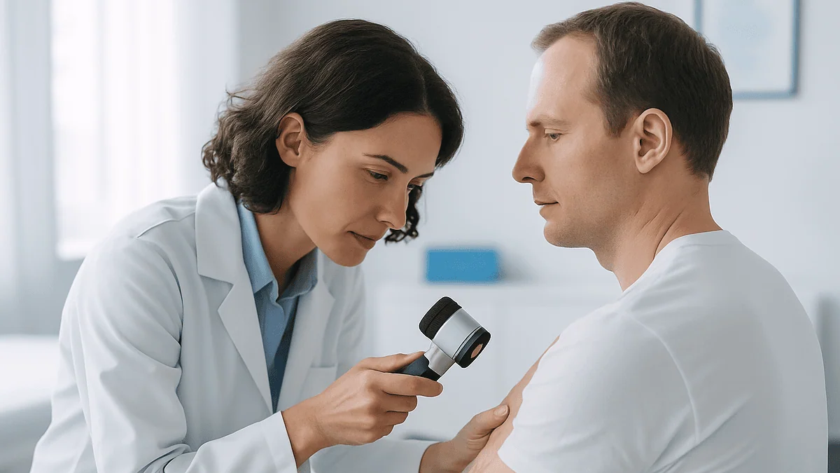
A dermatologist sorts this out with a few tools rather than a glance. The first is the story: when the color appeared, whether it is spreading, and what the original mole and its pathology report showed. The second is dermoscopy, a handheld magnifier with polarized light that reveals pigment patterns below the surface. Post-inflammatory darkening tends to be diffuse and uniform; a recurrent nevus often shows discrete pigment lines or dots confined within the borders of the scar; pigment reaching beyond the scar edge is the pattern that earns the closest attention.
Photographs help more than memory. Many clinics photograph a mole before removal and the site at follow-up, so a subtle change has a baseline. If you have your own phone photos of the original mole, bring them.
Hair is a common false alarm. A shaved facial mole that contained a coarse hair will often grow that hair again from the untouched follicle. Hair in a flat, skin-colored scar is not a mole returning, though it can be plucked or treated separately if it bothers you.
What a clinician will not do is decide from a description over the phone. The distinction between scar pigment and nevus regrowth rests on examination, and when examination leaves doubt, the tie-breaker is a small biopsy read by a pathologist who knows the history. That is where the decision belongs: with the treating team, not with a mirror.
What dermatologists do next when a mole grows back after removal
The first move is almost always paperwork. Before touching the skin, the dermatologist pulls the original pathology report. That single document shapes everything that follows. If it describes a benign nevus with cells extending to the base of the specimen, regrowth is the expected consequence of an incomplete shave and the mood in the room relaxes. If it describes an atypical mole or a melanoma with involved margins, regrowth is treated as a possible remnant of that lesion and the plan becomes prompt re-excision.
With a benign report and a typical dermoscopic picture, two paths are common. One is observation: photograph the site, note the size of the pigment, and re-check at an agreed interval. Recurrent pigment that stays inside the scar and stops changing is often left alone. The other is re-excision, chosen when the person wants the pigment gone for cosmetic reasons, when the pattern is unusual, or when the original report is missing. This time the clinician usually cuts full thickness with a margin so that the reservoir of cells is removed rather than trimmed again.
Whatever is removed goes to the laboratory with a clear note that this is a recurrent nevus from a prior excision, ideally with a request that the pathologist review the original slide alongside the new one. That pairing is the single most protective step in the whole process, because it prevents the benign irregularity of scar melanocytes from being misread.
When the original report cannot be found, many dermatologists lean toward re-excision and full examination rather than watching, simply because the reassurance of the first diagnosis is gone. The Mayo Clinic’s advice to see a doctor promptly if a removed mole grows back reflects exactly this logic: the visit is usually brief, and it turns an uncertain smudge into a documented, examined finding.
What the first days and weeks after mole removal usually look like
As the numbing wears off, most people describe soreness rather than pain. A shave site oozes a little on the first day and then forms a thin scab. A stitched excision looks like a neat line with slight swelling and bruising, more so near the eyelid or lip where the skin is loose.
Wound care is unglamorous and matters. Teams typically ask you to keep the area clean, apply a thin layer of plain ointment or the dressing they supplied, and leave the scab to lift on its own. Picking at a scab drags out healing and deepens the eventual scar; it also disturbs the bed where pigment cells are settling, which is the last thing you want if regrowth is a concern.
Stitches, if used, are removed at a follow-up visit. Facial stitches generally come out sooner than those on the back or legs, because facial skin heals quickly and prolonged sutures leave track marks; your team sets the exact timing. Dissolvable stitches under the skin need no removal.
The pathology report usually arrives around the time of the first follow-up. Ask for a copy. It is the document that will matter most if pigment ever reappears.
Over the following months the scar changes. It is often pink or slightly raised early on, then flattens and fades. Sun exposure during this period darkens both scars and any surviving pigment cells, so broad-spectrum sunscreen and shade on the site are the closest thing to regrowth prevention that daily habits offer. If a returning mole is going to appear, it tends to show itself during this maturing phase rather than years down the line, which is why some clinicians book a check once the scar has settled.
Does mole regrowth mean cancer? What the evidence actually says
Usually not, and the reasoning is straightforward. A mole that was benign under the microscope does not become malignant because part of it was left behind. The cells that regrow are the same benign cells, arranged messily in scar tissue. The NHS is explicit that most moles are harmless and that removal is often for cosmetic reasons or because a mole is irritated. Regrowth from such a mole is an inconvenience, not a transformation.
The anxiety around regrowth comes from two real issues, and neither is about the benign case. The first is misdiagnosis in the other direction: because a recurrent nevus can look like melanoma on a slide, there is a risk of being told a benign lesion is cancerous. The safeguard is the pathologist knowing the history and seeing the original slide.
The second issue is the incompletely removed atypical lesion. If the original mole was in fact an early melanoma, or a dysplastic nevus with unusual cells, and the report noted that cells reached the edge of the specimen, then any regrowth may represent residual disease. This is the scenario where come back promptly is not a formality. The NHS notes that the most common sign of melanoma is a new mole or a change in an existing one, and a change inside a scar at the site of a previously atypical lesion counts.
What separates the two situations is documentation, not appearance. Two regrown spots can look identical to the eye; one has a reassuring report behind it and one does not. That is the whole argument for pathology on every removed mole and for keeping the report. It is also the argument against destructive methods such as laser: a mole vaporized without examination leaves a scar that can never be fully reassuring, because no one knows what it was.
Can you prevent a mole from growing back? What helps and what does not
The most effective prevention happens before the mole comes off, in the choice of technique. Full-thickness excision with a margin removes the pigment reservoir; a shave, by design, does not. If your priority is never revisiting the site, say so during the consultation, and accept the linear scar that comes with it. If your priority is the smallest possible facial scar, a shave may be the right first choice with regrowth accepted as a known possibility. Neither is wrong; they answer different questions.
Confirming completeness matters as well. A pathology report stating that the lesion was removed with clear margins carries real information about the odds of regrowth. One that notes nevus cells extending to the deep margin is a heads-up that pigment may reappear.
After the procedure, sun protection is the one daily habit with a plausible mechanism. Ultraviolet light stimulates melanocytes to produce pigment and can darken both a healing scar and any residual nevus cells. Covering the site and using sunscreen once the wound has closed is standard advice from the NHS and the Mayo Clinic, quite apart from its broader role in skin cancer prevention.
What does not help: creams, oils or mole-fading products sold online. There is no evidence that any topical product removes nevus cells or prevents them from regrowing, and some cause chemical burns that scar worse than surgery. The NHS specifically warns against attempting to remove moles at home. Likewise, having a regrown mole shaved a second time at the same depth mostly resets the clock; if regrowth bothers you, the conversation with your team is usually about going deeper, not repeating.
None of this guarantees anything. Skin biology is variable, and a small number of full-thickness excisions with clear margins still show pigment later. The goal is to stack the odds and to make sure that if pigment does return, everyone knows exactly what it is.
Facial moles: why the face changes the calculation
The face is where regrowth is most visible and where the trade-offs are sharpest. A linear excision scar that would vanish on the back stays on show on a cheek. That pushes many dermatologists and their patients toward shave removal for benign raised facial moles, with the understanding that some pigment may return and can be dealt with later if it does.
Facial anatomy also feeds regrowth. The cheeks, chin and upper lip are dense with hair follicles and oil glands, and raised moles there often wrap around a follicle. Melanocytes cluster in the outer root sheath of hairs, so the untouched follicle beneath a shave is a natural source of returning pigment. It is also the reason a regrown facial mole often appears first as a fleck of brown beside a hair.
Healing on the face is quick and usually kind, thanks to rich blood supply, and facial scars often mature to a pale, barely visible mark. The exceptions are the jawline and the skin around the ears, where tension and a tendency to thicker scars make some clinicians cautious about long excisions. People with darker skin tones may see more post-inflammatory darkening in any facial scar, which can be mistaken for regrowth; the dermoscopy check described earlier sorts that out.
Sun exposure is hardest to avoid on the face, and it matters most there. A regrown mole and a sun-darkened scar can look alike, and both are more likely on skin that is outdoors daily without protection. A hat brim over a healing cheek is a low-tech intervention with a sound mechanism.
Finally, cosmetic motivation should not lower the bar for pathology. A facial mole removed for appearance still goes to the laboratory. The face is also a common site for a slow-spreading type of melanoma in older adults, and a lesion assumed to be an ordinary mole occasionally proves otherwise on the slide. The examination protects the person, not just the diagnosis.
What people often get wrong about a mole growing back
If it came back, the surgeon did a bad job. Regrowth after a shave is a known feature of the technique, not a failure of skill. The shave was chosen to minimize the scar; some pigment cells were always likely to remain. Regrowth after a full excision is less expected and does merit a conversation about margins, but even then it is not evidence of carelessness.
Regrowth means it was cancer all along. A benign mole regrows as a benign mole. Cancer is a question answered by the original report, not by the fact of regrowth.
Laser is the modern way and leaves nothing behind. Laser leaves nothing to examine, which is the problem. Guidance from the NHS and the Mayo Clinic steers away from laser for moles for exactly that reason, and destructive methods can leave scattered pigment cells just as a shave can.
A flat scar means the whole mole is gone. Flatness reflects the shave depth, not the depth of the nevus cells. Deeper cells can sit under a perfectly flat scar and stay invisible until they produce pigment.
Pathology is only for moles that look suspicious. Mainstream dermatology guidance recommends microscopic examination of any removed mole. Cases where a cosmetic mole turns out to be something else are uncommon but real, and they are the whole justification for the routine.
Hair in the scar means the mole is back. Hair comes from the follicle, which a shave leaves intact. Hair regrowth alone is not pigment regrowth.
A cream can fix a regrowth. No topical product removes nevus cells. Products marketed for this can cause burns and scarring, and the NHS advises against home removal attempts.
Once it regrows it will keep regrowing. A full-thickness re-excision removes the deeper reservoir; repeated regrowth after that is uncommon. The pattern is a matter of depth, not destiny.
Questions to ask your care team before and after mole removal
A good consultation answers most of these unprompted. Bringing the list anyway makes the conversation faster and the notes more useful later.
Before the procedure:
- Which technique are you planning, and why this one for this mole?
- How likely is pigment to return with that technique, and what would we do if it does?
- Will the tissue be sent for pathology, and how will I receive the report?
- Where exactly will the scar sit, and how is it likely to settle on this part of my face?
- Does anything in my history, such as blood-thinning medicines, previous keloids or pregnancy, change the plan?
- Can you photograph the mole before removal so we have a baseline?
After the procedure, or if pigment returns:
- What did the pathology report say, and did the cells reach the edge of the specimen?
- Is what I am seeing scar pigment, hair, or nevus cells returning?
- If we watch it, how will we know it has changed, and when should I come back?
- If we remove it again, will you go deeper this time, and what will the new scar look like?
- Will the pathologist see my original slide alongside the new sample?
- What should I notice at home that means I should call sooner?
Two habits round this out. Keep a copy of every pathology report in the same place you keep vaccination records; a report from years ago can spare you a second biopsy. Photograph the healed site once it has settled, in the same light you will use later, so any future change has a fixed reference. Neither habit replaces the dermatologist’s examination, but both make that examination sharper.
When to call your doctor about a removed mole or its scar
Most regrowth is unhurried, and most healing is uneventful. A short list of signs should move a routine follow-up to a prompt call. None of them are meant for self-diagnosis; they are prompts for an examination.
In the first days after removal, contact the team if bleeding soaks through a dressing and does not stop with firm pressure, if the area becomes increasingly red, hot, swollen or painful, if there is pus or a foul smell, or if you develop a fever. These point toward infection or a bleeding problem and are straightforward to treat early.
In the weeks and months afterward, the Mayo Clinic’s plain advice is to see a doctor promptly if a removed mole grows back. Beyond that baseline, arrange a sooner visit if the returning pigment spreads past the edge of the original scar into normal skin, if the site develops a raised lump, a nodule or a rough surface, if it bleeds, itches persistently or fails to heal, or if the color darkens quickly or shows several shades. The NHS describes a new or changing mole as the most common sign of melanoma, and a change at the site of a previous lesion belongs in that category.
Two situations warrant a call regardless of how the site looks. If your original pathology report described an atypical or dysplastic nevus or a melanoma, any pigment at the scar should be assessed without waiting for the next scheduled check. If you never received a report, or the mole was removed by laser or another method that left no specimen, mention that when you call; it changes how carefully the team will want to look.
Every decision about watching, biopsying or re-excising sits with the clinician who examines the skin. The role of this list is to get you into that room at the right time, not to settle the question in front of a mirror.
Frequently asked questions
Is a mole growing back after shave removal normal?
Yes, it is a recognized outcome of shave removal rather than a mistake. A shave takes the raised part of the mole and a thin layer beneath it, leaving deeper pigment cells and hair follicles in place. As the wound heals, those cells can migrate upward and produce color again inside the scar. The Mayo Clinic advises having any regrowth checked promptly so it can be documented and compared with the original pathology report.
How long after removal can a mole grow back?
There is no fixed interval backed by a large study. Case series describe regrowth appearing most often while the scar is still maturing, in the months after the procedure, rather than years later. Late reappearance is possible but less typical. Because the timing varies, dermatologists rely on examination and the original pathology report rather than the calendar to decide whether a returning spot needs monitoring or re-excision.
Does mole regrowth mean cancer?
Usually not. A mole that was benign on the original pathology report regrows as benign tissue; the cells do not change nature because part of the mole was left behind. Regrowth matters more when the original report described an atypical or dysplastic nevus or a melanoma, or when no tissue was ever examined, for example after laser. In those cases any pigment at the scar should be assessed promptly by the treating team.
What is a recurrent nevus, and is it the same as pseudomelanoma?
They are two names for the same finding: pigment returning within the scar of a previously removed mole. Pseudomelanoma is an older term that refers to how the regrown cells can look under the microscope, where their irregular arrangement in scar tissue can mimic melanoma. The name describes a diagnostic look-alike, not a dangerous lesion. Knowing the history allows the pathologist to interpret the cells correctly.
How can I tell if it is a mole removal scar or regrowth?
You usually cannot settle it reliably at home, and clinicians do not try to by phone. Scars pass through pink, brown and pale phases, and sun exposure can darken them, none of which is nevus tissue. A dermatologist uses dermoscopy, a lit magnifier, to look for discrete pigment structures, checks whether color stays within the scar, and compares the site with earlier photographs and the pathology report before deciding.
Will the dermatologist remove a regrown mole again?
Sometimes, but not always. If the original report was benign and the pigment sits quietly inside the scar, observation with photographs is a common plan. Re-excision is chosen when the pattern is unusual, the report is missing, the original lesion was atypical, or the person wants the pigment gone. A second removal is usually full thickness with a margin, and the specimen is labelled as a recurrent nevus for the pathologist.
Can a mole grow back after complete surgical excision?
It is uncommon but possible. A full-thickness excision with a margin removes most of the pigment cell reservoir, so regrowth is much less likely than after a shave. When it does happen, the dermatologist reviews the pathology report to see whether nevus cells reached the edge of the specimen, which would explain the return. Regrowth after an excision reported as clear is a reason for a careful re-examination rather than alarm.
Why do dermatologists avoid laser for removing moles?
Laser destroys the tissue instead of removing it, so nothing can be sent for microscopic examination. The NHS and the Mayo Clinic both note that laser and similar destructive methods are generally not used for moles for this reason. A lesion treated this way can never be confirmed as benign, and if pigment later reappears in the scar, the absence of an original report makes the assessment harder and often leads to a biopsy.
Does sunscreen help stop a mole coming back after removal?
It helps with the mechanism, though it is not a guarantee. Ultraviolet light stimulates melanocytes to produce pigment and can darken both a healing scar and any residual nevus cells. Protecting the site with clothing, shade and broad-spectrum sunscreen once the wound has closed is standard advice from the NHS and the Mayo Clinic. It also reduces post-inflammatory darkening that is often mistaken for regrowth on the face.
Is hair growing in the scar a sign the mole is back?
No. Raised facial moles often contain coarse hairs rooted in follicles that sit deeper than a shave reaches. After the surface is removed, the follicle continues to produce hair, which then emerges through the flat scar. Hair alone, without new pigment, is not a recurrent nevus. If the hair bothers you, it can be plucked or treated separately; if brown color appears beside it, have the site examined.
References
This article is for general information only and is not a substitute for professional medical advice. Please consult a qualified doctor about your individual situation.

More from the Blog

Mole Removal Healing: Dressing Changes, Stitch Removal and When Swimming Is Safe Again
Mole removal healing time depends on the technique. A shallow shave site usually closes at the surface within about two to three weeks, while…

Acne Scar Treatment Results: How Sessions Build and What Improvement Realistically Means
Acne scar treatment results usually build gradually over several sessions and months, because most procedures work by prompting the skin to remodel its collagen…
Healing After Reconstruction for Skin Disease: Graft Survival Checks and Scar Maturation
After reconstruction for skin cancer, a skin graft usually begins forming its own blood supply within about 36 hours and looks settled within 2–3…
Dermabrasion Results: What Scars and Fine Lines Realistically Look Like Once Healed
Once healed, dermabrasion typically leaves skin smoother in texture, with raised or shallow scars looking flatter and less sharply edged, and fine surface lines…

Suture Suspension (Thread) Lift vs Facelift: Where Threads Stop and Surgery Begins
A thread lift places absorbable barbed sutures under the skin to gently reposition mildly sagging tissue, usually under local anesthesia, with results that are…

Life After Disease Reconstruction: Activity Limits, Swelling and How the Area Settles Over Time
Life after reconstructive surgery usually unfolds in stages: several weeks of restricted lifting and reaching, months of gradual swelling reduction, and a year or…