

Can Arthroscopy Diagnose What an MRI Missed? When a Look Inside the Joint Is Used

Key Takeaways
- MRI builds pictures in slices a few millimeters thick, so thin cartilage damage, small loose fragments and problems that appear only under load can be missed while bone and larger soft-tissue injuries are shown well.
- Diagnostic arthroscopy lets a surgeon see joint surfaces in color, probe them for softness and test ligament stability by moving the joint, none of which a resting scan can do.
- Arthroscopy cannot see inside bone or beyond the joint capsule, so stress fractures, bone bruising and pain referred from the spine or hip remain imaging or clinical diagnoses.
- The NHS estimates serious complications at roughly 1 in 100 arthroscopies, and the risk applies equally whether or not the surgeon finds anything to treat.
- The NHS puts the procedure at 30 minutes to 2 hours and recovery anywhere from a few days to several weeks or months, driven mainly by whether a repair rather than a simple trim was performed.
- Arthroscopic washout for generalized osteoarthritis has been largely abandoned in guidelines, so worn joints are among those most often asked to wait or pursue other options.
Yes, in some cases. Diagnostic arthroscopy lets a surgeon look directly inside a joint with a thin camera, and it can reveal cartilage damage, loose fragments or ligament tears that an MRI showed poorly or not at all. It is usually reserved for people whose symptoms persist despite imaging and conservative care, because it is a surgical procedure with small but real risks.
The scan report was two paragraphs long and, as far as the radiologist could tell, unremarkable. Yet the knee still catches on the stairs every morning, still swells after a jog, still refuses to let its owner kneel to tie a shoe. Somewhere between the black-and-white slices of the MRI and the ache that will not leave, a gap has opened, and the person living in that gap wants to know what fills it.
That is often the moment a surgeon mentions diagnostic arthroscopy. The phrase can land like a small threat, since it means an operation, however minor. But it is really a question dressed as a procedure: if pictures taken from outside have not explained the pain, would a direct look from inside?
This article walks through what that look involves, what it can and cannot see better than a scan, who is usually offered it, and how to weigh the trade-offs with the people treating you.
What is diagnostic arthroscopy, in plain language?
Arthroscopy is keyhole surgery of a joint: a surgeon makes one or more small cuts, slides in a slender tube fitted with a light and a camera, and watches the inside of the joint on a screen. The tube is called an arthroscope, and the NHS describes it as roughly the size of a drinking straw. Sterile fluid is pumped in to expand the joint space so structures are not pressed together, which makes them easier to see.
When the purpose is mainly to find out what is wrong rather than to fix a known problem, the procedure is called diagnostic arthroscopy. In practice the boundary blurs. Surgeons who spot a torn piece of cartilage while looking will usually trim or repair it during the same session, using fine instruments passed through a second small cut. The NHS notes that the whole procedure typically takes between 30 minutes and two hours, depending on what is found and what is done.
Three things separate this from a scan. First, the surgeon sees real color and texture: a cartilage surface that looks smooth on MRI may be soft, frayed or peeling when seen up close. Second, the surgeon can probe. A small hooked instrument can press on a ligament to test whether it is taut or lax, or lift a flap of cartilage to see how far it extends. Third, the joint can be moved during the procedure, so problems that appear only in certain positions can be provoked and watched.
Those advantages come at a price that a scan does not charge: an anesthetic, small wounds, a short recovery and a low but real chance of complications. That balance is the whole story of when it is used.
Why can an MRI miss something in a joint?
Magnetic resonance imaging, or MRI, builds pictures of soft tissue by measuring how water molecules in the body respond to a strong magnet and radio waves. It is very good at showing bone marrow, ligaments, tendons and the menisci, the crescent-shaped cartilage pads in the knee. According to the NHS, a full scan usually lasts between 15 and 90 minutes, and it involves no radiation.

Its limits come from how it works. An MRI is a stack of slices, each a few millimeters thick, and anything smaller than a slice or lying awkwardly between two slices can blend into its surroundings. The thin smooth cartilage that lines the ends of bones is a common example: early softening or a small flap may not change the signal enough to be seen. A loose fragment floating in joint fluid can be hard to distinguish from the fluid itself.
Timing matters too. A scan taken while the joint is at rest cannot show what happens when a ligament is loaded or a kneecap tracks sideways during a squat. Some tears are stable when the leg is lying still and only gape under weight.
Image quality varies with the machine, the protocol and, above all, the person reading it. Motion during the scan, metal implants nearby and scarring from earlier surgery can all blur the picture or produce artefacts that look like disease. Old injuries leave changes that are hard to separate from new ones.
None of this makes MRI a poor test. Mayo Clinic lists it among the imaging studies surgeons typically order before considering arthroscopy, precisely because it answers most questions without surgery. The point is that a negative scan means the scan did not find a cause, not that there is no cause.
Arthroscopy vs MRI: what each one actually shows
Patients often imagine the two as rivals. They are better understood as different instruments: one paints the whole joint and the tissue around it from outside, the other inspects the inside surfaces at close range. The table below sets out where each tends to be stronger, drawing on how the NHS, Mayo Clinic and MedlinePlus describe the two tests.
| Feature | MRI | Diagnostic arthroscopy |
|---|---|---|
| What it is | Non-invasive scan using magnetism and radio waves | Keyhole surgery with a camera inside the joint |
| Anesthetic needed | None | General, spinal or local, chosen with the anesthetist |
| Typical duration | 15 to 90 minutes (NHS) | 30 minutes to 2 hours (NHS) |
| Sees bone marrow and swelling inside bone | Yes | No, only the surface |
| Sees surface cartilage texture | Limited for early or shallow damage | Directly, in color, and can be probed |
| Sees tissue outside the joint capsule | Yes | No |
| Can test stability by moving the joint | No | Yes |
| Can treat what it finds | No | Often, in the same procedure |
| Risks | Very few; not suitable with some implants | Infection, bleeding, clots, stiffness, nerve injury (NHS) |
| Recovery | None | Days to weeks or longer depending on what was done (NHS) |
Two rows deserve emphasis. Arthroscopy cannot see inside bone, so a stress fracture, a bone bruise or early bone death is an MRI diagnosis, not an arthroscopic one. And arthroscopy is blind to anything outside the joint capsule, including many tendon problems and referred pain from the hip or spine. A camera in the knee will not explain pain that is really coming from a pinched nerve in the back.
Can arthroscopy really diagnose what an MRI missed?
The honest answer is that it can, for a particular set of problems, and that it is not a magic second opinion on everything.
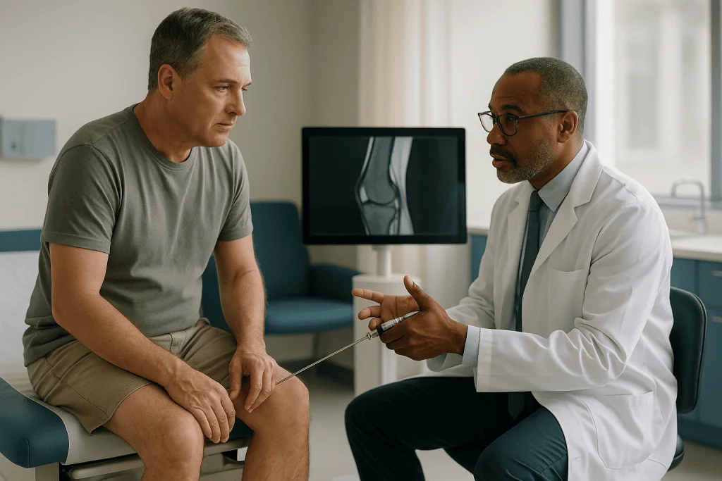
The categories where a direct look most often adds information are those that depend on surface texture, mechanical behavior or small size. Cleveland Clinic lists damaged or torn cartilage, loose fragments of bone or cartilage, inflamed joint lining and scar tissue among the conditions arthroscopy is used to identify. Early softening of the cartilage that caps the bone, sometimes called chondromalacia, is a classic example: the surface can look intact on a scan yet feel like wet cardboard under a probe. A partial ligament tear that is stable on a resting scan may show its true laxity only when the surgeon pulls on it.
There are also findings that look like disease on MRI but turn out to be normal variations or old healed changes when seen directly. Arthroscopy can therefore clarify a scan in both directions, ruling something in or ruling it out.
What it cannot do is invent a diagnosis where the pain has a source outside the joint. Nor does a clean arthroscopy guarantee the joint is healthy at a level the camera cannot reach, such as inside bone or in the deeper layers of cartilage.
Guidance from Mayo Clinic frames it this way: surgeons use arthroscopy to diagnose and treat joint conditions when imaging and examination have not given a clear answer, or when a known problem needs surgical treatment anyway. The word to hold onto is unexplained. A scan that already explains the symptoms rarely needs a camera to confirm it. A scan that leaves the story incomplete, in a joint that behaves as though something mechanical is wrong, is where a look inside earns its place.
Who is diagnostic arthroscopy usually for, and who is asked to wait?
Surgeons are cautious about operating to look, because the same small risks apply whether or not anything is found. Certain patterns make the case stronger.
The first is mechanical symptoms. Locking, where the joint sticks and will not straighten, catching, or a sense of something shifting inside all point to a physical obstruction such as a loose body or a displaced cartilage flap. These are exactly the findings MRI can underrate and arthroscopy can see and remove.
The second is persistence despite good conservative care. Most joint pain settles with rest, activity modification, physiotherapy and time. When months of appropriate rehabilitation have changed nothing, the balance tilts toward finding out why.
The third is a mismatch between examination and imaging. If a surgeon feels a clearly unstable ligament in clinic but the scan calls it intact, one of them is wrong, and a direct test under anesthetic settles the argument.
Other people are usually asked to wait, or steered elsewhere. Pain that is diffuse, present at rest, and unaccompanied by swelling or mechanical signs is more often inflammatory, referred or related to widespread wear, and arthroscopy tends to disappoint in those settings. Advanced osteoarthritis, where the cartilage is worn across the whole joint, is not a condition a camera can fix, and major guidelines in several countries have moved away from arthroscopic washout for it. Someone whose recent injury is still swollen and healing may be asked to let the acute phase pass first, because early swelling can itself limit what the surgeon can see.
Medical fitness matters as well. Uncontrolled diabetes, active skin infection near the joint, bleeding disorders and anticoagulant treatment all shape whether and when an anesthetic and surgery are sensible. Those conversations belong with the treating team, who weigh your particular history rather than a general rule.
What actually happens on the day of an arthroscopy?
Most arthroscopies are day-case procedures. The NHS notes that people usually go home the same day, often within a few hours of waking.
Before the operation you will meet the anesthetist. Options range from a general anesthetic, where you are asleep, to a spinal anesthetic that numbs the lower body, to a local anesthetic combined with sedation. The choice depends on the joint, the expected length of surgery, your health and your preference. The surgical site is marked, and you will be asked to confirm which side is being operated on more than once; the repetition is a safety check, not forgetfulness.
In the operating room the limb is cleaned with antiseptic and draped. For a knee, a tight band called a tourniquet may be inflated around the thigh to keep the field clear of blood. The surgeon makes a small cut, inserts the arthroscope, and fills the joint with sterile saline. Mayo Clinic describes the incisions as being about the size of a buttonhole.
The camera is then walked systematically around the joint, compartment by compartment, so nothing is skipped. A probe passes through a second cut to test what the camera sees. If something needs treating, the instruments for shaving, cutting, grasping or stitching pass through the same or an additional small cut. Photographs or video are usually recorded, and many surgeons show these to patients afterward, which is one of the more satisfying moments of the process for people who have spent months being told nothing is wrong.
At the end the fluid is drained, the cuts are closed with stitches or adhesive strips, and a padded dressing is applied. In the recovery area nurses check circulation and sensation in the limb, and a physiotherapist may show you how to move or, for a leg, how to manage crutches before you leave.
Which joints and which problems are commonly looked at?
The knee is the joint most often examined by arthroscopy, followed by the shoulder. The NHS lists the knee, shoulder, hip, wrist, elbow and ankle as joints where the technique is used. Smaller joints require thinner scopes and a surgeon with specific experience, so availability varies.
In the knee, the usual suspects are meniscal tears, damaged articular cartilage, loose bodies, problems with the ligaments that cross inside the joint, and inflamed or thickened joint lining. Kneecap tracking, the way the patella slides in its groove as the knee bends, can also be watched in real time.
In the shoulder, arthroscopy is used to inspect the rotator cuff tendons where they enter the joint, the labrum, a rim of cartilage around the socket, the biceps tendon anchor and the joint lining. Impingement, where tendons rub under the bony arch above the joint, is often assessed and treated this way.
Hip arthroscopy is more technically demanding because the joint is deep and tightly held, but it is used for labral tears and for bony shapes that cause the ball and socket to pinch. Wrist arthroscopy looks at the small ligaments between the wrist bones and at the cartilage disc on the little-finger side, structures that are notoriously hard to see well on imaging because of their size. Ankle arthroscopy addresses impingement, loose fragments and cartilage injuries after sprains.
Across all of these, the common thread is a structure that is small, thin, or defined by how it moves under load. Those are the qualities that make a direct look valuable and a scan fallible. Bone problems, deep tendon disease away from the joint, and pain referred from elsewhere fall outside the camera’s reach regardless of the joint involved.
What can be treated during the same procedure?
Part of the appeal of looking with a camera is that the same tools can act on what they find. MedlinePlus describes knee arthroscopy as a procedure used both to diagnose and to treat problems in the joint, and the two often happen in a single sitting.
Common treatments performed arthroscopically include trimming a torn piece of meniscus so it no longer catches, stitching a meniscal tear back together when the tissue is healthy enough to heal, removing loose fragments of cartilage or bone, smoothing frayed cartilage, releasing tight scar tissue, and removing inflamed joint lining. Ligament reconstruction and rotator cuff repair are more involved operations that are also done through keyholes, though these are usually planned in advance rather than discovered during a diagnostic look.
This flexibility has an important implication for consent. Before the operation your surgeon will typically discuss a range of possible findings and ask permission to treat some of them if seen, so that you do not wake up to be told a second operation is needed. It also means you may not know in advance exactly how long the procedure will take or how your recovery will look, because a joint that turns out to need a repair recovers differently from one that only needed trimming.
Ask what your surgeon would and would not do without waking you to discuss it. Some findings, such as a large cartilage defect that might warrant a specialized reconstructive procedure, are usually documented and discussed afterward rather than treated on the spot. Knowing where those lines sit beforehand turns an uncertain morning into a plan with branches you have already agreed to.
Whatever is found, the decision about treatment remains a shared one, guided by your goals as well as the surgeon’s view of what the tissue can support.
Knee arthroscopy risks: how common are complications?
Arthroscopy is among the lower-risk operations in orthopedics, but low is not zero, and a diagnostic procedure carries the same risks as a therapeutic one. The NHS estimates the overall risk of serious complications at around 1 in 100 procedures.
The complications listed by the NHS and Mayo Clinic include infection inside the joint, bleeding into the joint, blood clots in the deep veins of the leg that can travel to the lung, damage to nerves or blood vessels near the incisions, stiffness of the joint afterward, and problems related to the anesthetic. Numbness of a small patch of skin near a cut is relatively common and often improves over months, though it can be permanent.
Some risks are specific to how the procedure is done. Fluid pumped into the joint can leak into surrounding tissue and cause temporary swelling. A tourniquet left inflated for a long operation can cause thigh discomfort for a few days. Instruments can, rarely, break inside the joint. Cartilage can be scuffed by the scope itself.
A less tangible risk deserves naming: the possibility of a normal result. A proportion of diagnostic arthroscopies find nothing that explains the symptoms, and the person has then undergone an anesthetic and a recovery for information rather than relief. That is not a failure of the procedure, since excluding a mechanical cause redirects the search, but it is a genuine outcome to weigh beforehand.
Risk is not spread evenly. Smoking, diabetes, obesity, prior surgery on the same joint, and clotting disorders each raise the chance of particular problems, which is why the pre-operative assessment asks about them. Your surgeon should be able to tell you how your own profile shifts the general figures, and that conversation is more useful than any average.
What are the alternatives before a look inside?
A camera is rarely the first tool. Most guidelines and the sources cited here describe a ladder of investigations and treatments that usually precedes arthroscopy, and understanding the rungs helps you judge whether you have really climbed them.
Clinical examination remains the foundation. An experienced examiner can provoke pain from a torn meniscus, test ligament laxity by hand, and assess how the kneecap tracks. Findings that are consistent across visits carry weight even when a scan disagrees.
Plain X-rays show bone alignment, joint space narrowing and fractures. They are quick, cheap in resources and often ordered before MRI to rule out arthritis or bony causes.
MRI itself can sometimes be repeated with a different protocol, a stronger magnet, or the addition of contrast dye injected into the joint, a technique called arthrography that outlines surfaces and can highlight cartilage flaps and labral tears more clearly than a standard scan. Asking whether a repeat or enhanced scan would change the picture is a reasonable question before agreeing to surgery.
Ultrasound is useful for tendons and fluid around a joint and can be done while the joint moves, though it cannot see deep inside.
A diagnostic injection of local anesthetic into the joint under image guidance is another underused step. If the pain disappears for the duration of the numbing, the joint is very likely the source; if it does not, the search should look elsewhere before anyone operates.
Structured physiotherapy, activity modification and time remain treatments in their own right, and many joints that puzzle a scan settle with them. The case for diagnostic arthroscopy is strongest after these options have been genuinely tried, not merely mentioned.
Arthroscopy recovery time: what the following days and weeks look like
Recovery depends far more on what was done inside the joint than on the size of the cuts outside it. The NHS puts the range broadly: some people recover within a few days, others need several weeks or months, particularly if a repair rather than a simple trim was performed.
The first two or three days are about swelling and comfort. The joint is typically bandaged, elevated and iced, and the surgical team will advise on pain relief; that advice, including any medicines and how to take them, comes from your prescribing clinician rather than from an article. For a knee or ankle you may be given crutches, sometimes to protect a repair and sometimes simply for balance while the joint is sore. Dressings are usually kept dry until the wounds are checked.
During the first two weeks the small wounds heal and stitches, if used, are removed or dissolve. Gentle range-of-motion exercises usually begin early, because a joint that is not moved tends to stiffen, and stiffness is one of the more frustrating complications to reverse. Physiotherapy guidance, whether in person or as a home program, is tailored to what was found.
Driving is a common question. The NHS advises that you may not be able to drive for a while after arthroscopy, and the practical test is whether you can perform an emergency stop safely and are no longer affected by anesthetic or strong painkillers. Your insurer may have its own view.
Return to desk work often happens within the first week or two after a simple procedure, while manual work and sport take longer, and any ligament or tendon repair extends everything. Your surgeon’s plan will be specific to your operation, and it should be treated as a range rather than a deadline.
What people often get wrong about diagnostic arthroscopy
Several beliefs circulate in waiting rooms and search results, and most of them make the decision harder than it needs to be.
The first is that a normal MRI means nothing is wrong. As explained above, the scan is excellent for many structures and weaker for thin cartilage, small loose bodies and problems that appear only under load. A normal report means the scan found no cause, which is a different statement.
The second is the reverse: that arthroscopy is the gold standard for every joint problem and should replace imaging. It cannot see inside bone, cannot see outside the joint capsule, and cannot detect pain referred from the spine or hip. Surgeons order MRI first for good reason.
The third is that keyhole means trivial. The cuts are small, but the anesthetic, the fluid, the tourniquet and the internal work are real, and the NHS figure of roughly 1 in 100 for serious complications applies whether the surgeon finds a problem or not.
The fourth is that a clean joint means the pain is imagined. A negative arthroscopy is useful information that redirects attention toward soft tissue outside the joint, nerve-related causes or wider pain conditions. It does not mean the symptoms were not real.
The fifth is that arthroscopy is a routine treatment for worn joints. Washing out or smoothing an arthritic knee was once common; large trials and guidelines have since found it offers little for generalized osteoarthritis, and most surgeons no longer recommend it for that purpose.
The last is that recovery is measured by the size of the scar. A meniscal repair through three tiny cuts can take longer to rehabilitate than an open procedure elsewhere. Ask what was done, not how big the marks are.
Questions to ask your care team before agreeing
A good pre-operative conversation should leave you able to explain to a friend why this procedure, why now, and what happens in each likely scenario. These questions help get there.
- What do you think is causing my symptoms, and what would you expect to find? A surgeon who can name two or three specific suspects has a clearer purpose than one who hopes to see something.
- What did the MRI show and not show, and would a repeat scan, a scan with contrast, or a diagnostic injection change your mind about operating?
- Which of the possible findings would you treat during the same operation, and which would you photograph and discuss with me afterward?
- How would my recovery differ depending on what you find? Ask for the range for a simple trim and for a repair.
- What is your estimate of the chance that the arthroscopy finds nothing that explains my pain, and what would we do next in that case?
- Which anesthetic options are suitable for me, and why?
- How do my own health factors change the general risks?
- What signs after surgery should make me call you, and how do I reach the team out of hours?
- What happens if I choose not to have the procedure and continue with physiotherapy for another few months?
Write the answers down or bring someone to listen. It is entirely reasonable to take the information home and decide later, and a second opinion from another surgeon is a normal part of orthopedic care rather than a slight. The best decisions are made when both you and the team can describe the plan and its branches in the same words.
When to call your doctor after arthroscopy
Most recoveries are uneventful, and the most common complaints are mild swelling, bruising and soreness that improve day by day. Certain signs, however, point to a complication that needs prompt attention, and both the NHS and Mayo Clinic advise seeking help if they appear.
Call your surgical team, or urgent care if they are unreachable, if you notice any of the following in the days and weeks after the procedure:
- A fever, chills or feeling generally unwell, especially combined with a joint that is hot, red or increasingly painful, which can signal infection inside the joint.
- Pus, cloudy fluid or spreading redness at any of the wounds, or a wound that opens.
- Swelling that increases sharply rather than settling, or a joint that becomes tense and very painful, which may indicate bleeding inside.
- Pain, tenderness, warmth or swelling in the calf, or the whole leg becoming swollen, which can be a sign of a deep vein clot.
- Numbness, tingling or weakness in the limb that is new or worsening, or a foot or hand that is pale, cold or blue.
- Pain that is not controlled by the plan you were given.
Seek emergency help without delay for sudden shortness of breath, chest pain, coughing up blood, collapse or fainting. These can indicate a clot that has traveled to the lungs, which is a medical emergency.
You should also contact the team if the joint locks again, if you cannot bear weight when you were told you could, or if a repair you were asked to protect has been stressed by a fall. Uncertainty itself is a reason to call: nurses and surgeons would far rather reassure you over the phone than see a problem late.
What matters most when deciding on a look inside the joint
Strip away the technology and the decision comes down to a single judgment: does the joint behave as though something mechanical is wrong, and has everything short of surgery been genuinely tried?
If the answer to both is yes, a diagnostic arthroscopy is a well-established next step with a defined purpose. It can identify and often treat the small, thin, load-dependent problems that scans handle least well, and the NHS and Mayo Clinic describe it as a routine day-case procedure with a low but real complication rate. The information it produces, even when the finding is a normal joint, changes what happens next.
If the joint is diffusely painful, worn throughout, or the pain pattern points elsewhere, the camera is likely to add a recovery without adding an answer. That is where surgeons who say no are protecting you rather than dismissing you, and where asking about a repeat scan, a diagnostic injection or a fresh look at the spine and hip may be the more productive path.
Between those poles lies most real life, and the honest position is that the evidence supports arthroscopy as a targeted investigation rather than a general search. The surgeon who can tell you what they expect to find, what they will do about each possibility, and what a negative result would mean is offering you the procedure on the right terms.
Whatever you decide, the decision sits with you and the team caring for you. Bring your questions, expect specific answers, and remember that the goal is not a perfect picture of the joint but a body that lets you get back to the stairs, the run and the shoelace without thinking about it.
Frequently asked questions
What is diagnostic arthroscopy and how is it different from ordinary arthroscopy?
Diagnostic arthroscopy is keyhole surgery performed mainly to find the cause of joint symptoms, using a thin camera inserted through small cuts. Ordinary or therapeutic arthroscopy uses the same approach to treat a known problem. In practice the two overlap, because surgeons usually treat what they find during a diagnostic look, so consent discussions cover both possibilities beforehand.
Arthroscopy vs MRI: which is more accurate for a knee problem?
Neither is more accurate across the board. MRI is stronger for bone marrow, deeper soft tissue and anything outside the joint, and it carries almost no risk. Arthroscopy is stronger for surface cartilage texture, small loose bodies and testing stability under movement, but it is surgery. Surgeons generally use MRI first and reserve arthroscopy for unexplained mechanical symptoms.
If my MRI was normal, does that mean my knee is fine?
Not necessarily. A normal MRI means the scan did not find a cause, which is different from proving there is none. Early cartilage softening, small flaps and problems that only appear when the joint is loaded can be under-detected. If symptoms such as locking or catching persist, your clinician may consider a repeat scan, a diagnostic injection or arthroscopy.
How long does an arthroscopy take?
The NHS states that arthroscopy usually takes between 30 minutes and two hours, depending on the joint and whether anything is treated during the same procedure. Most people go home the same day. The time under anesthetic is often longer than the time spent inside the joint, because preparation, positioning and closing the wounds are included.
What is the typical arthroscopy recovery time?
Recovery varies widely. The NHS notes that some people recover within a few days, while others need several weeks or months, particularly after a repair rather than a simple trim. Desk work often resumes within the first couple of weeks after a straightforward procedure, whereas manual jobs, sport and driving depend on the joint, the operation and your surgeon’s advice.
What are the main knee arthroscopy risks?
The NHS estimates serious complications at around 1 in 100 procedures. Listed risks include infection inside the joint, bleeding, blood clots in the leg, damage to nearby nerves or blood vessels, joint stiffness and anesthetic problems. Small patches of skin numbness near the cuts are relatively common. Your own risk depends on health factors such as smoking, diabetes and previous surgery.
Can the surgeon fix the problem during a diagnostic arthroscopy?
Often, yes. Trimming or repairing a torn meniscus, removing loose fragments, smoothing frayed cartilage and releasing scar tissue can all be done during the same procedure with fine instruments. Larger reconstructions are usually planned separately. Before surgery your surgeon should explain which findings they would treat immediately and which they would discuss with you afterward.
What happens if the arthroscopy finds nothing wrong?
A normal arthroscopy is genuine information: it makes a mechanical cause inside the joint unlikely and shifts attention to tendons and soft tissue outside the capsule, nerve-related pain, or problems referred from the hip or spine. It does not mean the symptoms were imagined. Your team would typically review the diagnosis and recovery plan with you after the findings.
Is arthroscopy useful for arthritis?
For generalized osteoarthritis, where cartilage is worn across the joint, arthroscopic washout or smoothing has largely been abandoned because trials found little benefit, and guidelines now steer away from it. Arthroscopy may still be considered for a specific mechanical problem, such as a loose fragment causing locking, in a joint with mild wear. Your surgeon can explain which applies.
What type of anesthetic is used for arthroscopy?
Options include a general anesthetic, a spinal anesthetic that numbs the lower body, or a local anesthetic with sedation. The choice depends on the joint, the expected length of the operation, your health and your preferences, and is made with the anesthetist before surgery. Regardless of the type, you will need someone to take you home and stay with you afterward.
References
This article is for general information only and is not a substitute for professional medical advice. Please consult a qualified doctor about your individual situation.

More from the Blog
How Arthroscopy Works: The Camera, the Portals and What Surgeons Can Fix Through Keyholes
Arthroscopy works by passing a pencil-width tube with a light and a video camera into a joint through an incision a few millimetres long,…

Soft Tissue Injury Healing Stages: Why Sprains, Strains and Bruises Heal at Different Rates
Soft tissue injury healing time depends on what was damaged and how well it is supplied with blood. Bruises usually fade within about two…

What Happens Years After Meniscus Surgery? Cartilage Wear and Realistic Expectations
Years after meniscus surgery, the knee's future depends mainly on how much meniscus tissue was removed and how healthy the cartilage was beforehand. Partial…

Does a Sprain or Strain Need an MRI? When Imaging Adds to a Soft Tissue Injury Exam
Most sprains and strains do not need an MRI. Clinicians usually diagnose them by asking how the injury happened and examining the joint or…

Does Bursitis Come Back? Why Flares Recur and What Long-Term Results Usually Look Like
Yes, bursitis can come back. Most flares settle within a few weeks with rest, ice and reduced pressure on the area, but recurrence is…
Does a Torn ACL Always Need Surgery? Who Copes Without Reconstruction and Who Does Not
Not always. Many people with a torn ACL, particularly those who do not play pivoting sports, regain a stable, working knee through structured rehabilitation…