

Does Pregnancy Change Diabetic Retinopathy Care? Why Eye Checks Move Closer Together

Key Takeaways
- Pregnancy is a recognized risk factor for diabetic retinopathy progressing, which is why people with pre-existing diabetes are screened early in pregnancy and again around 28 weeks instead of every one to two years.
- An extra retinal check between 16 and 20 weeks is offered only when the first pregnancy screening shows retinopathy, so a clear baseline keeps the schedule to two visits.
- Gestational diabetes does not need pregnancy eye screening because it has not existed long enough to damage retinal vessels.
- The 4-2-1 rule defines severe non-proliferative retinopathy as hemorrhages in four quadrants, venous beading in two, or intraretinal microvascular abnormalities in one, and reaching it usually triggers a more active plan.
- Rapidly tightening glucose in early pregnancy can briefly unsettle the retina, but guidelines still ask for tight control and pair it with more frequent eye checks rather than looser targets.
- Retinal changes that start in pregnancy can continue for months after delivery, so a postpartum eye check is part of the plan, not an optional extra.
Yes. Pregnancy can speed up diabetic retinopathy in people who had diabetes before conceiving, so eye checks are usually scheduled closer together: one early in pregnancy, another around 28 weeks, and an extra visit in between if changes are already present. Gestational diabetes alone does not usually need this. Treatment choices, including laser, are made case by case by the eye and maternity teams together.
The positive test is still on the bathroom counter when the first practical thought arrives: who else needs to know? For someone who has lived with type 1 or type 2 diabetes for years, the answer includes a person most pregnancy books never mention, the eye specialist. That is not a scare. It is simple scheduling. Diabetic retinopathy in pregnancy behaves a little differently from retinopathy in any other nine months of a person’s life, and the care plan quietly reshapes itself around that fact.
Most people with well-managed diabetes sail through pregnancy with no change to their vision at all. Yet retinal doctors have learned, over several decades of careful follow-up, that the hormonal, circulatory and metabolic shifts of pregnancy can nudge tiny retinal blood vessels in the wrong direction, sometimes quickly. So the calendar changes: fewer long gaps, more short ones.
This explainer walks through why that happens, what each visit actually involves, which findings matter, and what tends to settle after the birth. It is written to inform the conversation with your care team, not to replace it.
Why diabetic retinopathy in pregnancy gets its own rulebook
Diabetic retinopathy is damage to the small blood vessels of the retina, the light-sensing layer at the back of the eye, caused by years of raised blood sugar. Outside pregnancy, it tends to creep. Screening every one to two years is enough to catch most changes before they threaten sight, which is why national programs such as the NHS diabetic eye screening service run on that rhythm.
Pregnancy compresses the timeline. Blood volume rises, the heart pumps harder, hormones that support the placenta also encourage new vessel growth, and blood sugar targets become tighter than at almost any other point in life. Each of those shifts is harmless on its own. Together they create a short window in which retinal vessels that were quietly leaking can start leaking more, and vessels that were stable can begin to sprout new, fragile branches.
Guideline writers responded with a simple principle: look more often. The NHS, following national guidance for diabetes in pregnancy, asks for a retinal check soon after the first antenatal appointment and another at around 28 weeks, with an additional visit between 16 and 20 weeks if the first examination shows any retinopathy. That is roughly three looks in nine months instead of one look in a year or two.
Note the word “pre-existing.” This rulebook applies to people who had diabetes before conceiving. It does not apply to gestational diabetes, the type that appears during pregnancy and usually resolves afterward, because gestational diabetes has not had the years needed to damage retinal vessels. The distinction matters, and it is the first thing a midwife or obstetrician will clarify when drawing up the plan.
What the extra appointments buy is time. Retinopathy that is caught at the “a few leaking spots” stage can usually be watched. Retinopathy that has quietly moved into new vessel growth needs a different conversation, and having that conversation at 20 weeks rather than after the birth is the entire point of the closer spacing.
How diabetic retinopathy actually happens inside the eye
Picture the retina as a thin sheet of nerve tissue lining the inside of the eye, fed by a lace of capillaries finer than a human hair. Those capillaries have a job that sounds simple: deliver oxygen and sugar, remove waste, and keep their walls sealed so fluid stays where it belongs.
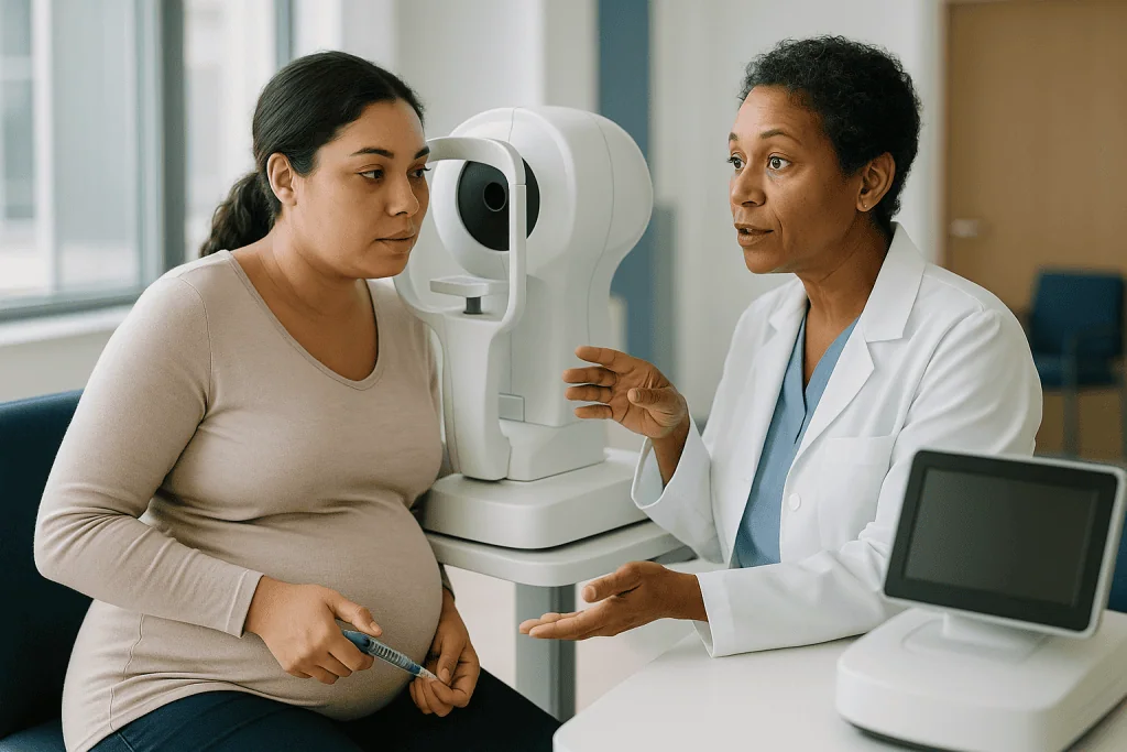
Years of high glucose stiffen and weaken capillary walls. Small outpouchings called microaneurysms form, like weak spots on a garden hose. Some leak fluid and fat into the surrounding retina. Others close off entirely, so patches of retina lose their blood supply. Eye specialists call this early phase non-proliferative diabetic retinopathy, meaning no new vessels have grown yet. Many people live with mild non-proliferative changes for years without any effect on vision.
Two developments turn a quiet problem into a sight-threatening one. The first is macular edema, swelling in the macula, the small central area of the retina responsible for reading and recognizing faces. The second is proliferative retinopathy, in which the starved retina releases a chemical signal called vascular endothelial growth factor, or VEGF, asking for new blood vessels. The new vessels that answer are brittle. They bleed into the clear gel that fills the eye, and the scar tissue they drag along can pull the retina out of position.
Pregnancy touches every step of that sequence. Higher blood volume raises pressure inside capillaries, encouraging leaks. Placental hormones and growth factors are, by design, pro-growth, and the retina does not know the difference. Rapid tightening of blood sugar, which is exactly what good pregnancy care asks for, can temporarily unsettle vessel function before it stabilizes. None of this guarantees that retinopathy will worsen. It explains why clinicians prefer to see the retina several times rather than guess.
Does pregnancy worsen diabetic retinopathy? What the evidence shows
The honest answer is: sometimes, in a minority, and most often in people whose retina already showed changes before conception. Mainstream guidance from the NHS and the Mayo Clinic lists pregnancy among the recognized risk factors for retinopathy getting worse, alongside long diabetes duration, poor glucose control, high blood pressure and high cholesterol. Those are the same risk factors that matter outside pregnancy; pregnancy simply adds another.
Long-term follow-up studies have consistently found a pattern that shapes modern screening. People who enter pregnancy with no retinopathy rarely develop sight-threatening disease during the nine months. People who enter with mild changes sometimes progress a stage. People who enter with moderate or severe non-proliferative disease are the group most likely to move toward new vessel growth, and they are the ones for whom the 16–20 week extra visit was designed.
Several specific drivers stand out in the research literature, and clinicians weigh each one when deciding how closely to watch:
- Starting glucose control: a high HbA1c (the three-month average blood sugar test) at conception raises the odds of progression.
- Speed of improvement: bringing glucose down quickly in early pregnancy has been linked to a short-term worsening that often settles later. Guidelines still ask for tight control, because the long-term benefit to mother and baby outweighs this temporary effect, but the eye team watches the retina while it happens.
- Blood pressure: hypertension before pregnancy, and pre-eclampsia (a pregnancy condition of high blood pressure with protein in the urine) during it, both add strain to retinal vessels.
- Duration: the longer someone has had diabetes, the more vessel damage has accumulated before the extra load of pregnancy arrives.
Precise percentages vary widely between studies depending on era, glucose control and the definition of “progression,” which is why this article does not quote a single headline number. The clinically useful message is stable across all of them: the retina at conception predicts the retina at delivery far better than any other factor.
Who needs extra eye checks in pregnancy, and who is usually asked to wait
The closer-spaced schedule is for people with diabetes that existed before the pregnancy began. Within that group, intensity scales with what the first examination finds.
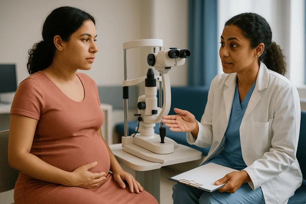
Someone with type 1 or type 2 diabetes and a clear retina at the first antenatal screening is usually asked to return once more, around 28 weeks, and then to rejoin routine screening after the birth. Someone whose first check shows any degree of retinopathy, even mild background changes, is typically offered the additional visit between 16 and 20 weeks, and the eye team may bring the 28-week check forward or add more if the picture is shifting.
People with moderate or severe non-proliferative disease, proliferative disease, or macular edema at the start of pregnancy move into a different category altogether. For them, care usually becomes a joint effort between the retinal specialist and the maternity diabetes team, with visits set individually rather than by the standard calendar. Some will be offered treatment during pregnancy; others will be watched closely with treatment held in reserve.
Who is usually asked to wait, meaning to follow the ordinary screening interval instead? Two groups. People with gestational diabetes, because the condition has not existed long enough to injure retinal vessels; NHS guidance explicitly notes they do not need pregnancy eye screening. And people with pre-existing diabetes who are planning a pregnancy but have not yet conceived, who are instead encouraged to have an up-to-date retinal examination as part of pre-conception care, so that any treatment can be completed beforehand.
One practical point that catches people out: screening in pregnancy uses dilating drops to widen the pupil. Those drops are considered acceptable in pregnancy, but they blur near vision for several hours. Arranging a lift home, or a companion, is worth doing before the first appointment rather than discovering the problem in the parking lot.
Diabetic retinopathy pregnancy screening: the typical timeline
Timelines below reflect NHS diabetic eye screening guidance for people with pre-existing diabetes. Every team adapts them to the individual, and a specialist can shorten any interval at any point.
| Stage of pregnancy | What usually happens | Why it is timed here |
|---|---|---|
| Before conception (planned pregnancy) | Up-to-date retinal examination; any needed treatment completed | Lets treatment finish before pregnancy adds vessel strain |
| Soon after the first antenatal appointment | Dilated retinal photographs or examination | Establishes the baseline that predicts the rest of pregnancy |
| 16–20 weeks | Extra check, only if retinopathy was found at the first visit | Catches early progression during the period of fastest glucose change |
| Around 28 weeks | Second routine check for everyone with pre-existing diabetes | Blood volume and hormonal effects peak in the third trimester |
| Third trimester to birth | Additional visits only if changes are progressing | Allows planning around delivery if treatment is needed |
| After the birth | Return to routine screening, or earlier follow-up if the eye team requests it | Changes that began in pregnancy can continue for months postpartum |
Reading the table, two features deserve a second look. The first is that the “extra” visit at 16–20 weeks is conditional. People with a clean baseline are not asked to attend it, which keeps the burden reasonable for the majority. The second is that the schedule does not end at delivery. The retina does not reset the day the baby is born, and the hormonal and circulatory changes of pregnancy unwind over weeks, so the team decides the first postpartum interval on the basis of the last pregnancy findings.
If a pregnancy was unplanned and the last eye screening was more than a year ago, the first antenatal check becomes doubly important: it is both the pregnancy baseline and the overdue routine exam. Telling the maternity team the date of your last screening at the booking visit lets them arrange it promptly.
What a diabetic eye exam during pregnancy involves
The examination itself is the same one used outside pregnancy. Nothing about it is unsafe for the baby, and it takes roughly half an hour including the wait for the drops to work.
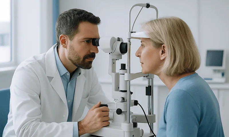
You will be asked to read a letter chart first, which records visual acuity, the standard measure of how sharply each eye sees. Then drops go in to dilate the pupil, so the examiner can see the whole retina rather than a keyhole view. The drops sting briefly and take fifteen to twenty minutes to work. Once the pupil is wide, a camera photographs the back of each eye, or an ophthalmologist examines it directly with a bright light and a hand-held lens. Some clinics add optical coherence tomography, or OCT, a painless scan that uses light to produce a cross-section of the retina and shows swelling in the macula that photographs can miss.
Photographs are graded according to a standard scale. In the NHS system, “R” grades describe background and proliferative retinopathy and “M” grades describe maculopathy, changes near the center of vision. Elsewhere, graders use the international scale of none, mild, moderate, severe non-proliferative, and proliferative. The letter or number on your result is what determines the next interval.
Afterward, expect blurred near vision and light sensitivity for four to six hours. Sunglasses help. Driving is not advised until the drops wear off. Breastfeeding is not relevant at this stage, but for postpartum checks the same drops are used routinely, and the clinician can discuss any concerns.
Results are usually sent to both the maternity team and the primary care clinician, which is what allows the joint planning described earlier. If the result letter arrives before you have heard from anyone, it is reasonable to ask the diabetes midwife to translate the grade into what it means for your own schedule.
What is the 4-2-1 rule in diabetic retinopathy?
The 4-2-1 rule is a shorthand that retinal specialists use to decide when non-proliferative retinopathy has become severe, meaning the eye is at high risk of moving on to new vessel growth. It comes from the grading system developed for the Early Treatment Diabetic Retinopathy Study, a landmark research program whose definitions still underpin most grading scales worldwide.
The retina is divided into four quadrants, like the face of a clock split into quarters. Severe non-proliferative retinopathy is diagnosed when any one of three findings is present:
- 4: extensive retinal hemorrhages (small bleeds) and microaneurysms in all four quadrants.
- 2: venous beading, where retinal veins take on a lumpy, sausage-like outline, in two or more quadrants.
- 1: intraretinal microvascular abnormalities, which are tangled, dilated capillaries that signal the retina is struggling for oxygen, in at least one quadrant.
Why does a counting rule matter to someone who is pregnant? Because the 4-2-1 threshold is the point at which clinicians stop simply watching and start discussing whether to treat before new vessels appear. Outside pregnancy, a person at this stage might be reviewed every few months. In pregnancy, where progression can be faster, reaching the threshold often triggers a more active plan: shorter intervals, closer liaison with the obstetric team, and sometimes laser treatment ahead of the third trimester.
The rule is a tool for graders, not a self-check. It cannot be applied without dilated examination and, usually, retinal photographs. Its value for a patient is in understanding the language on a results letter. If a report says “severe NPDR” or “pre-proliferative,” the 4-2-1 criteria are what the grader has applied, and asking which of the three findings was present is a perfectly reasonable question.
What are the signs of first-stage retinopathy? Usually none
People searching for the symptoms of early retinopathy are often hoping to monitor themselves at home between appointments. The most important fact about first-stage disease is that it produces nothing you can notice. Mild non-proliferative retinopathy sits in the retina silently, often for years. Vision stays sharp, colors stay true, and the eye feels entirely normal. This is why screening exists: there is no reliable warning that the retina has begun to change, and waiting for a symptom means waiting until damage has reached the macula or a vessel has bled.
The Mayo Clinic and the National Eye Institute make the same point in slightly different words. Both note that early diabetic retinopathy may cause no symptoms, and that by the time vision changes, the disease is usually well advanced. Pregnancy does not alter that silence; it only shortens the time it might take to end.
When symptoms do appear, they tend to belong to the later stages: blurring that comes and goes, dark strands or spots drifting across vision, a smudge in the center of what you are reading, or trouble with night vision. Fluctuating blur in pregnancy is also common for an entirely different reason, since fluid shifts can change the shape of the cornea and lens, and swings in blood sugar temporarily alter the lens as well. Distinguishing a harmless pregnancy blur from a retinal problem is not something anyone can do from the sofa, which is one more argument for keeping the scheduled examinations rather than relying on how the eyes feel.
The practical takeaway is reassuring in its way. Not noticing anything is the expected experience at every stage of a well-monitored pregnancy with diabetes. The job of noticing belongs to the camera and the grader. Your job is to turn up.
How to control diabetic retinopathy when you are pregnant
There is no treatment that reaches into the retina and repairs leaking capillaries. Control, in the sense people search for, means slowing the process by removing what drives it, and in pregnancy that comes down to three measurable things.
Blood glucose comes first. Both the NHS and the Mayo Clinic identify sustained high blood sugar as the strongest modifiable driver of retinopathy, and pregnancy guidance asks for tighter targets than usual to protect the baby. The paradox is that rapid improvement in early pregnancy can briefly unsettle the retina. Guidelines do not ask anyone to loosen control because of this; instead, they pair tight glucose management with the extra eye visits so that any transient worsening is seen and, if necessary, treated. The prescribing clinician sets the targets and adjusts insulin or other medicines; the eye team watches the result.
Blood pressure comes second. Pre-existing hypertension and pre-eclampsia both increase strain on retinal vessels. Routine antenatal blood pressure checks therefore do double duty, protecting the kidneys and placenta while also protecting the retina. Treatment choices for blood pressure in pregnancy sit with the obstetric team, who have a short list of medicines considered appropriate.
The third factor is attendance. Retinopathy that is seen can be managed; retinopathy that is missed cannot. Keeping each scheduled examination, and telling the midwife about any change in vision between them, is the single most effective action a patient controls directly.
Smoking cessation and attention to cholesterol complete the list, as they do outside pregnancy. What does not help, despite persistent folklore, is any supplement, eye drop or diet marketed for retinal health. No vitamin product has been shown to alter the course of diabetic retinopathy, and in pregnancy, unlisted supplements carry their own risks. Anything beyond standard antenatal vitamins is worth running past the maternity team before starting.
Treatment options for mild and more advanced diabetic retinopathy in pregnancy
For mild non-proliferative retinopathy, the treatment is observation. That is not a polite way of saying nothing is done. Observation during pregnancy means the extra 16–20 week visit, attention to glucose and blood pressure, and a lower threshold for repeat photographs if anything shifts. Most people with mild changes need nothing more, in pregnancy or afterward.
Moderate and severe non-proliferative disease, proliferative disease and macular edema bring active options into the discussion. Laser photocoagulation uses a focused light beam to seal leaking vessels or, in the panretinal form, to treat the oxygen-starved outer retina so that it stops sending out the VEGF signal that drives new vessel growth. Laser has a long track record, is delivered as an outpatient procedure in several short sessions, and is generally considered acceptable during pregnancy because its effect stays within the eye. The NHS describes it as the established treatment for new vessel growth; in pregnancy, specialists sometimes offer it earlier than they would otherwise, precisely because they expect faster progression.
Anti-VEGF injections, medicines placed into the gel of the eye to block the growth signal directly, are a mainstay of macular edema treatment outside pregnancy. Their use during pregnancy is more cautious. VEGF plays a role in placental and fetal development, small amounts of the medicine enter the bloodstream, and safety data in pregnancy are limited. Decisions about whether and when to use them are individual, weighing the threat to vision against the uncertainty, and belong to the retinal specialist and obstetrician together.
Steroid implants and surgery for bleeding or retinal detachment exist further along the pathway and are rarely needed during pregnancy itself.
Timing can also be a treatment decision. Where disease is advanced, teams sometimes discuss delivery planning with the retina in mind, because the straining of a long second stage of labor has historically been a concern in eyes with fragile new vessels. Current practice is individualized; it is a question to raise, not an assumption to make.
Retinopathy after pregnancy: what the following weeks and months look like
Birth changes the body’s hormonal and circulatory setting within days, but the retina responds more slowly. Changes that began during pregnancy can continue to develop over the following months before stabilizing, which is why the NHS asks people with retinopathy to continue follow-up after delivery rather than wait a full year for the next routine screening.
For the majority, who had no retinopathy or only mild changes, the postpartum story is uneventful. The eye team usually returns them to standard screening, with the first interval set according to the last pregnancy examination. Many of the mild changes that appeared during pregnancy regress on their own as blood volume and hormone levels fall, a pattern described in long-term follow-up research and reflected in guideline language about pregnancy-related worsening being frequently temporary.
Those who developed moderate or severe disease, or who had laser during pregnancy, are usually seen within a few months of the birth and then at intervals the specialist sets. If anti-VEGF treatment was deferred because of pregnancy, the decision is revisited at this point, with breastfeeding factored into the discussion.
Two practical realities of the postpartum period affect eye care in ways nobody warns you about. Sleep deprivation and the demands of a newborn make it easy to miss appointments, and glucose control often drifts in the early weeks as routines collapse and insulin needs change sharply after delivery. Both can affect the retina. Building the postpartum eye check into the same planning as the six-week maternal review, and asking a partner or relative to keep the date, is a small act with a real payoff.
For people planning a further pregnancy, the postpartum examination also serves as the pre-conception baseline for the next one. Completing any treatment the specialist recommends before conceiving again is the single most useful preparation, and the interval between pregnancies is the easiest time to do it.
What people often get wrong
Misunderstandings about diabetic retinopathy in pregnancy are common enough that it is worth correcting the main ones directly.
“Having retinopathy means I should not get pregnant.” Existing retinopathy is not a reason to avoid pregnancy. It is a reason to see the eye team before conceiving, complete any treatment, and expect closer monitoring. National guidance frames it exactly that way.
“Gestational diabetes needs the same eye checks.” It does not. Gestational diabetes has not existed long enough to damage retinal vessels, and the NHS specifically excludes it from pregnancy eye screening. People who later go on to develop type 2 diabetes join routine screening at that point.
“If my vision is fine, my retina is fine.” Early and even moderate retinopathy is silent. The absence of symptoms says almost nothing about the state of the retina, which is the entire reason screening is scheduled rather than symptom-driven.
“Tight glucose control is bad for my eyes, so I should ease off.” Rapid tightening can cause a short-term wobble, but guidelines still call for tight control because the long-term benefit for eyes, kidneys and baby is far larger. The answer is more eye checks, never looser glucose targets, and any change to a regimen belongs to the prescribing clinician.
“Laser is dangerous in pregnancy.” Laser treatment acts locally within the eye and is generally regarded as acceptable during pregnancy. The treatment with real uncertainty is anti-VEGF injection, and even there the decision is individual rather than a blanket prohibition.
“Dilating drops will harm the baby.” Standard dilating drops are used routinely in pregnancy screening. The main inconvenience is several hours of blurred near vision.
“Everything goes back to normal at delivery.” The retina lags the body. Follow-up after the birth is part of the plan, not an optional extra.
Questions to ask your care team
Appointments in pregnancy are busy and short. Having a few questions ready turns a results letter into a plan you understand. These are the ones that tend to matter most.
- What grade of retinopathy, if any, did my first pregnancy screening show, and in which eye?
- Based on that result, when is my next eye check, and will I need the 16–20 week visit?
- Who is coordinating between the eye clinic, the diabetes midwife and my obstetrician, and how do results get shared?
- Given my HbA1c at booking, how quickly are we aiming to bring glucose down, and does that change how often my eyes are checked?
- What is my blood pressure target in this pregnancy, and what would prompt a change in plan?
- If changes are found, what would be the likely next step: more frequent photographs, laser, or referral to a retinal specialist?
- Are there any treatments you would normally offer that are being deferred because I am pregnant, and when would they be reconsidered?
- Does my retinal picture have any bearing on plans for delivery?
- What vision changes should prompt me to call before my next scheduled visit, and which number do I use?
- When should my first eye check after the birth take place, and will it happen automatically or do I need to request it?
- If I plan another pregnancy, what should be done to my eyes beforehand?
Writing the answers down, or asking for them in the letter that goes to your primary care clinician, means they survive the fog of the third trimester and the newborn weeks. If different members of the team give answers that seem to conflict, say so; the resolution usually lies in a quick message between clinicians rather than in the patient having to choose.
When to call your doctor
Scheduled screening catches the slow changes. Sudden ones need a phone call the same day, to the maternity unit, the diabetes team or the eye clinic, whichever your care plan names as the first contact. Do not wait for the next appointment if you notice any of the following in one or both eyes:
- A sudden shower of dark spots, strands or “cobwebs” drifting across your vision, which can signal bleeding from new vessels into the gel of the eye.
- A curtain or shadow spreading across part of your visual field, a possible sign of retinal detachment.
- A rapid drop in vision, or distortion that makes straight lines look wavy or a blank patch in the center of what you are reading.
- Flashes of light that persist, particularly with new floaters.
- Eye pain with blurred vision, or a red, painful eye.
Some of these, especially distortion and central blur, can also overlap with symptoms of pre-eclampsia, which may cause visual disturbance alongside headache, swelling of the face and hands, and pain under the ribs. Pre-eclampsia is a maternity emergency in its own right, so any new visual symptom in the second half of pregnancy should be reported promptly to the maternity team even if the eyes themselves feel normal.
Mild, brief blurring that comes and goes, especially around meals or after a glucose swing, is common in pregnancy and usually reflects temporary changes in the lens rather than the retina. It is still worth mentioning at the next contact. The rule of thumb is simple: gradual and fluctuating can wait for the scheduled visit; sudden, persistent or worsening cannot.
After the birth, the same red flags apply, with one addition. If a postpartum eye appointment has not been arranged within the timeframe the specialist described, call and ask rather than assuming it will arrive. The team makes every decision about what to do next; your part is to make sure the retina is seen.
Frequently asked questions
Does pregnancy worsen diabetic retinopathy for everyone with diabetes?
No. Progression during pregnancy affects a minority, and the strongest predictor is the state of the retina at conception. People with no retinopathy at the first antenatal screening rarely develop sight-threatening disease in nine months. Those with moderate or severe changes at the start are the group most likely to progress, which is why their checks are spaced closest together and coordinated with the maternity team.
How often is a diabetic eye exam during pregnancy needed?
For pre-existing type 1 or type 2 diabetes, NHS guidance asks for a dilated retinal check soon after the first antenatal appointment and another around 28 weeks. If the first check shows any retinopathy, an extra visit between 16 and 20 weeks is added. Specialists can shorten any interval if changes are progressing, and follow-up continues after the birth.
What is the 4-2-1 rule in diabetic retinopathy?
The 4-2-1 rule identifies severe non-proliferative retinopathy, the stage just before new vessel growth. It is met when retinal hemorrhages appear in all four quadrants, venous beading in two or more quadrants, or intraretinal microvascular abnormalities in at least one quadrant. It is applied by graders using dilated examination or photographs, and in pregnancy, meeting it usually prompts closer monitoring or earlier treatment.
How do you control diabetic retinopathy during pregnancy?
Control means slowing the process rather than reversing it: keeping blood glucose within the targets set by the diabetes team, keeping blood pressure in range, not smoking, and attending every scheduled eye check so that changes are seen early. Any change to glucose or blood pressure medicines belongs to the prescribing clinician. No supplement or over-the-counter product has been shown to alter the course of retinopathy.
What are the treatment options for mild diabetic retinopathy in pregnancy?
Mild non-proliferative retinopathy is managed by observation: the extra 16–20 week visit, attention to glucose and blood pressure, and repeat photographs if anything shifts. Active treatment such as laser is reserved for more advanced disease or macular swelling. Most people with mild changes need nothing further during pregnancy, and many of those changes settle on their own after the birth.
What are the signs and symptoms of first-stage retinopathy?
Usually there are none. Early non-proliferative retinopathy causes no pain, no blur and no visible change, which is why it is found by screening rather than by symptoms. Vision changes such as floaters, central distortion or a dark shadow tend to appear only at later stages and need same-day contact with your care team rather than waiting for the next appointment.
Is laser treatment safe for the baby during pregnancy?
Laser photocoagulation acts locally within the eye and is generally regarded as acceptable during pregnancy. It is the established treatment for new vessel growth and is sometimes offered earlier in pregnant patients because progression can be faster. The decision to treat, and its timing, rests with the retinal specialist working alongside the obstetric team.
Why are anti-VEGF injections approached cautiously in pregnancy?
Anti-VEGF medicines block the growth signal that drives new vessels and macular swelling. Because VEGF also has a role in placental and fetal development, small amounts of the medicine reach the bloodstream, and pregnancy safety data are limited, specialists weigh their use individually. Treatment may be deferred until after delivery or offered where the threat to sight is judged to outweigh the uncertainty.
What happens to retinopathy after pregnancy ends?
The retina responds more slowly than the body. Changes that began in pregnancy can keep developing for months, so the eye team arranges follow-up after the birth rather than waiting for the next routine screening. Mild changes often regress as hormone levels and blood volume return to normal, while more advanced disease is reviewed within a few months and any deferred treatment reconsidered.
Does gestational diabetes need diabetic retinopathy pregnancy screening?
No. Gestational diabetes appears during pregnancy and has not existed long enough to damage retinal blood vessels, so NHS guidance excludes it from pregnancy eye screening. If someone later develops type 2 diabetes, they join the routine screening program at that point. Pre-existing type 1 or type 2 diabetes is the group the closer-spaced pregnancy schedule is designed for.
References
- NHS: Diabetic eye screening
- NHS: Diabetic retinopathy
- National Eye Institute (NIH): Diabetic retinopathy
- MedlinePlus: Diabetic eye problems
This article is for general information only and is not a substitute for professional medical advice. Please consult a qualified doctor about your individual situation.

More from the Blog

Farsightedness After 40: Why Reading Gets Harder and How Treatment Adapts
Farsightedness after 40 is most often presbyopia: the eye's natural lens gradually stiffens with age, so it can no longer bend enough to focus…
What Happens During Glaucoma Laser Treatment: Numbing Drops, the Lens and the Pressure Check
During a glaucoma laser treatment procedure, an eye specialist places numbing drops in the eye, rests a small contact lens on the surface to…

LASEK vs PRK: How the Two Surface Laser Techniques Differ and Why Surgeons Pick One
LASEK and PRK are both surface laser procedures that reshape the cornea without cutting a flap; the difference is what happens to the thin…
Is ILASIK Right for You? Corneal Thickness, Eye Pressure and Contact Lens Breaks
ILASIK candidate requirements generally include being at least 18 with a prescription that has been stable for a year or more, corneas thick and…

What Glaucoma Surgery Can and Cannot Do: Protecting the Vision You Still Have
Glaucoma surgery lowers pressure inside the eye to slow or stop further damage to the optic nerve. It cannot restore vision already lost, because…
Intraocular Lens Recovery: Eye Drops, Lifting Limits and When Vision Settles
Most people recover from intraocular lens (IOL) surgery over about 4 to 8 weeks, according to the NHS and Mayo Clinic, though everyday vision…