

Endoscopic Pituitary Tumor Surgery Explained: The Transsphenoidal Route and What It Avoids
Key Takeaways
- Transsphenoidal surgery reaches the pituitary through the nose and the sphenoid sinus, a natural air pocket directly beneath the gland, so the skull is not opened and the brain is not handled.
- The route spares the brain but not the neighbors: the carotid arteries and optic nerves sit millimeters away, and opening the dura creates the risk of a cerebrospinal fluid leak.
- Most pituitary tumors are noncancerous adenomas; surgery aims to relieve pressure on vision and normalize hormones rather than to treat a spreading cancer.
- Prolactin-producing tumors are usually managed first with dopamine agonist medication, while growth-hormone and cortisol-producing tumors and vision-threatening tumors are usually offered surgery first.
- Post-operative care revolves around fluid balance and sodium, because temporary diabetes insipidus and delayed low sodium are recognized complications in the first two weeks.
- Clear watery nasal drainage, fever with a stiff neck, extreme thirst with pale urine, and new vision change are red flags that warrant same-day medical contact.
Transsphenoidal surgery is an operation that reaches the pituitary gland through the nose and the sphenoid sinus, an air pocket behind the nasal cavity that sits directly beneath the gland. Because the route follows a natural corridor, surgeons avoid opening the skull or moving the brain aside. It is the usual operation for pituitary tumors that need removal, and the treating team decides whether it fits an individual case.
The scan report uses a word most people have never had to spell: sella. Somewhere behind the bridge of the nose, on a bony saddle no wider than a fingertip, a gland the size of a pea has grown a lump. The neurosurgeon says the operation goes “through the nose,” and the room goes quiet while everyone tries to picture how that could possibly work.
It works because of an accident of anatomy. The pituitary hangs from the base of the brain, but its floor is separated from a hollow, air-filled bone by a shell often thinner than a credit card. Get into that hollow from the nostril, and the gland is right there, without a single fold of brain in the way.
This is transsphenoidal surgery explained the way patients ask about it: what the surgeon actually does, what the route spares, how serious the operation is, what the nose feels like afterward, and which signs in the weeks that follow deserve a phone call rather than patience.
Transsphenoidal surgery explained: the route through the nose, step by step
Start with the name. “Trans” means across, and the sphenoid is a butterfly-shaped bone at the center of the skull base that contains an air pocket called the sphenoid sinus. The pituitary sits in a shallow bony cup on top of that sinus, the sella turcica, a Latin phrase for “Turkish saddle” that describes its shape. Transsphenoidal surgery simply means crossing that sinus to reach the saddle.
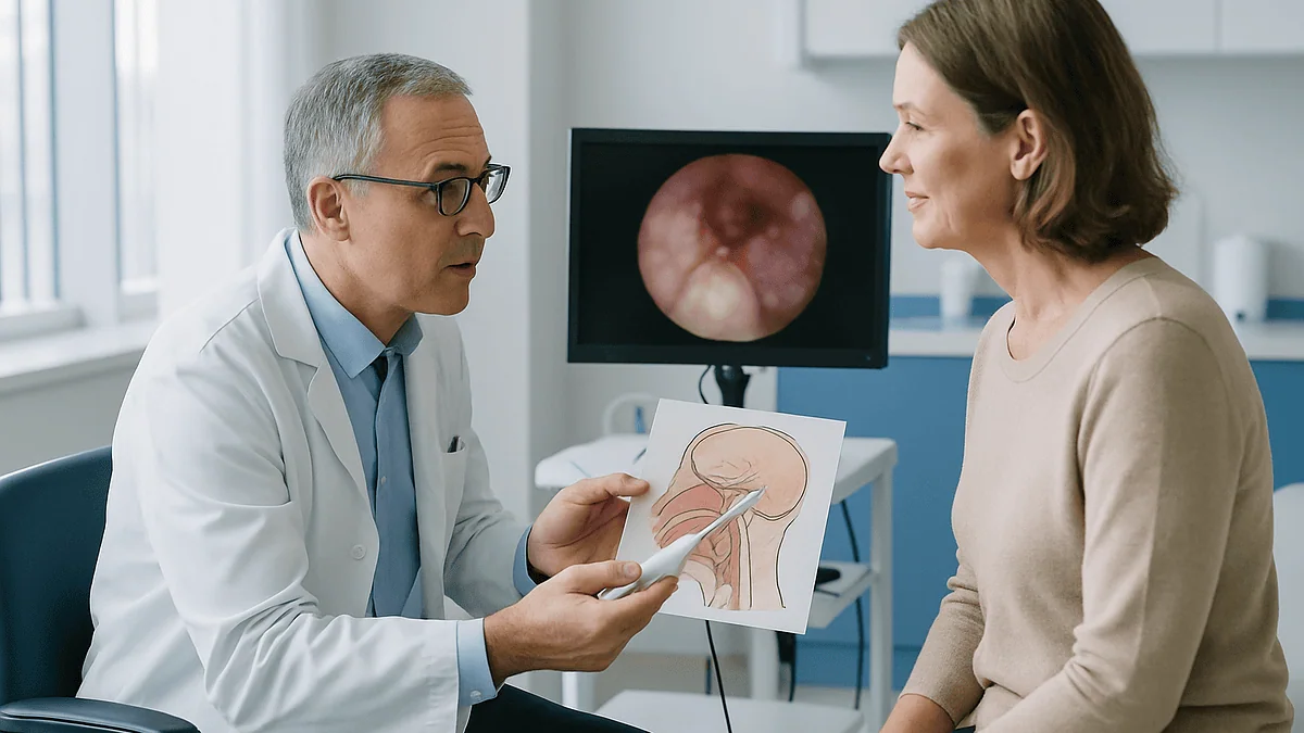
Under general anesthesia, the surgical team passes a thin lighted camera, an endoscope, into one nostril. No incision is made on the face. The camera travels back along the nasal passage to the natural opening of the sphenoid sinus, which is widened so instruments can follow. Some teams also open a small window in the septum, the wall dividing the nostrils, to work from both sides.
At the back of the sinus lies the floor of the sella. A small piece of that bone is removed, and beneath it is the dura, the tough membrane that lines the brain and its outposts. Once the dura is opened, the tumor is directly in view.
Most pituitary tumors are adenomas, meaning noncancerous growths that arise from the gland’s own hormone-making cells, and many are soft enough to be removed with gentle suction and small ring-shaped instruments. The surgeon works from within the tumor outward, trying to spare the normal gland pressed around it. Once the space is cleared, the opening is sealed, sometimes with a graft of the patient’s own tissue, and the nose is tidied. The whole thing typically takes a few hours according to Johns Hopkins and Mayo Clinic descriptions, though duration depends on the tumor.
What the transsphenoidal route avoids, and why that matters so much
The alternative way to reach a pituitary tumor is a craniotomy, an operation that removes a window of skull so the surgeon can approach from above. It remains a valid and sometimes necessary choice. Understanding what it involves explains why the nasal corridor became the default.

A craniotomy reaches the pituitary by passing alongside the frontal lobe and beneath it. To see the gland, the brain must be gently lifted and held, and the optic nerves, which run directly above the pituitary, sit between the surgeon and the target. The route through the nose comes from below instead, so the optic nerves are above the field rather than in front of it, and the brain itself is never touched.
Three things follow from that. First, there is no scalp incision and no bone flap, so there is no visible scar and no healing skull. Second, the risks that come from handling brain tissue, such as seizures and swelling, are largely sidestepped. Third, recovery tends to be shorter and less demanding, which Mayo Clinic and Cleveland Clinic both list among the reasons the endoscopic transsphenoidal approach is used for most pituitary tumors.
What the route does not avoid is worth stating plainly. It still opens the dura and enters the space where cerebrospinal fluid circulates. It still asks the surgeon to work millimeters from the carotid arteries, which run in the cavernous sinuses on either side of the gland. Avoiding the brain is not the same as avoiding risk; it is a trade that moves the risk to a smaller, more predictable set of structures. The next sections describe that set honestly.
Why a pea-sized gland causes so much trouble when it grows
The pituitary is small, but it behaves like the control tower for most of the body’s hormone traffic. MedlinePlus describes it as pea-sized, and from that footprint it releases signals that direct the thyroid, the adrenal glands, the ovaries and testes, growth, milk production, and the kidneys’ handling of water.
A tumor here causes problems in two distinct ways, and knowing which applies to you shapes every decision that follows. The first is pressure. The sella is a rigid cup with the optic nerves crossing just above it. A tumor that grows upward pushes into that crossing and typically erodes the outer edges of vision first, so people notice they keep bumping into door frames or missing cars in the side mirror before they notice anything wrong with reading. Headache is common but nonspecific. A large tumor can also squash the normal gland, leading to hormone shortfalls.
The second problem is overproduction. Some adenomas keep making the hormone their parent cells were designed for, but without any off switch. A prolactin-secreting tumor can stop periods or cause milk production unrelated to pregnancy. A growth-hormone tumor causes acromegaly, with gradual enlargement of hands, feet and facial features, as the NHS describes. A tumor driving cortisol production causes Cushing’s disease. Tumors that make no active hormone are called non-functioning, and they tend to be found late, when they are large enough to press on something.
According to Mayo Clinic, the great majority of pituitary tumors are noncancerous and do not spread elsewhere. That is genuinely reassuring, and it also explains why the goal of surgery is framed around relieving pressure and normalizing hormones rather than around survival.
Endoscopic versus microscopic: what the word “endoscopic” actually changes
For decades the standard version of this operation used an operating microscope. The surgeon looked down a narrow metal speculum held in the nostril, and light and vision traveled in a straight line to the sella. It worked well, and some surgeons still prefer it for particular cases.
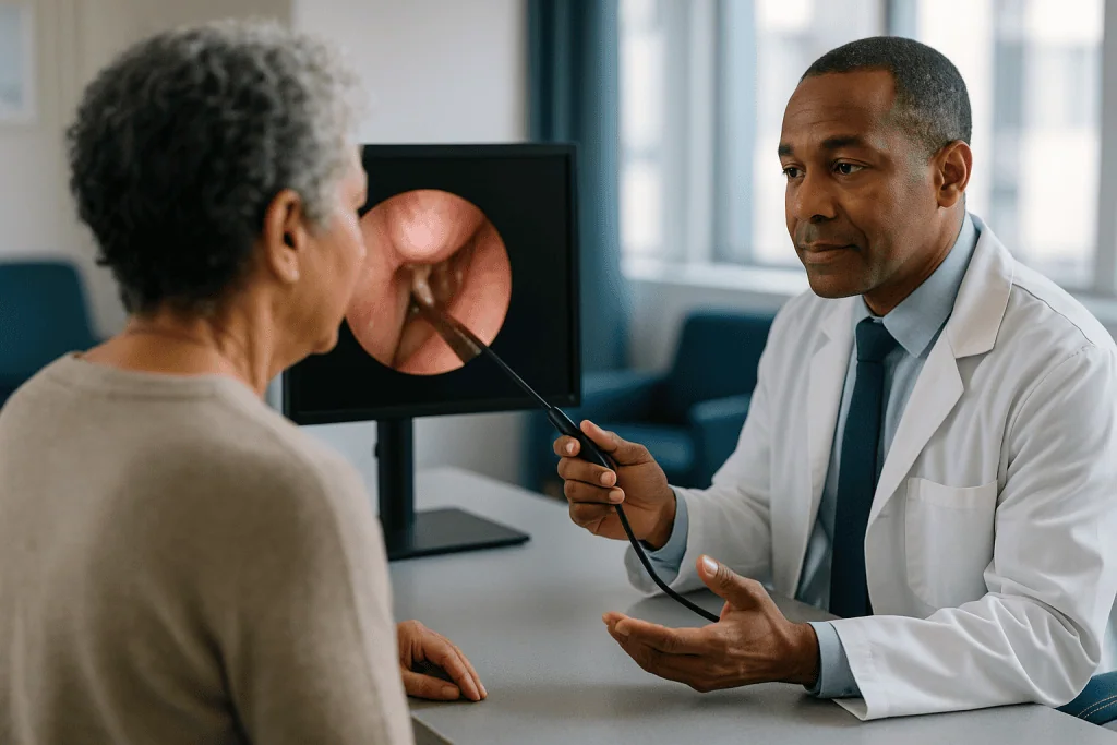
An endoscope is different in one important way: the camera and light source sit at the tip of the instrument, inside the sinus, rather than outside the nose. The picture appears on a screen, and the view is wide-angled instead of tunnel-shaped. Angled lenses let the team look around corners, into the folds of the sella where tumor can hide beside the carotid artery or tucked under the diaphragm of the gland.
The practical differences, drawn from Johns Hopkins and Cleveland Clinic descriptions, are these. The endoscopic approach usually needs less disruption of the nasal septum because no rigid speculum has to be wedged in place. The surgeon can see the edges of the tumor cavity directly rather than inferring them. Two surgeons often work together, one guiding the camera and one operating, which is why many centers pair a neurosurgeon with an ear, nose and throat specialist.
What the evidence does not support is a claim that one technique is universally superior. Outcomes depend heavily on the tumor’s size, consistency and whether it has spread sideways into the cavernous sinus, where no instrument can safely follow. A good conversation with the treating team is less about microscope versus endoscope and more about what the scan shows and what a realistic goal for the operation looks like in your case.
Who is usually offered surgery, and who is usually asked to wait
Not every pituitary tumor needs an operation, and the clearest example of that is also the most common hormone-producing type. Prolactinomas, tumors that release prolactin, generally respond to a class of medicines called dopamine agonists, which mimic the brain chemical that normally suppresses prolactin. Mayo Clinic and MedlinePlus both describe medication as the usual first approach for these tumors, with surgery reserved for people who cannot tolerate or do not respond to the medicine. Nobody should start or stop such a medicine without the prescribing clinician, but knowing that a pill is often the first-line option changes the conversation.
Surgery is usually the first-line treatment when a tumor is pressing on the optic nerves and threatening sight, when it is producing growth hormone (acromegaly) or driving Cushing’s disease, or when a non-functioning tumor is large and growing. The NHS notes that for acromegaly, surgery to remove the tumor is generally the recommended first step because normalizing growth hormone by medication alone is harder. For Cushing’s disease, removing the tumor is likewise the usual aim.
People who are commonly asked to wait fall into a few groups. Small non-functioning adenomas discovered by chance, sometimes on a scan taken for headaches or after a car accident, are often simply monitored with periodic imaging and hormone tests, because many never grow. A tumor that has wrapped around the carotid artery may be partly removed and the rest treated with radiation rather than chased surgically. And anyone with poorly controlled cortisol excess or untreated hormone deficiency may need those corrected first, since operating on someone with low cortisol carries its own dangers.
These are typical patterns, not rules. The endocrinologist and neurosurgeon weigh them together.
How the options compare: surgery, medicine, radiation and watchful waiting
Patients often meet these options one at a time, from different specialists, without anyone laying them side by side. The table below does that in neutral terms. It is not a ranking; each row suits a different situation, and many people end up with a combination over the years.
| Approach | What it does | Usually considered when | Main trade-offs |
|---|---|---|---|
| Endoscopic transsphenoidal surgery | Removes the tumor through the nose and sphenoid sinus | Vision is threatened; growth-hormone or cortisol excess; large or growing tumor | Fluid leak, temporary or lasting hormone changes, nasal recovery |
| Craniotomy | Removes the tumor through an opening in the skull | Tumor extends far above or beside the sella, out of reach of the nasal corridor | Brain handling, longer recovery, visible incision |
| Medication | Suppresses hormone output or shrinks certain tumors | Prolactinoma; residual hormone excess after surgery; when surgery is unsuitable | Ongoing treatment, side effects, does not remove the tumor |
| Radiation therapy | Slows or stops growth of remaining tumor over months to years | Residual or recurrent tumor; tumor in the cavernous sinus | Slow effect, gradual pituitary hormone loss over time |
| Observation | Regular scans and hormone checks | Small, non-functioning, symptom-free tumor | Requires follow-up discipline; some tumors eventually grow |
Two things stand out. Radiation, which Mayo Clinic describes as an option for tumor that cannot be fully removed, is slow by design and tends to reduce pituitary function gradually, so it is rarely used where quick relief of pressure is needed. And medication does not make a tumor disappear in most cases; it controls it. That is why surgery remains central for the tumors that cause pressure or dangerous hormone excess, and why the treating team, rather than the table, makes the final call.
How serious is transsphenoidal surgery? An honest account of the risks
It is brain-adjacent surgery under general anesthesia, so it is serious. It is also one of the more routine operations in neurosurgery, performed through a natural corridor with the brain untouched. Both statements are true, and the risks deserve to be named rather than softened.
The most talked-about complication is a cerebrospinal fluid leak. Cerebrospinal fluid, or CSF, is the clear liquid that cushions the brain, and once the dura is opened it can seep through the repair and drip from the nose. Cleveland Clinic and Johns Hopkins both list this as a recognized risk. Small leaks may seal on their own; others need a return to the operating room to reinforce the closure. An untreated leak matters because it gives bacteria a path to the lining of the brain, which is why fever and stiff neck after this surgery are never to be waited out.
The second group of risks involves hormones. Handling the gland can disrupt its water-balance hormone, causing diabetes insipidus, a condition in which the kidneys pass large volumes of dilute urine and thirst becomes relentless. It is unrelated to blood sugar despite the name. Mayo Clinic notes this is often temporary. The opposite problem, a drop in blood sodium, can also occur in the first couple of weeks. And any of the pituitary’s other hormones can fall, temporarily or permanently, a state called hypopituitarism that may need lifelong replacement.
Rarer but graver events include bleeding from the carotid artery, injury to the optic nerves, stroke and infection. Sinus problems, reduced smell and nasal crusting are more common and usually settle. How these odds apply to one person depends on tumor size and location, previous surgery and general health, which is exactly what the pre-operative consultation exists to weigh.
Nose packing after pituitary surgery: what the nose is really like afterward
People searching for this operation ask about the nose more than about the brain, and fairly so. The brain is asleep for the surgery; the nose is what you wake up with.
Practice varies. Some teams place soft dissolvable material or a small sponge in the nasal passage to control oozing and hold the sinus repair in place. Others use no packing at all, or a light dressing removed within a day or two, according to Johns Hopkins descriptions of the endoscopic technique. If a fat or tissue graft was taken from the abdomen or thigh to seal a fluid leak, there will be a small incision there too, and many patients report that spot is sorer than the nose.
The first week typically feels like a heavy cold that will not clear. Breathing through the nose is limited, the sense of smell may be dulled, and there is often a bloody or straw-colored discharge that gradually thins. Crusting inside the nose is normal and can persist for weeks while the lining heals. Cleveland Clinic notes that nasal congestion and stuffiness commonly last for several weeks after this surgery.
What helps, in the guidance most teams give: saline rinses or sprays as instructed to keep the passages moist, sleeping with the head raised, and humidified air. What hurts: picking at crusts, forceful sniffing, and anything that pushes air or pressure through the healing sinus, which is the subject of the “what not to do” section below.
Most people are surprised that the nose recovers as fully as it does. Septal perforation and lasting nasal deformity are uncommon with the endoscopic approach because far less of the internal architecture is disturbed than in older techniques. Lasting change in smell, though, is a real possibility and worth asking about beforehand.
Transsphenoidal surgery recovery time: the first days in the hospital
Recovery from this operation is measured less by pain than by fluid and hormones. Pain is usually moderate and managed with medicines chosen by the team. The real work of the first days happens on the nursing chart.
Every trip to the bathroom is measured. Nurses track how much you drink and how much urine you pass, because a sudden surge of pale urine with intense thirst is the signature of diabetes insipidus, the water-balance problem described earlier. Blood sodium is checked regularly, since it can swing high if too much water is lost or low if the body retains too much. Mayo Clinic and Cleveland Clinic both describe these hormone and electrolyte checks as standard after pituitary surgery. If diabetes insipidus does appear, a synthetic version of the missing hormone can be given by the team while the gland recovers.
Cortisol is the other headline. The pituitary tells the adrenal glands to make cortisol, the stress hormone the body cannot go without. Many teams give a steroid cover around the time of surgery and then measure morning cortisol to decide whether it can be stopped or needs to continue. This is a clinician’s decision at every step; never adjust a steroid on your own.
Vision is checked, especially if the tumor was pressing on the optic nerves. Some people notice the edges of their world return within days. Others improve over months, and some changes are permanent.
Hospital stays are typically short, on the order of a few days according to Johns Hopkins, provided sodium and fluid balance are stable and there is no sign of a fluid leak. Discharge usually comes with specific instructions about fluids, activity and a follow-up blood test, because the second week carries its own risk, described next.
What not to do after transsphenoidal surgery: the weeks at home
The repair at the back of the sinus is a small patch of tissue and glue holding back cerebrospinal fluid. Anything that spikes pressure inside the head pushes on that patch from behind; anything that spikes pressure in the nose pushes from the front. The home rules exist to protect it while it knits.
- Do not blow your nose. Dab or let it drip until the team clears you, which is usually a matter of weeks.
- Sneeze with your mouth open. It sounds undignified and it diverts the pressure away from the sinus.
- Avoid straining, whether lifting anything heavy, bending with effort, or pushing on the toilet. Ask about a stool softener if constipation is a problem.
- Do not drink through a straw or use a CPAP machine unless specifically told it is safe, because both force air or suction through the nasal passages.
- Skip swimming and flying until the team gives the go-ahead; pressure changes and water in the nose are both unwelcome early on.
Fluids need a middle path. Because sodium can fall in the first couple of weeks as the gland readjusts, some teams ask patients to keep fluid intake moderate rather than “flushing the system,” and to have a sodium blood test around the end of the first or second week. Cleveland Clinic and Johns Hopkins both flag delayed low sodium as a recognized post-operative issue. Follow the specific instruction you were given rather than general advice.
Fatigue is the complaint that surprises people most. Hormone levels are settling, sleep is disrupted by a blocked nose, and the body has had a general anesthetic. Cleveland Clinic suggests full recovery commonly takes several weeks, with return to desk work often sooner than return to heavy exertion. Walking daily is encouraged; heroics are not.
Hormones after surgery: replacement, testing and the long follow-up
Surgery ends in a few hours. Endocrine follow-up can last a lifetime, and understanding why prevents the sense of being abandoned once the surgical wounds heal.
In the first weeks, the questions are about function lost. If cortisol production has not recovered, a replacement glucocorticoid, the class of steroid that stands in for the body’s own cortisol, is continued and reviewed. Thyroid hormone is checked, since a sluggish pituitary can starve the thyroid of its signal, and levothyroxine, the synthetic thyroid hormone, may be needed. Sex hormones, growth hormone and the water-balance hormone are each assessed in turn. MedlinePlus and Mayo Clinic describe this pattern of replacement for deficient hormones after pituitary treatment. Every decision about whether to start, continue or stop any of these rests with the endocrinologist; the timelines vary widely between people.
For hormone-producing tumors, the questions are about excess controlled. After surgery for acromegaly, growth hormone and a related marker called IGF-1 are measured over subsequent months to judge whether the tumor’s output has normalized. After surgery for Cushing’s disease, a very low cortisol in the first days is often taken as an encouraging sign, though it also means replacement is needed until the body’s own axis wakes up, which the NHS notes can take many months.
Imaging follows. A first post-operative MRI is usually delayed until swelling and packing material have settled, typically within the first several months according to Johns Hopkins guidance, so that scar is not mistaken for tumor. After that, scans are spaced out over years. Some tumors leave a remnant that is simply watched. Others regrow slowly and are treated again with surgery, medication or radiation. The follow-up schedule is individual, and it is a fair question to ask what yours will look like.
What people often get wrong about pituitary tumor surgery
Misconceptions cluster around this operation because the anatomy is unfamiliar and the word “tumor” carries weight it does not always deserve here.
“A pituitary tumor is brain cancer.” The pituitary is not brain tissue, and Mayo Clinic notes that the vast majority of these tumors are noncancerous adenomas that do not spread. Cancer of the pituitary is rare. The concern is pressure and hormones, not metastasis.
“Surgery through the nose means it is minor.” No incision on the face does not mean no risk. The corridor passes within millimeters of the carotid arteries and optic nerves and opens the lining of the brain. It is less invasive than a craniotomy, not trivial.
“Once the tumor is out, the hormones fix themselves.” Sometimes they do. Often the gland needs weeks or months to recover, and some people need permanent replacement of one or more hormones. Others still have residual hormone excess that needs medication or radiation. Endocrine follow-up is part of the treatment, not an afterthought.
“Everyone gets diabetes insipidus and it is permanent.” It is a recognized risk, and in most cases it is temporary, as Mayo Clinic describes. Persistent diabetes insipidus is the less common outcome.
“Vision comes back the moment the pressure is off.” Some people notice improvement within days. For others the optic nerves recover slowly over months, and long-standing compression may leave permanent loss. Duration of pressure before surgery matters more than the operation itself.
“Medicine can always replace surgery.” For prolactinomas, medication usually comes first. For growth-hormone and cortisol-producing tumors, and for anything pressing on vision, the NHS and Mayo Clinic describe surgery as the usual first step. The right sequence depends on the tumor type, which is why a diagnosis comes before a treatment plan.
Questions to ask your care team before endoscopic pituitary surgery
A good consultation leaves you knowing not just what will be done but why, and what the realistic goal is. These questions tend to open the most useful conversations. Bring someone to take notes; hormone talk is dense.
- What type of pituitary tumor do I have, and is it producing a hormone? The answer determines whether surgery, medicine or observation comes first.
- Is the aim to remove all of it, or to remove enough to relieve pressure and control hormones? Tumors in the cavernous sinus often cannot be fully removed safely, and knowing that beforehand prevents disappointment.
- Will the operation be done with an endoscope, and will an ear, nose and throat surgeon be part of the team?
- Might I need a tissue graft from my abdomen or thigh, and will there be packing in my nose?
- Which hormones are already low, and which might fall after surgery? Will I go home on a steroid, and how will you decide when it stops?
- What will you be watching for in the first two weeks, particularly around thirst, urination and sodium, and when is my first blood test after discharge?
- How will my vision be checked, and what recovery is realistic given how long the nerves have been under pressure?
- What activities are restricted, and for how long? Who do I call, at any hour, if I notice clear fluid from my nose?
- What is the plan if some tumor is left behind or comes back? When is the first follow-up MRI?
- Who coordinates my long-term hormone care, and how often will I be seen?
None of these questions has a one-size answer, and a team that welcomes them is telling you something about how they will handle the months that follow.
When to call your doctor after transsphenoidal surgery
Most recovery days are dull in a good way: a stuffy nose, some tiredness, a gradual return of energy. A handful of signs are different, because they point to complications that get worse with waiting. Every team gives its own contact instructions; use them, and if in doubt, call.
Seek help the same day, or go to an emergency department, for any of the following, which Cleveland Clinic, Johns Hopkins and Mayo Clinic each list among post-operative warning signs:
- Clear, watery fluid dripping from the nose, especially when leaning forward, or a persistent salty or metallic taste at the back of the throat. This can indicate a cerebrospinal fluid leak.
- Fever, a stiff or painful neck, severe headache worsened by light, or unusual drowsiness and confusion. These can signal meningitis, an infection of the brain’s lining, or dangerously low sodium.
- Sudden, extreme thirst with large volumes of pale urine, or the opposite: nausea, headache, muscle cramps and feeling unwell without obvious cause, which can accompany low sodium in the second week.
- Any new or worsening change in vision, including loss of side vision, double vision or a drooping eyelid.
- A heavy nosebleed that does not stop with gentle pressure and sitting upright, or bleeding that returns repeatedly.
- Vomiting, dizziness on standing, profound weakness or collapse, particularly if a steroid was reduced or missed. Low cortisol is a medical emergency.
- Redness, swelling or discharge at any graft site on the abdomen or thigh.
None of these is a reason for panic in itself. Each is a reason to be seen promptly rather than to see how the night goes. The treating team would far rather hear about a false alarm than about a leak or a sodium crash that was tolerated at home for three days.
Frequently asked questions
How serious is transsphenoidal surgery?
It is a serious operation performed under general anesthesia that opens the lining of the brain, but it avoids opening the skull or moving brain tissue. The main risks are cerebrospinal fluid leak, temporary or lasting hormone changes, nasal effects and, rarely, bleeding or injury to the optic nerves or carotid artery. How those risks apply to you depends on tumor size, location and health, which the surgical team assesses individually.
Do they pack your nose after pituitary tumor surgery?
Sometimes. Some teams place dissolvable material or a small sponge to control bleeding and support the sinus repair; others use no packing or remove a light dressing within a day or two. Practice varies by surgeon and by whether a fluid leak was sealed during the operation. Expect a stuffy, cold-like nose and some discharge for days to weeks regardless of whether packing is used.
What not to do after transsphenoidal surgery?
Avoid blowing your nose, sneezing with your mouth closed, straining, heavy lifting, drinking through straws, swimming, flying and using a CPAP machine until your team clears each one. These activities raise pressure across the healing sinus repair. Follow specific fluid-intake instructions, since sodium can drop in the second week, and never adjust a steroid or other hormone medicine without the prescribing clinician.
How long are you in recovery after transsphenoidal surgery?
Hospital stays are typically a few days, provided fluid balance and sodium are stable, according to Johns Hopkins and Cleveland Clinic descriptions. Nasal congestion and fatigue commonly last several weeks, and return to desk work often comes before return to strenuous activity. Hormone recovery is slower and individual; some people need months of monitoring or ongoing replacement. Your team’s timeline will reflect your tumor type and how the operation went.
What is the transsphenoidal surgery recovery time for vision?
It varies widely. Some people notice their side vision returning within days as pressure on the optic nerves is relieved; others improve gradually over months. Long-standing compression before surgery can leave permanent loss. Formal visual field testing after surgery tracks the change. How much recovery is realistic depends mainly on how long and how hard the nerves were pressed, not on the operation itself.
Is endoscopic pituitary surgery the same as transsphenoidal surgery?
Endoscopic pituitary surgery is one version of transsphenoidal surgery. Transsphenoidal describes the route, through the nose and sphenoid sinus. Endoscopic describes the tool, a thin camera with the light at its tip that gives a wide, angled view on a screen. The older version used an operating microscope through a speculum in the nostril. Both follow the same corridor; the endoscope changes how the surgeon sees.
Will I need hormone replacement after pituitary surgery?
Possibly, and it may be temporary or permanent. Handling the gland can reduce cortisol, thyroid hormone, sex hormones, growth hormone or the water-balance hormone. Many people need a steroid around the time of surgery while cortisol is tested; some need long-term replacement of one or more hormones. Your endocrinologist decides what to start, continue or stop based on repeated blood tests over weeks and months.
Can a pituitary tumor come back after transsphenoidal surgery?
Yes, some can. Tumors that extend into the cavernous sinus beside the carotid artery often cannot be fully removed safely, and a remnant may regrow slowly over years. That is why follow-up MRI scans and hormone tests continue long after the nose has healed. Regrowth can be treated with repeat surgery, medication or radiation therapy, a decision made by the treating team.
Why is diabetes insipidus a risk after pituitary surgery?
The pituitary releases the hormone that tells the kidneys to hold onto water. Handling the gland can disrupt that signal, so the kidneys pass large volumes of dilute urine and thirst becomes intense. It has nothing to do with blood sugar despite the name. Mayo Clinic notes it is often temporary; the team monitors urine output and sodium closely and can give a synthetic version of the hormone if needed.
Is medication an alternative to surgery for a pituitary tumor?
For prolactin-producing tumors, medication is usually the first approach, and surgery is reserved for those who cannot tolerate or do not respond to it. For growth-hormone and cortisol-producing tumors, and for tumors pressing on the optic nerves, surgery is generally the first step, with medication or radiation used for any remaining hormone excess. Which sequence applies depends on your tumor type and is a decision for your endocrinologist and surgeon.
References
This article is for general information only and is not a substitute for professional medical advice. Please consult a qualified doctor about your individual situation.

More from the Blog
Questions to Ask Your Endocrine Surgeon Before a Parathyroidectomy
Before a parathyroidectomy, ask your endocrine surgeon which gland or glands are thought to be overactive, whether a focused or four-gland operation is planned,…

Surgery, Medication or Radiotherapy for Acromegaly: How Do Specialists Combine the Three?
Specialists usually start with transsphenoidal surgery to remove the pituitary tumor when it can be reached safely, then add medicines such as somatostatin analogs,…
Total or Partial Thyroidectomy: How Do Surgeons Decide Whether the Whole Gland Comes Out?
Surgeons choose between total and partial thyroidectomy mainly on what the gland is doing and why it needs surgery. Removing one lobe (hemithyroidectomy) is…

How Is Androgen Insensitivity Syndrome (AIS) Diagnosed? Karyotype, Hormone Tests and Imaging
Androgen insensitivity syndrome is diagnosed by combining three lines of evidence: a karyotype (chromosome test) that typically shows 46,XY, blood hormone tests showing testosterone…

Living With Thyroid Disease: Monitoring, Medication Routines and Symptoms Worth Reporting
Living with thyroid disease usually means a steady routine rather than constant crisis: taking prescribed medicine consistently, having thyroid blood tests about every 6–8…

Who Is a Longevity Program Designed For? Age, Family History and Risk Factors Considered
A longevity program is generally designed for adults who want structured, evidence-based prevention: typically people in midlife or beyond, anyone with a strong family…