

Fat Grafting Complications: Oil Cysts, Lumps and Asymmetry, and How Each Is Managed

Key Takeaways
- Oil cysts and fat necrosis both come from the same event: grafted fat cells dying and releasing oil that the body either walls off as fluid or replaces with firm scar tissue.
- Placing fat in thin threads through healthy tissue, rather than in one large pool, is the technique factor most consistently linked to better graft survival and fewer lumps.
- Any persistent new breast lump after fat grafting follows the standard pathway of imaging and, if needed, needle biopsy, because history explains a lump but does not diagnose it.
- Fat embolism, the one potentially fatal complication, is concentrated in gluteal grafting and is minimized by injecting only into the fatty layer above the muscle.
- Fat transfer and implants carry different complications rather than more or less risk; neither the Mayo Clinic nor the NHS ranks one as safer overall.
- Surgeons generally wait until swelling has fully settled, which the NHS notes can take up to six months, before judging asymmetry or planning a second session.
Fat grafting complications most often include oil cysts, firm lumps from fat necrosis, uneven volume or asymmetry, bruising, numbness and infection, with fat embolism a rare but serious risk, especially in deep buttock grafting. Many small oil cysts and lumps soften or are watched with ultrasound; persistent ones may be drained or excised, and asymmetry is usually addressed only after swelling has fully settled, with the treating surgeon deciding timing.
Two weeks after her procedure, a patient runs her fingers along the area where fat was placed and stops. There it is: a firm spot, about the size of a chickpea, that was not there yesterday. She is not in pain. Nothing is red. But the question arrives anyway, usually at night: is this one of the fat grafting complications the consent form mentioned, or just a body doing the slow work of healing?
Fat grafting has a reputation for being the gentle option in body contouring, because the material is your own tissue rather than a silicone device. That reputation is partly earned. It is also incomplete. Transferred fat is living tissue that has to find a new blood supply, and not all of it does. What happens to the cells that fail is where most problems begin.
This explainer walks through the complications patients actually ask about, what the evidence says about each, and how surgeons typically manage them.
What actually happens during fat grafting?
Fat grafting, also called fat transfer or autologous fat transfer (autologous simply means tissue taken from your own body), is a three-step procedure. First, a surgeon removes fat with liposuction from a donor area such as the abdomen, flanks or thighs. Second, the harvested fat, known as lipoaspirate, is processed to separate usable fat cells from blood, fluid and oil released by damaged cells. Third, the fat is injected in small amounts into the target area: the breast, the buttocks, the face, or an area left hollow by surgery or trauma.
That third step is where technique matters most. A grafted fat cell has no blood supply of its own on the day it is moved. It survives for the first few days by soaking up nutrients from surrounding tissue, then depends on new capillaries growing into the graft. Cells placed in thin threads, spread across healthy tissue, sit close to a blood supply and have a reasonable chance. Cells packed into a single large pool do not; the center of the pool is too far from any vessel to be reached.
Surgeons describe the goal as placing fat like layers of a lattice rather than filling a balloon. It is slower, and it limits how much volume can be added in one session, but it is the single most important factor in reducing the problems described below. The Mayo Clinic overview of liposuction describes the harvesting side of the procedure, including the small incisions, the fluid injected to make fat easier to remove, and the thin tube (cannula) used to suction it out.
Everything that follows in this article traces back to that biology: fat that lives becomes soft, natural volume; fat that dies becomes oil, scar tissue or calcium.
Why do fat grafting complications happen at all?
If you understand one mechanism, understand this one. When grafted fat cells die, they do not vanish. Each cell is essentially a thin membrane wrapped around a droplet of oil. The membrane breaks down, the oil leaks out, and the body has to decide what to do with a substance it cannot absorb quickly.
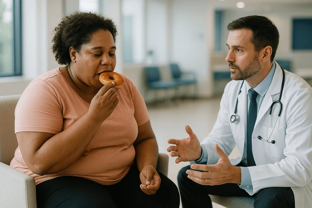
Three outcomes follow, and they explain nearly every lump a patient feels afterwards:
- The body walls the oil off inside a thin capsule. That is an oil cyst.
- Immune cells move in, the area becomes inflamed, and the dead fat is gradually replaced by firm scar tissue. That is fat necrosis (necrosis means tissue death).
- Over months to years, calcium salts settle into the scarred area. These calcifications are harmless but visible on imaging, which matters particularly in the breast.
Why do cells die in the first place? Partly from the mechanical stress of harvesting and processing, and partly from being placed too far from a blood supply, as explained above. Smokers, patients with poorly controlled diabetes and people with heavy scarring from prior radiotherapy or surgery tend to have poorer tissue blood flow, which is why they are often counseled about higher risk.
Technique-related problems are a separate category. Liposuction itself can leave contour irregularities, pockets of fluid called seromas, bruising under the skin called hematomas, and temporary or occasionally persistent numbness; the Mayo Clinic and NHS both list these among recognized liposuction risks. Injecting fat into or below muscle, rather than in the fatty layer under the skin, raises the risk that fat enters a blood vessel. That rare event, fat embolism, has its own section below.
The honest summary is that fat grafting complications are usually biological, sometimes technical, and only occasionally dangerous.
Oil cysts after fat transfer: what they are and how they are managed
An oil cyst feels different from what most people expect. Rather than a hard pebble, it is often a smooth, slightly mobile, sometimes squishy bump beneath the skin, like a small grape under a tablecloth. Because it is fluid, it can grow slowly as more oil collects, then stabilize.
Many oil cysts cause no symptoms and are found only on imaging. In the breast they can look like a well-defined round shadow on ultrasound (imaging that uses sound waves) and, over time, may develop a rim of calcium that gives them a characteristic appearance on a mammogram. Radiologists who know a patient has had fat grafting can usually recognize them. That is one reason surgeons ask patients to tell every future imaging team about the procedure.
Management is graded rather than automatic:
- Small, painless, stable cysts are usually observed. Some soften or shrink as the oil is slowly absorbed.
- Cysts that are large, tender, visible or distressing can be aspirated: a needle is passed into the cyst, often under ultrasound guidance, and the oily fluid is drawn out. Cysts can refill, so aspiration is sometimes repeated.
- Cysts that keep returning, become infected, or cannot be confidently distinguished from other lumps may be surgically removed.
In the breast, a new lump after any procedure is not assumed to be benign on the basis of history alone. Standard practice is to image it, and if imaging is not conclusive, to sample it with a needle biopsy. This is not a sign that something has gone wrong; it is the same pathway any new breast lump follows, and it is how clinicians avoid missing a problem that happens to coincide with the surgery.
Patients often ask whether massaging a cyst will disperse it. There is no good evidence that it does, and vigorous pressure on healing tissue can increase inflammation. The treating surgeon is the right person to decide what, if anything, should be done.
Lumps and fat necrosis: when a firm area needs a scan
Fat necrosis is the hard cousin of the oil cyst. Instead of a pocket of liquid, the body builds dense scar tissue around dead fat, producing a firm, often irregular lump that is fixed in place and sometimes tender. The skin above it can occasionally dimple or look slightly thickened, which understandably frightens people because those are also features clinicians check for in other breast conditions.
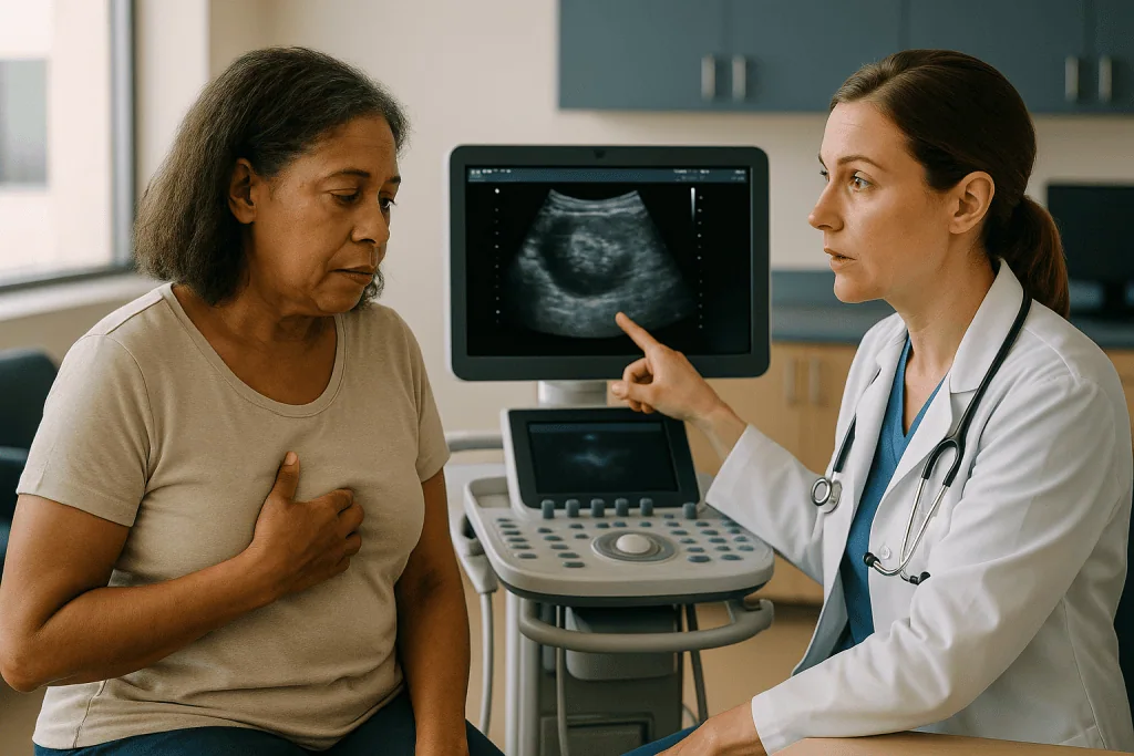
Timing helps distinguish early normal healing from a true lump. In the first few weeks, areas of firmness are common and often reflect swelling and early scar formation; much of this softens as the tissue matures. A lump that persists after swelling has otherwise settled, grows, or appears months later is the kind that earns an ultrasound.
Imaging is the key management step. Ultrasound and mammography can often identify fat necrosis with reasonable confidence because of its typical features, including fat density and, later, coarse calcifications. When the picture is unclear, a needle biopsy gives a definitive answer. Treatment then depends on symptoms. Painless, stable fat necrosis is generally left alone; the body may remodel it over one to two years, although some areas remain permanently firm. Painful or cosmetically troubling areas can be excised, which does mean a small scar and a loss of volume that may itself need correction later.
Patients with breast reconstruction after cancer deserve a specific word. Fat grafting is widely used to refine reconstructed breasts, and lumps in that setting are especially anxiety-provoking. Current mainstream evidence does not show that fat grafting increases the risk of cancer recurrence, but any new lump in a treated breast is investigated promptly rather than attributed to the graft. The oncology and surgical teams decide the pathway together.
What matters most here is not the lump itself but the willingness to image it early and to accept a biopsy if imaging cannot settle the question.
Asymmetry and volume loss: why one side can look different
Asymmetry after fat grafting has two quite different causes, and the fix depends on which one is at work.
The first is unequal survival. Even with identical technique, the two sides of a body are not identical. One breast or buttock may have slightly better blood flow, less scar tissue, or simply a different starting shape, so a different proportion of grafted fat survives. The result is a difference that was not visible on the operating table and only emerges over the following months as the non-surviving fat is absorbed.
The second is pre-existing asymmetry that the procedure was never going to erase. Most people have one side larger, higher or differently shaped than the other, and a surgeon adding similar volume to both sides preserves that difference. Good preoperative planning often involves deliberately placing more fat on one side; even so, perfect symmetry is not a realistic promise for any body-contouring procedure.
Management follows a patient timeline. Surgeons generally decline to judge asymmetry, let alone correct it, while swelling is still resolving, because swelling itself is often uneven. Once the result has stabilized, the options are to add fat to the smaller side in a further session, to remove a small amount from the larger side with liposuction, or to accept a modest difference. The NHS notes that fat transfer results are not always permanent because some of the fat is reabsorbed, and that the procedure may need to be repeated; this is the clinical basis for planning staged sessions rather than one large one.
A small, honest point: volume loss is not a complication in the strict sense. It is an expected feature of the procedure that becomes a problem only when it is uneven or greater than the patient was prepared for. Clear counseling beforehand prevents most of the distress that follows.
What is the fat graft survival rate, honestly?
This is the question patients want a single number for, and it is the question where a single number would mislead. Published studies report widely different proportions of transferred fat surviving long term, because they measure different body areas, different harvesting and processing methods, different volumes, different follow-up periods and different imaging tools. A figure quoted from one series of facial grafts tells you little about buttock grafting, and vice versa.
What mainstream guidance agrees on is the direction of travel. Some fat is always reabsorbed, the loss happens mostly over the first several months, and the volume that remains at around six months is a reasonable guide to the long-term result. The NHS patient information on breast enlargement states plainly that with fat transfer, results may not be permanent and the procedure may need repeating. That is a more useful sentence than any percentage.
Several factors are consistently associated with better graft take in the surgical literature, even if their exact weight is debated:
- Gentle harvesting with low suction pressure and minimal handling of the fat.
- Placing fat in small aliquots spread through healthy, well-vascularized tissue, rather than in one pool.
- Not overfilling. Tissue has a limit to how much graft it can nourish; beyond that, more fat simply means more necrosis.
- Recipient tissue that has not been damaged by radiotherapy or heavy scarring.
- Avoiding smoking and nicotine, which constrict small blood vessels.
Patients sometimes hear that weight gain after grafting will make the transferred fat grow. The cells that survive do behave like the fat of their donor site, so meaningful weight change in either direction can alter the result. Stable weight is the realistic goal.
If a clinician gives you a precise survival figure, a fair question is: measured how, in which body area, and at what follow-up?
Fat transfer side effects at the donor site: infection, bleeding and numbness
Discussions of fat grafting complications often forget that two areas were operated on, not one. The donor site has had liposuction, and liposuction carries its own recognized risks.
The Mayo Clinic lists contour irregularities, fluid accumulation, numbness, infection, internal puncture, fat embolism, kidney and heart problems related to fluid shifts, and toxicity from the local anesthetic used in the injected fluid among the possible risks of liposuction. The NHS adds lumpy or uneven results, bleeding under the skin, persistent numbness that can last weeks or months, changes in skin color, and blood clots in the lungs. These are not predictions; most patients experience only bruising and soreness. They are the recognized menu from which a problem, if one occurs, is likely to come.
Bruising and swelling at donor sites are near universal and are managed with compression garments, which the NHS describes as typically worn for several weeks. A seroma, a pocket of straw-colored fluid under the skin, may be aspirated with a needle if it is large. A hematoma, a collection of blood, is handled the same way or occasionally drained in theater if it is expanding.
Infection at either the donor or the recipient site is uncommon but real. The classic signs are increasing rather than decreasing redness, warmth, pain, swelling, discharge from an incision, or fever. Treatment usually involves antibiotics chosen by the clinician and, if a collection has formed, drainage. Decisions about antibiotics sit entirely with the treating team; this article does not suggest any specific agent.
Numbness deserves a calmer framing than it often gets. Small sensory nerves in the skin are bruised by the cannula and recover slowly. Most altered sensation improves over weeks to months, although a minority of patients report permanent patches of changed feeling, and the NHS flags this as a known risk.
Fat embolism and deep gluteal grafting: the rare, serious complication
Fat embolism means fat entering a blood vessel and traveling with the bloodstream to the lungs, heart or brain, where it can block circulation. It is rare across fat grafting as a whole, but it has been reported disproportionately with gluteal (buttock) fat grafting, and it is the reason this procedure is treated with more caution than fat transfer to the face or breast.
The mechanism is anatomical. Large veins run through and beneath the gluteal muscles. If a cannula tears one of these veins while fat is being injected under pressure, fat can be pushed directly into the venous system. Fat placed in the fatty layer under the skin, above the muscle, is far from these vessels. Fat injected into or below the muscle is not.
Surgical societies in several countries have responded with guidance that converges on the same points: inject only into the subcutaneous fat layer, never into or beneath the gluteal muscle; use a cannula wide enough not to flex into deeper planes; keep the cannula tip angled away from the pelvis; use continuous motion while injecting; and ensure the operating surgeon, not an assistant, controls placement. Many surgeons now use ultrasound during the procedure to confirm the depth of the cannula.
For patients, the practical implications are two. First, ask any surgeon proposing gluteal grafting exactly which tissue plane they inject into and how they confirm it. Second, know the warning signs in the hours after surgery: sudden shortness of breath, chest pain, a racing heart, confusion, or a rash of tiny red or purple spots. These require emergency care, not a phone call to the clinic in the morning.
The Mayo Clinic lists fat embolism among liposuction risks because harvesting can also, rarely, release fat into vessels. In practice, the gluteal injection phase is where the concern concentrates, and where technique has the greatest effect.
Fat transfer vs implants safety: a neutral comparison
People searching for which is safer, implants or fat transfer, usually hear a confident answer in one direction. The evidence does not support confidence. The two approaches carry different risks rather than more or less risk, and the better choice depends on anatomy, goals and health.
| Consideration | Fat grafting | Implants |
|---|---|---|
| Material | Your own fat | Silicone or saline device |
| Typical volume per session | Modest; limited by what tissue can nourish | Larger, chosen in advance |
| Predictability of final size | Lower; part of the fat is reabsorbed | Higher |
| Characteristic complications | Oil cysts, fat necrosis, calcifications, asymmetry from uneven take, donor-site liposuction risks | Capsular contracture (scar tightening), rupture, rippling, malposition, a rare implant-associated lymphoma |
| Serious rare event | Fat embolism, mainly in deep gluteal grafting | Device-related complications requiring removal |
| Future surgery | Possible repeat sessions; excision of problem lumps | Implants are not lifetime devices; replacement or removal is expected eventually |
| Imaging | Can produce calcifications and cysts that imaging teams need to know about | Can obscure breast tissue; may need extra mammogram views |
| Second operated area | Yes, the donor site | No |
The Mayo Clinic notes that breast implants are not guaranteed to last a lifetime and that further surgery may be needed, and lists capsular contracture, rupture and changes in sensation among implant risks. The NHS describes fat transfer as an option that may need repeating. Neither source ranks one as safer overall.
Some patients are offered both: an implant for volume with fat grafted over it to soften edges. Each component then brings its own risk profile. The treating surgeon weighs these against the individual in front of them, which is the only comparison that counts.
Who is fat grafting usually for, and who is usually asked to wait?
Fat grafting tends to suit people who want a moderate change in volume or contour, who have enough donor fat to harvest, and who accept that the result may need a second session. It is widely used to refine breast reconstruction, to fill hollows after injury or tumor removal, to restore facial volume, and in buttock contouring where the fat is placed in the subcutaneous layer.
Surgeons usually ask certain patients to wait or to reconsider:
- People who smoke or use nicotine. Nicotine narrows the small vessels that grafted fat depends on. Most teams ask for a nicotine-free period before and after surgery, with the length set by the surgeon.
- People with poorly controlled diabetes, since high glucose impairs wound healing and raises infection risk.
- People who are very lean, because there may not be enough fat to harvest, and over-aggressive liposuction of a thin donor area raises the chance of contour irregularities.
- People planning significant weight loss or gain, or pregnancy, because the transferred fat will change with them.
- People with a history of blood clots, bleeding disorders, or medicines that affect clotting, who need a specific plan from the prescribing clinician. Nothing should be stopped or started without that clinician’s instruction.
- People with active infection anywhere, or with skin disease at the operative sites.
- People whose expectations center on a dramatic, single-session change, who may be better served by discussing alternatives frankly.
Age alone is rarely the deciding factor; tissue quality and overall health matter more. For those with a personal history of breast cancer, the decision is shared between the surgical and oncology teams, and surveillance imaging is planned around the knowledge that grafting has been done.
None of these are rules that a patient applies to themselves. They are the considerations a surgeon brings to a consultation, and the final decision rests with the treating team after examination and a full history.
What the days and weeks after fat grafting usually look like
The first week is mostly about the donor site. Expect bruising that darkens before it fades, swelling, and a deep ache described by many as similar to the soreness after an unusually hard workout. The grafted area is swollen too, often looking fuller than the final result will be. Compression garments over the liposuctioned areas are typically worn for several weeks according to NHS guidance, and the surgeon will say when they can come off.
Weeks two to six are the deflation phase. Swelling recedes, the grafted area shrinks toward its true volume, and patients often feel firmness as early scar forms. This is when the chickpea from the opening of this article tends to appear, and when most of the anxious phone calls happen. Bruising at donor sites can take weeks to clear; the NHS notes that some swelling can take up to six months to settle fully, and the Mayo Clinic describes a leaner appearance emerging over several months as swelling subsides.
Patients ask what to avoid. The common instructions, which vary by surgeon and body area, include avoiding direct pressure on the grafted area for a period, which in gluteal grafting means not sitting directly on the buttocks for a defined time; avoiding strenuous exercise until cleared; avoiding smoking and nicotine entirely; avoiding hot tubs and swimming until incisions have healed; and avoiding tight clothing over the grafted area that could compress it. Walking is encouraged early, because gentle movement reduces the risk of blood clots in the legs.
By around three to six months the result is largely what it will be, and this is usually when a surgeon will first discuss whether a second session or any correction makes sense. Asking for revision earlier is understandable but rarely productive, because the target is still moving.
What people often get wrong about fat grafting complications
Myth: Because the fat is my own, my body cannot react to it. Your body does not reject your own fat the way it might reject a foreign material, but it does react to dead fat with inflammation, scarring and calcification. Oil cysts and fat necrosis are the proof.
Myth: A lump after fat grafting is always fat necrosis, so it does not need checking. Most lumps are benign and graft-related. A new lump still follows the standard pathway of examination, imaging and, if necessary, biopsy, particularly in the breast. History explains a lump; it does not diagnose it.
Myth: More fat injected means a bigger result. Beyond what the tissue can nourish, more fat means more necrosis, more oil cysts and more lumps, not more volume. Experienced surgeons stage large changes over more than one session.
Myth: Fat transfer is the safe alternative to implants. It is a different set of risks, including the one serious rare event, fat embolism, that implants do not carry. The comparison table above sets out the trade-offs without a winner.
Myth: Massaging lumps will break them up. There is no reliable evidence for this, and pressure on inflamed tissue can make it worse. Ask the surgeon before touching anything that worries you.
Myth: Grafted fat is permanent once it survives. Surviving fat behaves like living fat. It shrinks with weight loss, enlarges with weight gain, and ages with the rest of the body.
Myth: Pain tells you whether something has gone wrong. Oil cysts and fat necrosis are often painless. Infection and embolism, by contrast, announce themselves. Pain that increases after the first few days, rather than fading, is more informative than pain on day one.
The thread connecting these corrections is simple: fat grafting is biology, not sculpture, and biology has its own timetable.
Questions to ask your care team before fat grafting
A good consultation answers most of these before you ask. If it does not, ask.
- Which tissue layer will you inject into, and how do you confirm the depth during the procedure? For buttock grafting, is the fat placed only above the muscle?
- How much volume do you plan to graft in this session, and why that amount for my tissue?
- What proportion of patients in your practice have needed a second session, and how is that decided?
- How do you process the fat after harvesting, and does the method change my risks?
- Which donor areas will you use, and what are the specific liposuction risks for those areas on my body?
- If I develop a lump, what is your pathway: who examines it, what imaging do you order, and who performs any biopsy?
- How will this affect future mammograms or other imaging, and what should I tell future imaging teams?
- What will the first two weeks feel like, what pain management plan do you use, and who prescribes it?
- What should I avoid, for how long, and what are the specific instructions on sitting, exercise, compression and nicotine?
- What are your warning signs for infection, bleeding and fat embolism, and who do I contact at three in the morning?
- How many of these procedures do you perform, and how do you audit your complications?
- If the result is asymmetric, when would you consider correction, and what would it involve?
- Is fat grafting the right option for my goal, or would an implant, a lift, or doing nothing serve me better?
Write the answers down. A surgeon who welcomes these questions, gives specific rather than reassuring replies, and volunteers the limits of the procedure is giving you useful information about more than the operation. Any decision to proceed, delay or choose a different approach belongs to you and the treating team together.
When to call your doctor after fat grafting
Most of what you feel after fat grafting is expected: bruising, swelling, soreness, firmness and temporary numbness. The signs below are different. They are the reasons to contact the surgical team the same day, or to seek emergency care immediately where stated.
Seek emergency care immediately for:
- Sudden shortness of breath, chest pain, coughing up blood, a racing heartbeat, fainting or confusion. These can indicate fat embolism or a blood clot in the lungs and cannot wait.
- A rash of tiny red or purple pinpoint spots on the chest, neck or in the eyes appearing in the first days after surgery.
- Heavy bleeding that soaks dressings and does not slow with firm pressure.
Contact the surgical team the same day for:
- Pain in one calf or thigh with swelling, warmth, or skin that looks red or darker than usual. The NHS describes these as signs of deep vein thrombosis, a clot in a leg vein, which needs prompt assessment.
- Redness, warmth or swelling around an incision or grafted area that is increasing after the third day rather than fading, especially with fever, chills or discharge.
- A rapidly enlarging, tense, painful swelling at any operated site, which may be a collecting hematoma.
- Skin over the grafted or donor area that turns pale, dusky, purple or black, or an open area of skin that will not heal.
- Severe pain not controlled by the plan your clinician gave you.
Book a review, not an emergency visit, for:
- A new lump that persists once swelling has otherwise settled, or any lump that grows.
- Numbness or altered sensation that is not improving after several weeks.
- Asymmetry or volume loss that concerns you after the early months.
When in doubt, call. Surgical teams would rather hear about a bruise that turned out to be a bruise than miss an infection that was left for the weekend. Any treatment, including antibiotics or drainage, is decided by the clinician who examines you.
Frequently asked questions
Which is safer, implants or fat transfer?
Neither is safer overall; they carry different risks. Fat transfer avoids a foreign device and the capsular contracture, rupture and device-related problems implants can bring, but it adds donor-site liposuction risks, oil cysts, fat necrosis, less predictable volume and, in deep buttock grafting, a rare risk of fat embolism. The right choice depends on your anatomy, goals and health, and is a decision for you and your surgeon.
What are the most common fat transfer side effects?
Bruising, swelling, soreness and temporary numbness at both the donor and grafted sites are the most common and usually resolve over weeks to months. Less common are seromas, hematomas, infection, oil cysts, firm areas of fat necrosis and uneven volume. Serious events such as fat embolism or blood clots are rare but require emergency care, which is why surgeons give specific warning signs before discharge.
What should I avoid after fat grafting?
Typical instructions include avoiding direct pressure on the grafted area for a period set by your surgeon, avoiding strenuous exercise until cleared, avoiding all nicotine because it narrows the small vessels grafted fat depends on, and avoiding hot tubs or swimming until incisions have healed. Walking early is usually encouraged to reduce clot risk. Follow your own team’s written instructions over any general list.
How bad does fat grafting hurt?
Most people describe the donor site as the sorer area, with a deep, bruised ache similar to intense muscle soreness that peaks in the first few days and then fades. The grafted area is typically more tender than painful. Pain that increases after the first few days rather than easing is a reason to call your team. Pain relief is planned and prescribed by your clinician.
What is the fat graft survival rate?
There is no single reliable figure. Published studies report widely varying proportions of surviving fat depending on the body area, technique, volume and how survival was measured. What is consistent is that some fat is always reabsorbed, mostly in the first several months, and the NHS notes the procedure may need repeating. The volume remaining at around six months is a reasonable guide to the long-term result.
How are oil cysts after fat transfer treated?
Small, painless, stable oil cysts are usually observed, and some shrink as the oil is slowly absorbed. Larger or tender cysts can be drained with a needle, often under ultrasound guidance, although they may refill and need repeat aspiration. Cysts that keep returning, become infected or cannot be distinguished from other lumps on imaging may be surgically removed. Your surgeon decides which approach fits.
Will fat grafting affect my mammograms?
It can. Oil cysts, fat necrosis and calcifications from grafted fat are visible on mammograms and ultrasound. Radiologists who know you have had fat grafting usually recognize these features, but unexplained findings may prompt extra views or a biopsy. Always tell every imaging team about the procedure, and ask your surgeon whether a baseline scan after healing is advisable.
Why is one side bigger than the other after fat grafting?
Usually because a different proportion of fat survived on each side, or because a pre-existing difference was preserved. Blood supply, scar tissue and starting shape vary between sides, so uneven take is common. Surgeons generally wait until swelling has fully settled before judging asymmetry, then may add fat to the smaller side, remove a little from the larger side, or recommend accepting a modest difference.
Can grafted fat cause cancer or hide it?
Mainstream evidence does not show that fat grafting causes breast cancer or increases recurrence in people treated for it. The practical concern is that lumps and calcifications from the graft can resemble other findings on imaging, which is why any persistent new lump is imaged and, if necessary, biopsied rather than assumed to be graft-related. Surveillance is planned jointly with the oncology team where relevant.
How long do fat grafting lumps take to go away?
Early firmness from swelling and new scar tissue often softens over the first few months. Established oil cysts and fat necrosis may remodel over one to two years, shrink partially, or remain permanently as small firm areas. Lumps that persist, grow or cause symptoms should be assessed with ultrasound; management ranges from observation to drainage or excision, decided by your surgeon.
References
- NHS: Liposuction, recovery and risks
- NHS: Breast enlargement, including fat transfer
- NHS: Deep vein thrombosis (DVT), symptoms and when to get help
This article is for general information only and is not a substitute for professional medical advice. Please consult a qualified doctor about your individual situation.

More from the Blog
HD Liposculpture vs Standard Liposuction: What Sculpting Around Muscle Lines Adds
HD liposculpture is a variant of liposuction in which the surgeon removes fat selectively, in layers, to reveal the outlines of underlying muscles such…

Can Fat Grafting Improve Scars and Contour Defects? When Surgeons Recommend It
Fat grafting can improve some scars and contour defects. A surgeon removes a small amount of a person's own fat by liposuction, processes it,…

How Thigh Lift Surgery Is Performed: Incision Lines, Tissue Removal and Closure
Thigh lift surgery removes loose skin and some fat from the inner or outer thigh through planned incisions, most often along the groin crease…
What Pectoral Augmentation Results Look Like Over Time: Realistic Expectations for Men
Pectoral augmentation results change for months after surgery. In the first week the chest usually looks swollen, high and tight; over 6 to 12…
What Are the Risks of Aesthetic Genital Surgery? Infection, Wound Healing and Sensation
Aesthetic genital surgery carries the general risks of any operation, including bleeding, infection and reactions to anesthesia, plus risks specific to this area: wound…
How Arm Liposuction Is Performed: Incision Points, Cannula Passes and Time Under Anesthesia
Arm liposuction removes fat from the upper arm through two or three small skin openings, usually near the elbow crease and the back fold…