

FibroScan: The Liver Stiffness Test Explained
Key Takeaways
- FibroScan reports liver stiffness in kilopascals, and a value under about 8 kPa makes advanced scarring unlikely while 12 kPa and above makes it probable, with cut-offs adjusted for the underlying disease.
- A reliable scan needs at least ten valid measurements with an interquartile range no more than 30 percent of the median; anything less should be repeated before interpretation.
- Eating within three hours, active inflammation, heart congestion, bile obstruction and the wrong probe can all push the stiffness number falsely high.
- Guidelines recommend a blood-based FIB-4 score first, reserving FibroScan for the roughly one-third of screened adults whose FIB-4 lands between 1.3 and 2.67.
- FibroScan detects cirrhosis with roughly 90 percent accuracy against biopsy but is noticeably weaker at separating mild from moderate fibrosis, its indeterminate middle band.
- In biopsy-based studies of fatty liver disease, losing 10 percent or more of body weight was associated with fibrosis improving by at least one stage in about 45 percent of people within a year.
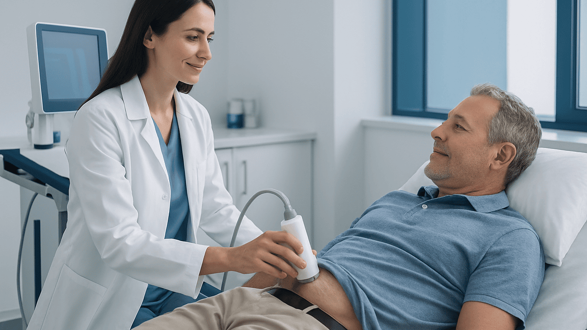
FibroScan is a painless, non-invasive scan that uses a brief vibration and ultrasound to measure how stiff the liver is, reported in kilopascals (kPa), and how much fat it contains, reported as a CAP score. Stiffness tracks with scarring (fibrosis). It takes about 10 minutes, needs roughly three hours of fasting, and is interpreted by a clinician alongside blood tests, because several factors other than scarring can raise the number.
The nurse presses what looks like an electric toothbrush between two ribs on your right side, asks you to breathe normally, and you feel a tap. Ten taps later, a number appears on a screen. That number, a liver stiffness reading, is the reason fibroscan is one of the most-searched medical tests of the moment.
The surge has a clear cause. Since the first prescription medicine for metabolic dysfunction-associated steatohepatitis (MASH, the inflamed form of fatty liver disease) was approved in the US in March 2024, with a second approval following in 2025, clinicians have needed a quick, repeatable way to decide who has enough scarring to benefit. Treatment eligibility in trials leaned heavily on non-invasive stiffness tests, and FibroScan is the one most people meet first. As of early 2026, searches for the test and its results have climbed alongside those approvals.
What follows is what the number means, what it cannot tell you, and where the evidence is firm versus merely promising.
What is a FibroScan and why is everyone searching for it?
FibroScan is the brand name of a device that performs vibration-controlled transient elastography, a technique that measures liver stiffness by timing how fast a gentle mechanical wave travels through the organ. The word elastography simply means imaging or measuring how elastic, or stiff, a tissue is. Because a scarred liver is stiffer than a healthy one, the speed of that wave becomes a stand-in for fibrosis, the medical term for scar tissue laid down after repeated injury.
The appeal is obvious once you know the alternative. For decades, the only way to stage liver scarring was a biopsy: a needle through the skin, a sliver of tissue, a day of soreness and a small risk of bleeding. FibroScan takes about ten minutes, involves no needle, and can be repeated every year or two to watch for change. Most readings are done in a clinic room by a trained nurse or technician rather than in a radiology suite.
Interest has broadened well beyond hepatology. Roughly one in four adults worldwide has fat in the liver, according to the NIDDK and WHO-aligned estimates, and most never know it. Type 2 diabetes, obesity, high triglycerides and heavy alcohol use all push the odds higher. Primary care clinicians now use a two-step approach, a blood-based score first and then a stiffness scan for anyone who lands in the gray zone, so the test has moved from specialist tool to routine screening step.
One caution belongs up front. FibroScan measures stiffness, not scarring directly. Inflammation, a recent meal, heart congestion and bile blockage can all stiffen the liver temporarily. The number is a strong clue, never a verdict, and that distinction shapes everything that follows.
How does a fibroscan work, step by step?
Picture tapping a block of soft cheese and then a block of hard cheese. The tap travels faster through the firmer block. FibroScan does something similar with the liver, using physics rather than fingers.
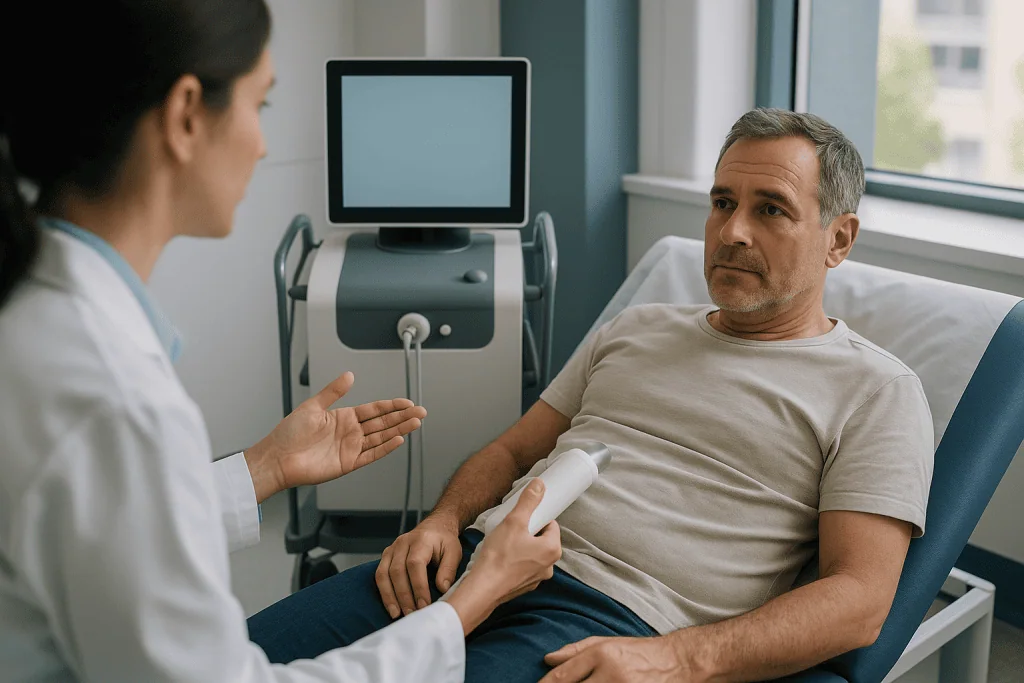
The probe contains a small piston that delivers a low-frequency vibration, about 50 hertz, which is why you feel a light flick against your skin. That vibration sends a shear wave, a sideways ripple, into the liver. Immediately afterward, the same probe fires rapid ultrasound pulses that track the ripple as it moves through roughly four centimeters of tissue. The device calculates the wave speed and converts it into a stiffness value expressed in kilopascals (kPa), the standard unit of pressure. Faster wave, higher kPa, stiffer liver.
At the same moment, the machine measures how much the ultrasound signal weakens as it passes through the tissue. Fat scatters and absorbs sound, so a fatty liver dampens the signal more. This attenuation is reported as the controlled attenuation parameter, or CAP, in decibels per meter (dB/m). One scan therefore yields two numbers: stiffness for scarring and CAP for fat.
Each session collects at least ten valid measurements, and the result reported is the median, the middle value, rather than the average, so a single odd reading cannot drag the whole result up or down. The device also reports the interquartile range, a spread measure; a spread of 30 percent or less of the median is considered reliable.
Two probes exist. The standard M probe suits most adults. The XL probe emits a lower-frequency, deeper-reaching pulse for people with a thicker chest wall or higher body weight, and modern devices select automatically based on the skin-to-liver distance. Choosing the wrong probe is a classic source of a falsely high number, which is one reason operator training matters as much as the hardware.
What changed recently: dates that explain the search spike
Three developments in under three years turned a niche hepatology tool into a household search term.
In June 2023, international liver societies retired the terms non-alcoholic fatty liver disease (NAFLD) and NASH in favor of metabolic dysfunction-associated steatotic liver disease (MASLD) and MASH. The NIDDK and Cleveland Clinic pages now carry both names. The new definition is positive rather than exclusionary: fat in the liver plus at least one cardiometabolic risk factor such as high blood sugar, high blood pressure, excess waist circumference or abnormal lipids. That shift pulled millions of people with diabetes or obesity into a group where guidelines suggest a fibrosis check, and the check most often means a FibroScan.
In March 2024, the US Food and Drug Administration approved the first medicine specifically for MASH with moderate to advanced fibrosis. The pivotal trial used biopsy to confirm scarring, but the labeling permits clinicians to establish eligibility with non-invasive tests, and liver stiffness measurement is the one named most often in practice guidance from US liver societies. A second medicine, already in use for weight management and diabetes, received a MASH indication in 2025 on the strength of a trial showing fibrosis improvement on biopsy.
Behind both approvals sits the 2023 practice guidance from the American Association for the Study of Liver Diseases, which formalized a pathway: calculate a blood-based FIB-4 score first, then send intermediate or high scores for stiffness measurement, with values under 8 kPa considered reassuring and values at or above 12 kPa prompting specialist referral. That pathway, not any single viral post, is why family physicians began ordering the scan in large numbers.
The honest summary: the technology is not new, the first devices appeared in Europe around 2003, but for the first time a reassuring or worrying stiffness number can change what a clinician prescribes.
What will a FibroScan tell you? Reading the kPa and CAP numbers
A FibroScan report usually shows two headline values, and knowing which is which saves a lot of unnecessary worry.
The liver stiffness measurement, in kPa, is the fibrosis signal. A healthy adult liver typically reads between about 2 and 6 kPa. Below roughly 8 kPa, advanced scarring is unlikely; large studies show this threshold misses very few people with serious fibrosis. Between 8 and 12 kPa, the result is indeterminate: some people in this band have moderate scarring, others have inflammation or another temporary cause. From 12 kPa upward, advanced fibrosis becomes probable, and above 15 to 20 kPa, depending on the underlying disease, cirrhosis is likely. Cirrhosis is the stage at which widespread scar tissue begins to distort the liver’s structure and impair its function.
Those cut-offs are not universal. Hepatitis C, hepatitis B, alcohol-related disease and MASLD each stiffen the liver somewhat differently at the same biopsy stage, so a specialist will interpret the kPa against the diagnosis rather than a single chart.
The CAP value, in dB/m, estimates fat. Values under about 248 dB/m suggest minimal fat, while readings around 280 dB/m and above point to fat in a majority of liver cells. CAP is less precise than stiffness and its cut-offs are debated; the NHS and NIDDK describe it as a supportive measure rather than a diagnostic one.
The report also lists the probe used, the number of valid shots, the success rate and the interquartile range. If the spread exceeds 30 percent of the median or fewer than ten shots succeeded, the scan is technically unreliable and should be repeated, ideally fasted and by an experienced operator, before anyone draws conclusions.
What the scan will not tell you: the cause of the scarring, whether inflammation is active, or how you will feel in five years. Those answers need blood tests, history and sometimes a biopsy.
What are bad results from a FibroScan? Thresholds in context
People ask this question bluntly, so here is a blunt but careful answer: no single FibroScan number is “bad” on its own, but certain values carry enough weight that guidelines recommend action.
| Liver stiffness (kPa) | What it usually suggests | Typical next step in guidelines |
|---|---|---|
| Under 8 | Advanced fibrosis unlikely | Address risk factors; repeat in 2–3 years if risk persists |
| 8–12 | Indeterminate; possible moderate scarring or a confounder | Repeat fasted, add blood markers, consider specialist review |
| 12–15 | Advanced fibrosis probable | Hepatology referral; treatment eligibility assessed |
| 15–20 | Cirrhosis likely | Confirm; begin liver cancer surveillance |
| 20–25 and above | Clinically significant portal hypertension possible | Assess for varices; closer monitoring |
The lower rows deserve explanation. Portal hypertension is raised pressure in the vein that carries blood from the gut to the liver, and it drives complications such as enlarged veins (varices) in the esophagus. International consensus from the Baveno VII meeting, summarized in Mayo Clinic and NIH material on cirrhosis, uses a “rule of five”: a stiffness of 15 kPa or less combined with a normal platelet count makes significant portal hypertension unlikely, while 25 kPa or more makes it likely.
A high number is a reason for follow-up, not for despair. A fair proportion of readings above 12 kPa fall when the scan is repeated after a proper fast, after an alcohol-free stretch, or once a liver enzyme flare has settled. Equally, a very low number in someone with cirrhosis already visible on imaging does not undo the diagnosis.
For CAP, a value above roughly 280 dB/m is often labeled severe steatosis, yet fat alone, without stiffness or inflammation, is the component most responsive to weight loss and alcohol reduction. The clinician who ordered the test, not the printout, decides what counts as bad.
Fibroscan vs ultrasound: what is the difference from a liver ultrasound?
Both use sound waves, both are painless, and that is roughly where the similarity ends.
A conventional liver ultrasound creates a picture. A sonographer sweeps a gel-covered probe across the abdomen and the screen shows the liver’s size, outline, texture and blood vessels, along with the gallbladder, bile ducts and nearby organs. It is excellent for spotting gallstones, cysts, tumors, blocked ducts, fluid in the abdomen and the lumpy, shrunken contour of advanced cirrhosis. It also detects fat, which makes the liver look brighter than the adjacent kidney, but only once fat occupies around a third of liver cells or more. For scarring short of cirrhosis, standard ultrasound is close to blind; a liver with moderate fibrosis can look entirely normal.
FibroScan does not produce an anatomical image. Its screen shows a wave trace and numbers. It cannot find a tumor, a stone or a cyst. What it does is quantify stiffness in a cylinder of tissue roughly one centimeter wide and four centimeters long, a sample about 100 times larger than a biopsy core, and convert that into a fibrosis estimate with reasonable accuracy across the whole range from mild to severe.
Some radiology departments now offer shear wave elastography built into a regular ultrasound machine, which combines a picture with a stiffness map. The physics differs slightly and the kPa values are not interchangeable with FibroScan’s, so comparisons over time should use the same method.
In practice, the two tests answer different questions. Ultrasound asks “what does the liver look like, and is anything else going on?” FibroScan asks “how much scarring is probably present?” Many people need both: ultrasound every six months for cancer surveillance once cirrhosis is established, and FibroScan periodically to track stiffness. Neither replaces the other, and neither replaces blood tests.
FibroScan vs liver biopsy: is the scan good enough?
Liver biopsy remains the reference standard for staging fibrosis, which is a careful way of saying it is the best imperfect test we have. A pathologist grades scarring from F0 (none) to F4 (cirrhosis) under a microscope, and also sees inflammation, cell injury and fat, information no scan can match.
Biopsy has well-documented limitations, though. The needle samples about one fifty-thousandth of the organ. Scarring is patchy, so two cores from the same liver disagree on stage in roughly a quarter of cases, and two pathologists reading the same slide can disagree too. Complications are uncommon but real: significant pain in up to a fifth of people, bleeding requiring intervention in well under one percent, and a very rare risk of death. Nobody wants to repeat it yearly.
FibroScan was validated against that imperfect standard. Across large pooled analyses, its ability to detect cirrhosis is high, with an area under the curve around 0.9 or above, meaning it correctly ranks a person with cirrhosis above a person without it roughly nine times out of ten. For distinguishing mild from moderate fibrosis it performs less well, closer to 0.8, because the stiffness ranges overlap. The test is therefore strongest at the extremes: confidently ruling out advanced disease at low values and strongly suggesting cirrhosis at high ones.
Current US and European guidance treats biopsy as a tool for specific situations rather than a routine step: when non-invasive tests conflict, when another liver disease is suspected alongside fat, when a trial requires it, or when the result would change management and the scan sits stubbornly in the indeterminate band.
The fair conclusion is that FibroScan has not made biopsy obsolete, but it has made biopsy unnecessary for most people most of the time, and that is a genuine advance.
Who should have a fibroscan? The two-step screening pathway
Not everyone with a fatty liver on ultrasound needs a stiffness scan straight away, and guidelines are explicit about the order of operations.
Step one is risk. The AASLD, the American Diabetes Association and European societies recommend fibrosis assessment for adults with type 2 diabetes, for people with obesity plus at least one other metabolic risk factor, for anyone with persistently raised liver enzymes, for people with hepatitis B or C, and for those drinking alcohol at hazardous levels. The NHS takes a similar view for its fatty liver pathway.
Step two is a blood-based score, usually FIB-4, which combines age, two liver enzymes (AST and ALT) and the platelet count into a single number. A FIB-4 below 1.3 (or below 2.0 in people over 65) carries a low probability of advanced fibrosis, and guidelines suggest simply repeating it in a few years. A value above 2.67 is high risk and warrants specialist referral regardless of scanning. The middle band, 1.3 to 2.67, is where roughly a third of screened people land, and that is where FibroScan earns its place.
The logic is efficiency and honesty about test limits. FIB-4 alone leaves too many people in limbo; FibroScan alone would mean scanning millions whose risk is already low. Together, in studies of primary care populations, the sequence reduced unnecessary specialist referrals by more than half while catching the great majority of advanced fibrosis.
Some situations call for the scan outside this pathway. People with hepatitis C who have completed antiviral treatment are often scanned to reassess stiffness. Those with known cirrhosis may be scanned to estimate portal pressure. Children are scanned in specialist centers with pediatric-specific thresholds.
The decision always belongs to the clinician who knows your history. A scan requested without a clear question tends to produce a number without a clear answer.
How to prepare for a FibroScan and what the appointment feels like
Preparation is simple but not optional, because the single most common cause of a misleading result is a recent meal.
Eating sends extra blood to the liver through the portal vein, which can raise stiffness by one to several kPa for up to three hours. Most centers therefore ask for no food and no drinks other than water for at least three hours beforehand; some request longer. Routine medicines can usually be taken as prescribed unless the clinic says otherwise. Alcohol in the preceding days can also inflate the number, so an honest account of recent drinking helps the interpreter.
On arrival you lie on your back with your right arm raised behind your head, which spreads the ribs. The operator applies a little gel over the lower right chest and positions the probe between two ribs. You breathe normally; there is no breath-holding. Each measurement produces a brief tap or flick, often described as a flicked rubber band, and most people find it painless. The device signals a valid shot with a tone, and the operator continues until at least ten valid measurements are banked. Including positioning, the whole process rarely exceeds 10 to 15 minutes.
Results appear on the screen immediately, but the operator may be instructed not to interpret them. Interpretation by the ordering clinician, with blood tests alongside, usually follows within days.
Certain circumstances limit or defer the scan. Fluid in the abdomen (ascites) blocks the shear wave entirely. Pregnancy is listed by the manufacturer as a situation in which the device has not been evaluated. Older implanted cardiac devices were once a listed precaution, though current labeling has relaxed this; the clinic will ask. A very narrow gap between ribs or a high body weight may require the XL probe or an alternative method.
Children’s appointments follow the same steps with a smaller probe.
What can make a liver stiffness measurement falsely high or low?
Stiffness is not scarring. It is the sum of everything that makes liver tissue resist deformation at the moment of the scan, and several of those things have nothing to do with fibrosis.
Inflammation is the biggest confounder. During an active flare of hepatitis, whether viral, alcoholic or metabolic, swollen liver cells push the kPa reading upward, sometimes dramatically. Studies in hepatitis B show that stiffness can overstate fibrosis by a full stage or more when ALT is several times normal. Many specialists wait until enzymes settle before trusting a borderline result.
Blood flow matters too. Right-sided heart failure backs blood up into the liver, producing congestion that mimics advanced fibrosis. Blocked bile ducts (cholestasis) have the same effect, which is why a scan done while someone is jaundiced from a gallstone is largely meaningless until the obstruction clears.
Technical factors are common and preventable. The recent meal already discussed. Operator inexperience, defined in validation studies as fewer than about 500 scans. Using the M probe in someone who needed the XL probe, which can add stiffness artificially. Scanning through a thick layer of fat or fluid, which weakens the signal and widens the spread.
Falsely low readings are rarer but occur. A liver with large regenerative nodules and uneven scarring may yield a low number if the sampled cylinder happens to miss the worst tissue. Very early cirrhosis in some people reads under the usual cut-offs.
The practical implication is that one indeterminate or surprising result should prompt a repeat scan under better conditions before it prompts anything else. The test is highly reproducible when conditions are controlled, with the same operator obtaining results within about 10 percent on repeat, but reproducibility only helps if the first conditions were right. A high number three hours after a sandwich is not a diagnosis; it is a scheduling error.
What the evidence actually says, graded by strength
Enthusiasm for a test is easy; weighing its evidence is the useful part. Here is where FibroScan stands, graded in the plain terms clinicians use.
Strong evidence (large meta-analyses of diagnostic accuracy studies against biopsy). For detecting cirrhosis across viral hepatitis, alcohol-related disease and MASLD, pooled sensitivity and specificity both sit in the mid-80s to low-90s percent range, with over 10,000 participants studied. For ruling out advanced fibrosis at a threshold near 8 kPa, negative predictive values above 90 percent are consistent. This is the test’s firmest ground, and it is why guidelines endorse it.
Moderate evidence (observational cohorts). Liver stiffness predicts future events. In cohorts followed for several years, people with higher baseline kPa had substantially higher rates of liver decompensation, liver cancer and death, and a rise in stiffness over time tracked with worse outcomes. These studies cannot prove that stiffness causes harm, only that it marks risk, but the consistency across populations is persuasive.
Moderate evidence for CAP. CAP distinguishes a fatty from a non-fatty liver reasonably well, but its ability to grade fat severity is modest and thresholds vary between studies. Magnetic resonance measurement of fat is more accurate when precision matters.
Weaker or emerging evidence (expert opinion and early trials). Using a fall in kPa as proof that a treatment is working is attractive but not yet validated against hard outcomes; stiffness can drop because inflammation settles rather than because scar has regressed. Whether population-wide screening with FIB-4 plus FibroScan improves survival has not been tested in a randomized trial, although modeling studies suggest it is worthwhile in people with diabetes.
The balanced reading: FibroScan is an excellent instrument for sorting people into low- and high-probability groups and a reasonable one for tracking trends, with its weakest performance in the middle of the fibrosis range and in grading fat. Clinicians who know this use the number as one input among several.
FibroScan-AST (FAST) score and other combined scores explained
Because stiffness alone cannot say whether the liver is actively inflamed, researchers have combined FibroScan outputs with blood tests to sharpen the picture. The FAST score is the best known.
FAST stands for FibroScan-AST. It merges liver stiffness, CAP and the blood level of AST, an enzyme released from injured liver cells, into a single probability between 0 and 1. It was designed in a 2020 international study published in The Lancet Gastroenterology & Hepatology to identify people with MASH who have both meaningful inflammation and at least moderate fibrosis, in other words the group most likely to progress and most likely to qualify for treatment trials. A score of 0.35 or below makes that combination unlikely, a score of 0.67 or above makes it likely, and values in between remain undetermined. In the derivation and validation cohorts, about 60 to 70 percent of people fell outside the gray zone, which means FAST gives a usable answer for most but not all.
Two newer scores, Agile 3+ and Agile 4, add age, sex, diabetes status, platelet count and the AST-to-ALT ratio to the stiffness value. They aim to reduce the indeterminate band for advanced fibrosis and cirrhosis respectively, and early validation suggests they shrink it by roughly a third compared with stiffness alone.
Blood-only alternatives also exist. The Enhanced Liver Fibrosis test measures three proteins involved in scar formation and is recommended by the NHS as a first-line fibrosis test in England. Proprietary panels combining several routine markers are used elsewhere.
These scores matter to patients mainly because clinicians may quote one of them when discussing treatment eligibility. Each is a probability, not a stage; each is strongest at its extremes; and each should be interpreted by the person who ordered it. The evidence base for FAST and the Agile scores is rated moderate, resting on large but mostly retrospective validation rather than outcome trials.
Can liver fibrosis be reversed? What the data show
The question people type is “can liver fibrosis be cured,” and the most accurate reply is that fibrosis is not a fixed state. Scar tissue in the liver is biologically active; it is laid down and broken down continuously, and when the injury driving it stops, the balance can tip toward removal. The evidence for this is now strong for some causes and moderate for others.
Hepatitis C offers the clearest demonstration. After antiviral treatment clears the virus, repeat biopsies and repeat stiffness scans over several years show fibrosis regressing in a majority of people, including some who began with cirrhosis. Stiffness typically drops sharply in the first year as inflammation resolves and then more slowly as true scar remodels.
In MASLD, weight loss is the lever with the most data. In a prospective study of nearly 300 people with biopsy-proven disease followed for a year, those who lost 10 percent or more of body weight saw fibrosis improve by at least one stage in about 45 percent of cases, and inflammation resolve in the large majority. Smaller losses, around 5 to 7 percent, reliably reduced fat but less often scar. Randomized trials of bariatric surgery and of the newly approved MASH medicines have shown fibrosis improvement on biopsy in a meaningful minority of participants over 12 to 18 months compared with placebo.
Alcohol-related fibrosis also improves with sustained abstinence, though cohort data are less tidy.
Two caveats keep this honest. Cirrhosis, once the liver’s architecture is distorted, is far harder to reverse than earlier stages, and complications such as portal hypertension may persist even when stiffness falls. And a declining kPa reading, while encouraging, is not proof that scar has gone; some of the drop reflects reduced inflammation.
What this means in practice: fibrosis can be managed and often improved, the earlier the better, and the actions that work, treating the underlying cause, reducing alcohol, and sustained weight loss where relevant, are decisions to make with your clinician rather than from a search result.
Common myths about FibroScan, corrected
Viral explainers have attached several confident claims to this test. Most need trimming.
Myth: a normal FibroScan means your liver is healthy. A low kPa makes advanced scarring unlikely; it says nothing about tumors, gallstones, bile duct problems or inflammation without scarring. A person with active hepatitis and no fibrosis can have an unremarkable scan.
Myth: a high number means cirrhosis. Readings above 12 kPa raise the probability substantially, but inflammation, a recent meal, heart congestion, bile obstruction and probe choice all inflate stiffness. Guidelines call for confirmation, not a diagnosis from a single scan.
Myth: FibroScan replaces blood tests and ultrasound. It complements them. Blood tests reveal inflammation and function, ultrasound reveals anatomy, FibroScan reveals stiffness. Each covers a blind spot in the others.
Myth: you can “pass” the test by fasting longer or avoiding alcohol for a week. Fasting is required for accuracy, not for flattery, and a short alcohol-free spell lowers stiffness only by removing a temporary artifact. The underlying scar does not change in a week. Hiding recent drinking from the operator leads to a less useful result, not a better liver.
Myth: the CAP number is the important one. Fat is the most reversible component and, on its own, carries less risk than scarring. The stiffness value drives decisions; CAP supports the picture.
Myth: it is a cancer screening test. It is not. People with cirrhosis need ultrasound every six months to look for liver cancer precisely because FibroScan cannot see a mass.
Myth: results are interchangeable between machines. FibroScan values are not directly comparable with shear wave elastography from a standard ultrasound unit or with MR elastography. Trends should be followed on one method.
The thread through all of these is the same: the test is a probability instrument, excellent when used as designed and misleading when read like a verdict.
When to see a doctor about liver health and FibroScan results
Every FibroScan result, whether reassuring or worrying, should be discussed with the clinician who ordered it. That person has your blood tests, your history and the context the printout lacks. The notes below are about timing, not self-diagnosis.
Book a routine appointment to ask about liver assessment if you have type 2 diabetes, obesity with another metabolic risk factor, raised liver enzymes on a recent blood test, hepatitis B or C, or you drink at levels above recommended limits. Guidelines from US and UK bodies all suggest fibrosis assessment in these groups, and the conversation often starts with a simple FIB-4 calculation from blood you may already have had drawn.
Arrange prompt follow-up if a scan has returned an indeterminate (8–12 kPa) or high (12 kPa and above) stiffness value, if your clinic has recommended a repeat and it has not happened, or if you have been told you have cirrhosis and have not been offered six-monthly ultrasound surveillance.
Seek urgent medical care, the same day, for any of the following red-flag signs, which can indicate a liver complication regardless of what any scan showed:
- Yellowing of the skin or whites of the eyes (jaundice)
- Vomiting blood or passing black, tarry stools
- A rapidly swelling abdomen or new leg swelling with breathlessness
- Sudden confusion, drowsiness or personality change
- Severe pain in the upper right abdomen with fever
- Easy bruising or bleeding that will not stop
Never stop, start or adjust a prescribed medicine on the basis of a FibroScan reading. Some drugs used for other conditions affect liver enzymes, and only the prescriber can weigh that against your overall picture. Equally, the newly approved medicines for MASH have specific eligibility criteria and monitoring requirements; whether one is appropriate is a decision for a treating clinician, not a scan report.
If you are unsure who ordered the test or how to reach them, your primary care clinician can access the result and coordinate the next step.
Frequently asked questions
What will a FibroScan tell you about your liver?
It tells you how stiff the liver is, in kilopascals, which estimates the amount of scar tissue, and how much fat it contains, reported as a CAP score in dB/m. It does not identify the cause of liver disease, show tumors or gallstones, or measure inflammation directly. Your clinician combines the numbers with blood tests and history to decide whether further assessment or treatment is needed.
What are bad results from a FibroScan?
There is no single bad number, but stiffness of 12 kPa or more makes advanced fibrosis probable, values above about 15 to 20 kPa suggest cirrhosis, and 25 kPa or more raises concern for portal hypertension. Any of these should prompt specialist review. Readings between 8 and 12 kPa are indeterminate and are often repeated fasted before conclusions are drawn.
What is the difference between a liver ultrasound and a FibroScan?
Ultrasound makes a picture of the liver and surrounding organs and is good at finding masses, stones, fluid and the distorted shape of advanced cirrhosis, but it cannot detect early or moderate scarring. FibroScan produces no image; it measures stiffness to estimate fibrosis across the whole range. Many people with liver disease need both tests for different reasons.
Can liver fibrosis be reversed or cured?
Fibrosis can improve and in many people regresses when the underlying injury stops; this is well shown after hepatitis C treatment and after sustained weight loss of 10 percent or more in fatty liver disease. Cirrhosis is far harder to reverse and some complications can persist. Clinicians describe fibrosis as manageable and often improvable rather than something that is simply removed.
How accurate is a liver stiffness measurement compared with biopsy?
Pooled studies show FibroScan identifies cirrhosis with sensitivity and specificity in the mid-80s to low-90s percent and rules out advanced fibrosis with a negative predictive value above 90 percent at a threshold near 8 kPa. Its accuracy for distinguishing mild from moderate scarring is lower. Biopsy itself has sampling error, so neither test is perfect.
What does a CAP score mean for fatty liver?
CAP, the controlled attenuation parameter, estimates liver fat by measuring how much the ultrasound signal weakens as it passes through tissue. Values under about 248 dB/m suggest little fat and values around 280 dB/m or higher suggest fat in most liver cells. CAP cut-offs are debated and it grades fat severity only moderately well, so it is treated as supportive information.
Do I need to fast before a FibroScan?
Yes. Most clinics ask for no food and no drinks other than water for at least three hours, because a meal increases blood flow to the liver and can raise stiffness by one or more kPa for up to three hours. Prescribed medicines can usually be taken as normal unless the clinic advises otherwise. Recent alcohol should be mentioned to the operator.
Who should get a fibroscan according to guidelines?
US and European liver societies recommend fibrosis assessment for adults with type 2 diabetes, obesity plus another metabolic risk factor, persistently raised liver enzymes, hepatitis B or C, or hazardous alcohol use. Usually a FIB-4 blood score is calculated first and FibroScan is reserved for intermediate results. The ordering decision rests with your clinician.
Can FibroScan results be wrong?
They can be misleading when conditions are poor. Eating beforehand, active inflammation, heart failure, blocked bile ducts, ascites, an inexperienced operator or the wrong probe can all distort the number, usually upward. A scan with fewer than ten valid shots or a wide spread is unreliable. A surprising result is generally repeated under controlled conditions before it changes management.
What is the FAST score on a FibroScan report?
FAST combines liver stiffness, CAP and the blood enzyme AST into a probability from 0 to 1 that someone with fatty liver disease has both significant inflammation and at least moderate fibrosis. Scores of 0.35 or below make this unlikely and 0.67 or above make it likely; values between are indeterminate. It is a research-derived tool that clinicians may use when discussing treatment eligibility.
References
- NIDDK: Diagnosis of NAFLD and NASH (MASLD and MASH)
- Cleveland Clinic: Fatty Liver Disease (MASLD)
- NHS: Non-alcoholic fatty liver disease (NAFLD)
- MedlinePlus: Fatty Liver Disease
This article is for general information only and is not a substitute for professional medical advice. Please consult a qualified doctor about your individual situation.

More from the Blog
Celiac Testing: The Blood Tests, the Biopsy, and Why You Must Not Go Gluten-Free First
Celiac disease is diagnosed in two steps: a blood test that looks for antibodies, most often tissue transglutaminase IgA (tTG-IgA), followed, for most adults,…

Calprotectin Stool Test: How It Separates IBS From Inflammatory Bowel Disease
Calprotectin is a protein released by white blood cells into the gut when the bowel lining is inflamed. A stool test measures it in…
The PSA Test: What Levels Mean by Age, and Why Context Is Everything
There is no single normal PSA. Commonly used age-referenced upper limits are roughly 2.5 ng/mL for men in their 40s, 3.5 in their 50s,…
The FIT Test: Home Stool Screening That Actually Works
The fecal immunochemical test (FIT) is a yearly at-home colorectal cancer screening that uses antibodies to detect hidden blood in a small stool sample.…

Gut Microbiome Tests: What the Science Supports and What It Does Not, Yet
At-home gut microbiome tests can describe which bacteria appear in a stool sample, but current evidence does not show they can diagnose disease, predict…

EKG Explained: What the 12 Leads Show and What ‘Abnormal’ Usually Means
An EKG (electrocardiogram, also written ECG) records the heart's electrical activity for about ten seconds using ten electrodes that produce twelve views, or leads,…