

Flap or Graft? How Reconstructive Surgeons Choose Between the Two Ways to Rebuild Tissue
Key Takeaways
- A graft has no blood supply when placed and must be fed by the wound bed, which is why it cannot survive on bare bone, tendon, cartilage, implants or heavily irradiated tissue.
- Full-thickness grafts keep their color and shrink far less than split-thickness grafts, so they are favored on the nose, eyelids and ears despite being harder to nourish.
- Local flaps borrow skin from a few centimeters away, which is why they usually match facial skin better than any graft.
- Free flaps can move skin, fat, muscle and bone together with reconnected vessels, but require general anesthesia, hours of surgery and hospital monitoring of the flap's circulation.
- Bleeding under the graft, infection and movement against the bed are the recognized reasons grafts fail; tension, kinking or a clot at the vessel join are the reasons flaps fail.
- MedlinePlus advises protecting a new graft from trauma and heavy stretching for about 2 to 3 weeks, and scars from either technique keep maturing for a year or more before revision is judged.
A skin graft is tissue detached completely from one site and laid onto a wound, where it must grow a new blood supply from the wound bed. A flap is tissue moved with its own blood vessels intact or reconnected by microsurgery. Surgeons choose based on whether the wound bed can nourish a graft, how much depth and bulk are missing, and how the result needs to look and function.
The dermatologist has just finished reading the pathology report aloud. The skin cancer on the side of the nose is gone, margins clear, and now there is a dime-sized hole where it used to be. The patient, still numb from the local anesthetic, asks the only question that matters to them: what goes in the gap?
The surgeon sketches two options on a paper towel. One is a patch of skin borrowed from behind the ear and simply set into place. The other is a tongue of neighboring cheek skin, cut on three sides and swung across, still attached at its base. Same hole, two very different repairs. That sketch is the flap vs graft reconstruction decision, and it is made thousands of times a day, on noses and eyelids, on burned hands, on breasts after mastectomy, on legs where bone shows through.
What follows is how reconstructive surgeons actually think through it: not which option is superior, because neither is, but which one the wound in front of them can support.
What is the difference between a flap and a graft?
Both techniques move living tissue from a place where the body can spare it to a place where tissue is missing. The difference is what the tissue brings with it.
A graft arrives with nothing. The surgeon lifts a piece of skin, sometimes with a little fat, from a donor site, cuts it entirely free, and places it onto the wound like a patch on a pair of jeans. Every blood vessel that once fed it has been severed. For the first few days the graft is, in effect, on life support, absorbing fluid from the wound bed while the body decides whether to accept it. If the bed cannot feed it, it dies.
A flap arrives with its own plumbing. The tissue is moved while still attached by a stalk of skin, fat or muscle that carries an artery and a vein; surgeons call that stalk the pedicle. In the most advanced version, the free flap, the pedicle is cut and the vessels are stitched to vessels near the wound under a microscope. Either way, the flap is being fed from minute one. It does not depend on the wound bed for survival, which is why it can cover bone, tendon, cartilage, metal implants and tissue damaged by radiation, none of which will support a graft.
A gardener’s comparison helps. A graft is a cut flower placed in a vase: it can look wonderful if the water is good and nothing disturbs it. A flap is a shrub transplanted with its root ball intact. It is heavier work to move, it leaves a bigger hole where it came from, but it does not need the new soil to be perfect.
MedlinePlus and the NHS both describe grafts and flaps as complementary tools in the same kit rather than competing treatments, and that framing is closer to how surgeons talk in the operating room than any question of which is better.
How a skin graft actually takes: what happens in the wound bed
Surgeons rarely say a graft healed. They say it took, and the word is precise. Taking is a three-stage rescue.

In the first day or two the graft has no circulation at all. It survives by soaking up plasma from the raw surface beneath it, a process called imbibition, which is simply the tissue drinking. Next, tiny vessels in the wound bed begin lining up with the severed vessel stumps on the underside of the graft and reconnecting; this is inosculation, and it is the reason a graft must lie perfectly still against the bed. Finally, new capillaries grow into the graft and it becomes living, bleeding skin again. Anything that lifts the graft off the bed during that window, a pocket of blood, a pocket of pus, a shearing slide of the dressing, breaks the fragile connections and the graft fails in that spot.
Two kinds of graft are in common use. A split-thickness graft takes the top layer of skin and part of the second layer; it is shaved off with a special blade, often from the thigh, and the donor site heals on its own like a graze because deeper skin cells are left behind. A full-thickness graft takes the entire skin down to fat and the donor wound is stitched closed, so the graft can only be as large as a wound the surgeon can sew shut, which is why full-thickness grafts tend to come from behind the ear, the neck or the groin crease.
The trade-off matters for the face. Split-thickness grafts take more reliably but shrink as they heal and tend to look shiny and pale. Full-thickness grafts are more demanding to nourish but keep their color, texture and thickness far better, which is why they are the graft of choice around the eyelids and nose. Cleveland Clinic’s overview of skin grafting describes this thickness trade-off in patient-friendly terms.
How a flap keeps itself alive
A flap succeeds or fails on one question: is blood getting in and getting out? Everything about flap design is an answer to it.
The simplest flaps are local. The surgeon cuts a piece of skin next to the wound, leaves it attached along one edge, and advances, rotates or transposes it into the gap. Blood travels through the attached base by way of the fine network of vessels that runs just beneath the skin. Because the tissue comes from a few centimeters away, it matches in color, thickness and hair pattern almost perfectly, which is the whole appeal of a local flap on the face.
Regional or pedicled flaps travel further. Tissue is raised on a named artery and vein and tunneled or swung to a wound some distance away, still tethered. The classic forehead flap used to rebuild a nose is one example: forehead skin is swung down on a vessel that runs beside the eyebrow, left connected for a few weeks while it establishes new blood supply at the nose, then divided in a second operation.
Free flaps break the tether completely. A block of skin, fat, muscle or even bone is removed with its supplying artery and vein, moved to the wound, and the vessels, often narrower than a matchstick, are sewn to local vessels under an operating microscope. Johns Hopkins describes this kind of microsurgical transfer for breast, head and neck, and limb reconstruction. The payoff is that surgeons can bring large volumes of exactly the right tissue from anywhere in the body. The price is that the entire flap now depends on two hand-sewn joins staying open.
Whatever the type, the enemy is the same: kinking, pressure, a clot, tight sutures, low blood pressure or nicotine narrowing the vessels. That is why patients with new flaps are watched so closely and asked so firmly not to smoke.
Flap vs graft reconstruction: the questions surgeons ask first
Watch a reconstructive surgeon examine a fresh wound and you will see them run a mental checklist that has little to do with size. The order goes roughly like this.
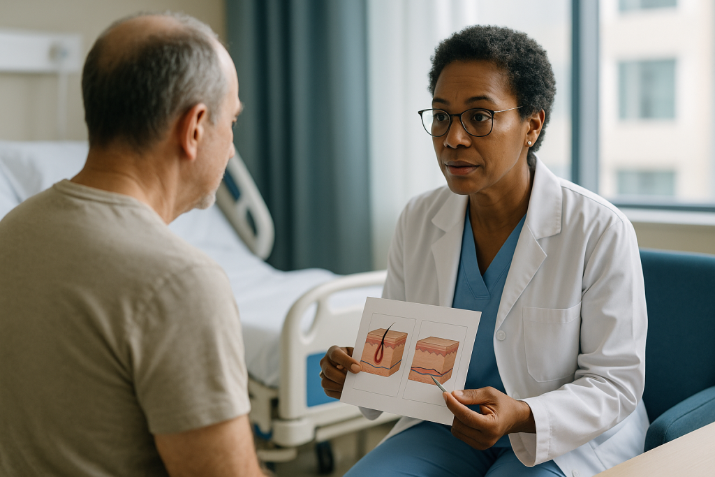
Can the bed feed a graft? A wound floored by healthy muscle, fat or the deep layer of skin bleeds readily and will nourish a graft. A wound with exposed bone stripped of its membrane, bare tendon, bare cartilage, a joint or a metal plate will not; those need a flap, full stop. Tissue that has been irradiated for cancer is scarred and poorly supplied and is treated with similar caution.
How much is missing? A graft replaces a surface. It cannot fill a hollow. If the wound is deep, if cheek fat or the bulk of a breast or the muscle over a shin has gone, a graft laid into the crater will simply sit at the bottom of it and the contour will stay sunken. Flaps carry fat and muscle and can rebuild volume.
Where is it, and what does it need to do? The face is divided by surgeons into aesthetic subunits, the nose tip, the ala, the cheek, the upper lip, each with its own texture and shadow. Reconstruction that respects those borders looks natural; reconstruction that ignores them reads as a patch. Skin over a joint must stretch. Skin on a lower eyelid must not pull the lid down. Skin on the sole must bear weight.
Who is the patient? Age, circulation, diabetes, smoking, medicines that thin the blood or suppress healing, and frankly how much surgery a person wants to go through all feed in. A ninety-year-old with a scalp defect may be better served by a quick graft under local anesthetic than by a long flap procedure, and the reverse can be true for a younger patient who will live with the result for decades.
Only after those answers does the flap vs graft reconstruction choice become obvious, and often it is a mixture: a flap to cover the exposed structure, a graft to close the donor site the flap left behind.
Who usually gets a skin graft, and who is usually asked to wait
Grafts earn their place wherever a large area of surface needs covering quickly over a bed that can support it. Burns are the textbook example: split-thickness grafts can resurface a limb in a single operation, and the same donor site can be harvested again once it heals. Grafts also close the wounds left after a surgeon removes a skin cancer on the scalp, temple or lower leg, cover the raw area where a flap was taken, and resurface chronic ulcers once infection is controlled and healthy tissue has filled the base.
On the face, full-thickness grafts are common for the nose, the inner corner of the eye and the ear, particularly when a wound is too broad for a local flap or when the surgeon wants to avoid distorting a nearby feature. A well-matched graft from behind the ear can be almost invisible on the side of the nose.
Not everyone is grafted the day the wound appears. Surgeons will often ask a patient to wait when the bed is not ready. An infected wound is cleaned and treated first; a graft placed on infected tissue is a graft that will lift and fail. A wound that is still bleeding or draining is allowed to settle. After cancer removal, some teams delay reconstruction until the pathologist confirms the edges are clear, so that a graft never has to be taken back up to chase cancer left behind. NHS guidance on reconstructive plastic surgery describes this staged, wound-first approach.
Patient factors trigger delays too. Poorly controlled blood sugar, ongoing smoking and some blood-thinning regimens are commonly addressed before surgery, with any medicine changes decided by the prescribing clinician rather than by the surgical team alone. A person whose swelling or nutrition is poor may be sent to a wound clinic for weeks to get the bed healthy first. Waiting is not indecision; it is the surgeon refusing to gamble a graft on a bed that is not ready to feed it.
When a flap is usually the better fit
Some wounds announce their need for a flap the moment the dressing comes off. Bone visible in the shin after a fracture and infection. Cartilage showing through the rim of the ear or the side of the nose. A tendon gleaming at the back of the hand. Radiation-damaged chest skin that has broken down. In each case there is nothing for a graft to feed on, and a flap that brings its own circulation is the only reliable way to close the wound and protect what lies beneath.
Flaps are also chosen for the face when appearance and movement are at stake, even where a graft would technically take. The nose is the clearest example. Its skin is thick, oily and pored, and a thin graft from behind the ear sits on it like a postage stamp on a leather bag. A local flap borrowed from the adjacent cheek or nasal skin matches. Larger nasal defects are commonly rebuilt with the staged forehead flap described earlier, precisely because forehead skin resembles nasal skin more closely than anything else on the body.
The lower eyelid teaches a different lesson. Any repair there must resist downward pull; a graft that contracts as it heals can drag the lid away from the eye, a condition called ectropion, which leaves the eye watering and exposed. Surgeons often favor flaps or carefully sized full-thickness grafts supported by a temporary suture to prevent this.
Volume is the third trigger. After mastectomy, after removal of a large tumor from the cheek or jaw, after trauma that has taken muscle from a limb, only a flap can restore the missing bulk. Mayo Clinic describes autologous flap breast reconstruction, using the patient’s own abdominal or back tissue, as one of the two broad routes to rebuilding a breast, alongside implants.
Finally, flaps suit wounds that must bear weight or endure friction, such as the heel, the buttock in a person who uses a wheelchair, or the palm. Padded, durable, sensate tissue is worth the extra surgery there.
Is a free flap better than a skin graft?
The honest answer is that the question compares a scalpel with a chisel. They are different instruments for different jobs, and a good surgeon reaches for the one the wound demands.
A free flap is the most complex tool in reconstruction. It can deliver skin, fat, muscle, nerve and bone in a single living block, rebuild a jaw with a piece of shin bone, resurface a whole cheek with thigh skin, or reconstruct a tongue after cancer surgery. Nothing else can do those things. But it requires general anesthesia, several hours of operating time, a microsurgical team, a hospital stay with round-the-clock monitoring of the flap’s circulation and a second wound at the donor site. Johns Hopkins describes free flap procedures as major operations reserved for large or complex defects.
A skin graft cannot rebuild a jaw. What it can do is resurface a clean, well-vascularized wound in under an hour, often under local anesthetic, with a donor site that heals on its own. For a shallow wound on the temple of an older patient with heart disease, a graft is not the lesser option; it is the safer and more sensible one.
Where wounds genuinely could be closed either way, the surgeon weighs the graft’s simplicity and lower risk of a long operation against the flap’s better color match, durability and contour. The published comparisons that rank one above the other tend to concern very specific defects in very specific patients, and their conclusions do not transfer to a different wound on a different person.
What patients usually mean by better is will it look natural and last. For deep, load-bearing or radiated wounds, flaps generally offer more durable coverage. For thin, well-perfused surface wounds, a full-thickness graft can look every bit as good with far less surgery. The choice sits with the treating team, who can see the bed, feel the tissue and know the patient’s health in a way no general rule can.
Skin graft vs skin flap at a glance
The table below compresses the decision into its working parts. Treat it as a map of how surgeons think, not a formula; real wounds mix these features, and the treating team weighs them together.
| Feature | Skin graft | Local or regional flap | Free flap |
|---|---|---|---|
| Blood supply | None at first; must grow in from the wound bed | Carried in on an attached pedicle | Vessels reconnected by microsurgery |
| Can cover bare bone, tendon, cartilage or implants | No | Yes | Yes |
| Replaces missing volume | Surface only | Modest bulk (skin plus fat) | Large bulk, including muscle or bone |
| Color and texture match on the face | Good with full-thickness from nearby; poorer with split-thickness | Excellent (neighboring skin) | Variable; distant skin often differs |
| Typical anesthesia | Often local; general for large burns | Local or general depending on size | General |
| Operating time | Short | Short to moderate | Long, often several hours |
| Hospital stay | Frequently none | Often none or brief | Several days with flap monitoring |
| Donor site | Heals alone (split) or small stitched scar (full) | Scar adjacent to the wound | Separate scar elsewhere on the body |
| Main early failure mode | Blood or fluid lifting the graft; shear; infection | Tension or kinking cutting off the pedicle | Clot at the vessel join |
| Tendency to shrink over time | Marked with split-thickness | Minimal | Minimal |
Two patterns stand out. First, the more the wound needs, the further right on the table the answer moves, and the more the operation asks of the patient in return. Second, no column is free of risk; each simply fails in a different way, which is why aftercare instructions differ so much between a graft and a flap. MedlinePlus and Cleveland Clinic patient pages on skin grafts, and Johns Hopkins material on flap surgery, describe these trade-offs from the patient’s side.
Is flap surgery a major surgery?
It can be either the smallest procedure a plastic surgeon does all day or the largest, and the word flap alone does not tell you which.
At one end sits the local flap for a facial skin cancer defect. The patient is awake, numbed with local anesthetic, and the surgeon cuts, lifts and swings a neighboring piece of skin into place in a single sitting. The whole visit may be shorter than a dental crown. There is a scar in a wrinkle line, a week or two of care, and a follow-up appointment. Calling that major surgery would surprise most of the people who have had it.
At the other end sits free flap reconstruction after removal of a head and neck cancer, or after mastectomy. Johns Hopkins and Mayo Clinic both describe these as lengthy operations under general anesthesia, frequently running several hours, sometimes involving two surgical teams working at once, one preparing the wound while the other harvests the flap. Patients stay in hospital for days, the flap is checked frequently for color, warmth and capillary refill, and a return to the operating room may be needed if circulation falters. There is a second wound where the tissue came from, and recovery is measured in weeks. By any definition that is major surgery.
Pedicled regional flaps, such as the staged forehead flap for the nose, sit between the two. Each operation is moderate in itself, but the patient lives with a bridge of tissue connecting forehead and nose for a few weeks before the second stage divides it. That period is more disruptive socially than medically, and surgeons spend time preparing people for it.
The practical question to put to the team is not whether the procedure is major but what it involves for you: what anesthesia, how long on the table, how many nights in hospital, how many stages, and how long before you can drive, work and lift. Those answers vary far more by flap type and by patient than by the label.
What are the downsides of flap surgery?
Flaps solve problems grafts cannot, and they bring problems grafts never have. Reconstructive surgeons discuss these openly because a patient who understands them is a patient who reports trouble early.
The most feared is loss of the flap. If the pedicle is kinked or compressed, or a clot forms at a microsurgical join, blood stops flowing and the tissue dies within hours. Total loss of a free flap is uncommon in experienced hands but it is not zero, and when it happens it means another operation and often a fallback plan such as a graft or a second flap. Partial loss, where the far tip of a local flap blanches and scabs, is more frequent and usually heals with dressings, though it may leave a thicker scar.
Bulk is the second issue. Because a flap carries fat, it can sit proud of the surrounding skin, particularly on the nose and around the mouth. Round flaps sometimes develop a raised, pincushioned look as the scar around them contracts. Many patients need a small revision months later to thin the flap or refine the edges, and it is worth knowing that from the start rather than discovering it as a disappointment.
There is always a second scar. A local flap lengthens the incision beyond the original wound; a regional or free flap leaves a scar on the forehead, thigh, forearm or abdomen. Numbness over the flap and around the donor site is common and can be permanent. Hair-bearing skin moved to a hairless area will grow hair. Skin from the forearm or thigh placed in the mouth or on the face may never match in color.
Operating time and anesthesia carry their own risks, higher for long free flap cases: bleeding, infection, chest and clot complications, and the strain of a long anesthetic on the heart and lungs. Cleveland Clinic and Johns Hopkins patient material lists these general surgical risks alongside the flap-specific ones. None of this argues against flaps; it argues for choosing them when the wound truly needs them.
What a skin graft can and cannot do: limits and downsides
Grafts look like the gentle option, and in terms of surgery they usually are. Their downsides show up later and lie in appearance and behavior rather than in the operating room.
The first is that a graft can fail. MedlinePlus lists bleeding under the graft, infection and movement of the graft against the bed as the common reasons a graft does not take. A collection of blood between graft and wound is the classic culprit: it lifts the graft off its food supply. Surgeons counter this by meshing or pie-crusting the graft so fluid can drain, by stitching a padded bolster dressing over it, and by asking patients to keep the area still and elevated. When a graft fails, the wound is usually allowed to heal with dressings or regrafted once the bed is clean.
The second is shrinkage. Split-thickness grafts in particular contract as they mature. Over a joint that can restrict movement; on an eyelid it can pull the lid away from the eye; on the neck it can produce a band. Full-thickness grafts contract far less, which is why they are preferred on the face despite being harder to nourish.
The third is appearance. A graft placed into a deep wound leaves a dip because it adds no volume. Grafted skin often looks paler or, in some skin tones, darker than its surroundings, has a smoother or shinier surface, and lacks the pores and fine hair of the original. It has no sweat glands of its own in the split-thickness form and can feel dry. Sensation returns slowly and incompletely.
The donor site is the fourth. A split-thickness donor area on the thigh is often described by patients as more painful than the grafted wound itself in the first days, and it may heal with a lighter or darker patch. MedlinePlus advises protecting the new graft from trauma and heavy stretching for about 2 to 3 weeks, and shielding grafted skin from the sun for months, because it burns and discolors more easily than native skin.
What the first days and weeks usually look like
Recovery from graft and flap surgery differs less in duration than in what you are asked to watch for.
After a graft, the most important days are the first several. The graft is usually covered by a firm dressing that the surgeon leaves undisturbed until the first check, and patients are asked to keep the area elevated and still: no bending a grafted knee, no rubbing a grafted cheek against a pillow, no vigorous exercise. MedlinePlus patient instructions for skin flap and graft self-care describe keeping the dressing dry and intact until the team removes it, and protecting the site from bumps and stretching for roughly 2 to 3 weeks while the new blood supply matures. When the dressing comes off, a healthy graft looks pink or dusky and slightly swollen; a purple, black or lifting graft is a call to the team. The split-thickness donor site is dressed like a graze and typically re-epithelializes over a couple of weeks, often with more soreness than the graft itself.
After a local flap, care is similar but tension is the enemy rather than shear. Patients are asked to avoid movements that pull on the flap, to keep sutures clean, and to report any tip of the flap turning pale or dark. Sutures on the face are typically removed within about a week; elsewhere they may stay longer.
After a free flap, the early period happens in hospital. Nurses check the flap’s color, temperature and refill frequently, sometimes with a small ultrasound probe listening for the pulse in the reconnected vessels. Patients are kept warm, well hydrated, off nicotine and often positioned so nothing presses on the pedicle. Johns Hopkins describes discharge once the flap is stable and the patient is mobile, with activity built back over several weeks.
For every type, the visible result keeps changing long after the wound has closed. Swelling settles over weeks, redness fades over months, and scars soften and flatten over a year or more. Surgeons usually wait for that maturation before judging whether any revision is worthwhile.
What people often get wrong
A few beliefs come up so often in clinic that they deserve direct correction.
A graft is the easy option. It is quicker to perform, but it is not easier to heal. A graft is the more fragile of the two in the first week, and it asks more of the patient in terms of stillness and elevation than a small local flap does. Ease in the operating room and ease in recovery are not the same thing.
A flap always looks better. Local flaps on the face usually do match better than grafts, but a bulky flap on the nose or a hairy flap on the cheek can look worse than a well-chosen full-thickness graft. Match depends on where the tissue comes from, not on whether it is called a flap.
Free flap means top of the range. It means complex, not superior. Choosing a free flap for a wound a graft could close would expose a patient to hours of anesthesia and a second major scar for no benefit. Surgeons match the tool to the wound.
If a graft fails, the surgeon did something wrong. Grafts fail for reasons that are often outside anyone’s control: a small bleed under the dressing, an unnoticed low-grade infection, a patient who could not keep the area still. Cleveland Clinic and MedlinePlus both list these as recognized risks rather than errors. A failed graft is usually redone or allowed to heal by dressings.
Grafted skin comes from someone else. In reconstructive surgery it almost never does. Grafts and flaps are autologous, taken from the patient’s own body, which is why there is no rejection and no need for anti-rejection medicine. Donor or manufactured skin substitutes exist as temporary coverage in burns, but they are a different subject.
Once the wound has closed, the job is finished. Scars mature for months, grafts and flaps shrink and settle, and revisions are a normal part of reconstruction rather than a sign of failure. The result at two weeks tells you little about the result at one year.
Questions to ask your care team
Consultations for reconstruction move quickly, and the sketch on the paper towel can be hard to remember afterward. These questions help turn a technical plan into something you can picture and plan around. Write the answers down or ask permission to record them.
- Why does this particular wound need a flap rather than a graft, or a graft rather than a flap? What about the bed, the depth or the location drove the choice?
- Where will the tissue come from, and what will that donor site look and feel like once it heals?
- Is this a single operation or a staged one? If staged, how many stages are anticipated and roughly how far apart?
- What anesthesia is planned, how long will the operation take, and will I stay in hospital?
- What exactly should I avoid doing in the first two weeks, and for how long after that should I protect the area?
- What would a struggling graft or flap look like in the first days, and who do I call, at any hour, if I see it?
- If the graft or flap does not survive, what is the fallback plan?
- How will the color, texture and thickness compare with the surrounding skin once it settles, and is a later revision likely?
- How will this affect movement, sensation, or in the case of the face, expression and symmetry?
- What in my health, medicines or habits should be addressed before surgery, and who manages those changes?
The last question deserves emphasis. Surgeons routinely ask patients to stop smoking and all nicotine before flap surgery, because nicotine constricts the very vessels the flap depends on, and they may coordinate with the prescribing clinician about blood-thinning or immune-suppressing medicines. Any change to a prescribed medicine is a decision for the clinician who prescribes it, made with the surgeon’s input, never something to attempt on your own.
When to call your doctor
Grafts and flaps declare trouble early and visibly, and early action is what turns a threatened reconstruction into a saved one. Your team will give specific instructions; these are the signs that should prompt a call the same day, or a trip to urgent care or the emergency department if you cannot reach them.
For a flap, color is the language. A flap that turns pale or white, or cool to the touch, may have lost its arterial inflow. A flap that turns dusky, purple or blue, swells rapidly or oozes dark blood from the edges may have blocked venous outflow. Both are emergencies in the first days, particularly after free flap surgery, because the window to restore circulation is measured in hours. Do not wait until morning.
For a graft, watch for the dressing becoming soaked or slipping, a graft edge lifting, fluid or blood collecting under the graft, or the graft turning black. A little dark crusting at the edges can be normal; a spreading dark area is not.
For either, call promptly about fever, spreading redness or warmth around the wound, increasing rather than settling pain, thick or foul-smelling discharge, a wound that opens, or bleeding that does not stop with gentle pressure. Numbness, tingling or coldness in a limb beyond the surgical site, especially under a tight dressing, also needs attention.
Call emergency services for chest pain, sudden shortness of breath, or a painful swollen calf after any operation, because these can signal a blood clot. After head and neck reconstruction, any difficulty breathing or swallowing, or bleeding into the mouth, is an emergency.
MedlinePlus self-care guidance for skin flaps and grafts lists these warning signs and stresses that patients should contact their surgeon rather than adjust dressings themselves. Decisions about re-exploring a flap, redressing a graft or starting treatment for infection rest with the treating team, and they would far rather hear from you about a false alarm than not hear from you about a real one.
Frequently asked questions
What is the difference between a flap and a graft?
A graft is tissue cut completely free from a donor site and placed on a wound, where it must grow a new blood supply from the wound bed. A flap is tissue moved with its own blood vessels either still attached or reconnected under a microscope. Grafts resurface clean, well-supplied wounds; flaps cover exposed bone, tendon or cartilage and replace missing bulk.
Is a free flap better than a skin graft?
Neither is better; they do different jobs. A free flap can rebuild large, deep or radiated defects with skin, fat, muscle or bone, but involves hours of surgery and a hospital stay. A skin graft cannot restore volume but can resurface a healthy wound quickly, often under local anesthetic. Surgeons choose based on what the wound bed can support and what is missing.
Is flap surgery major surgery?
It depends entirely on the flap. A local flap for a facial skin cancer defect is often done awake under local anesthetic in under an hour. A free flap for head and neck or breast reconstruction is a major operation under general anesthesia, frequently lasting several hours, with several days in hospital for monitoring. Ask your team what your specific procedure involves.
What are the downsides of flap surgery?
Flaps can lose their blood supply and die, partially or completely, requiring further surgery. They leave a second scar at the donor site, can look bulky or raised and may need a revision to thin them, and often cause numbness over the flap. Distant flaps may not match facial skin in color or may carry hair. Longer operations also carry higher anesthetic and clot risks.
What is the difference between a skin graft and a skin flap on the face?
On the face, a graft is usually full-thickness skin from behind the ear or the neck laid into the wound, while a flap is neighboring cheek, nose or forehead skin moved across with its blood supply attached. Flaps generally match color, thickness and pores better; grafts avoid extending scars into surrounding features and suit wounds too broad for a local flap.
Why did my surgeon say a graft would not work on my wound?
The most common reason is the wound bed. A graft can only survive on tissue that bleeds and can grow vessels into it, such as healthy fat, muscle or deep skin. Exposed bone without its covering membrane, bare tendon or cartilage, a joint, a metal implant or heavily irradiated tissue cannot feed a graft, so a flap that brings its own circulation is needed instead.
How long does a skin graft take to heal?
A graft establishes its new blood supply over the first week or so, which is why the initial dressing is left undisturbed and the area kept still. MedlinePlus advises protecting the graft from trauma and heavy stretching for about 2 to 3 weeks. The split-thickness donor site heals like a graze over a couple of weeks. Color and texture keep changing for months afterward.
What does a failing skin graft or flap look like?
A failing graft may turn dark purple or black, lift at the edges, or have blood or fluid collecting beneath it. A failing flap turns either pale and cool, suggesting blocked inflow, or dusky, blue and swollen, suggesting blocked outflow. Any of these in the first days, especially after free flap surgery, should be reported to the surgical team immediately, not left until the next appointment.
Does grafted or flap skin come from another person?
In reconstructive surgery it almost always comes from your own body, which is why there is no rejection and no anti-rejection medicine. Split-thickness grafts are commonly shaved from the thigh, full-thickness grafts taken from behind the ear or the neck, and flaps moved from adjacent skin or from the forehead, forearm, thigh or abdomen. Donated or manufactured skin is used only as temporary cover in some burns.
Will I need more surgery after a flap or graft?
Often, and it is not a sign of failure. Staged flaps such as the forehead flap require a planned second operation to divide the pedicle. Bulky flaps are frequently thinned once swelling settles, and depressed or discolored grafts can be revised. Surgeons typically wait for scars to mature, which takes many months, before judging whether a refinement is worthwhile. The decision is made together with your treating team.
References
- MedlinePlus: Skin graft
- MedlinePlus: Skin flaps and grafts, self-care
- NHS: Plastic surgery, what happens
- Cleveland Clinic: Skin graft
This article is for general information only and is not a substitute for professional medical advice. Please consult a qualified doctor about your individual situation.

More from the Blog
Recovery After Xanthelasma Removal: Eyelid Swelling, Ointments and Stitch Care Week by Week
Xanthelasma surgery recovery usually follows a predictable arc: eyelid swelling and bruising peak in the first two to three days and largely settle within…
Acne Scar Treatment for Darker Skin: Why Pigment Risk Changes the Method and the Prep
Acne scar treatment for darker skin usually starts by calming active acne and fading post-inflammatory hyperpigmentation before any resurfacing, because melanin-rich skin reacts to…

Realistic Results From Cellulite Treatment: Softer Texture, Not a Different Body
Realistic cellulite treatment results mean a softer, smoother skin texture and shallower dimples, not a different body and not permanent removal. Cellulite comes from…
Neck Laxity Surgery: Real Options for a Sagging Neck, From Lifts to Non-Surgical Care
Surgery for a sagging "turkey neck" usually means a neck lift (lower rhytidectomy), often combined with platysmaplasty to tighten the paired neck muscles, or…
Why Skin Grafts Sometimes Fail: Pressure, Infection, Smoking and the Signs Your Team Watches
Skin grafts fail most often because something separates the graft from the wound bed before new blood vessels can grow into it: a pocket…
Caring for the Skin After Cryotherapy: Dressings, Showering and Leaving the Crust Alone
After cryotherapy, keep the treated spot clean, let any blister flatten on its own, and leave the crust in place until it separates by…