

Heart Monitoring During Cancer Treatment: When Oncology Refers to Cardiovascular Toxicology

Key Takeaways
- Cardio-oncology guidance defines treatment-related heart damage as a fall of ten or more percentage points in ejection fraction to below the normal range, which is why a personal baseline scan matters more than a population average.
- A relative drop of more than fifteen percent in heart muscle strain on echocardiogram is treated as an early warning even when overall pumping strength still looks normal.
- HER2-targeted therapy typically prompts an echocardiogram about every three months while the drug is given, and the weakening it causes usually recovers once the drug is paused.
- Immune checkpoint inhibitor myocarditis is rare, affecting roughly one in a hundred people or fewer, but clusters in the first weeks to months, so troponin blood tests are emphasized early.
- Radiation to the chest raises the risk of coronary and valve disease five to fifteen years or more after treatment, making lifelong cardiovascular prevention part of survivorship.
- No supplement has shown protection against chemotherapy heart damage in people; controlled blood pressure and regular physical activity have far stronger evidence.
Heart monitoring during chemotherapy means checking the heart's pumping strength and stress markers before, during, and after treatments known to affect it. Oncologists typically order a baseline echocardiogram and sometimes blood tests such as troponin, then repeat them at set intervals. If results change, they refer to a cardio-oncology or cardiovascular toxicology team, which advises on protecting the heart while cancer treatment continues wherever possible.
The appointment was supposed to be about the tumor. Instead, the oncologist spent ten minutes talking about the heart: a scan before the first infusion, a blood test drawn alongside the usual counts, and a possible referral to a specialist whose title, cardiovascular toxicology, sounded like something from a laboratory rather than a clinic. Many people leave that conversation with the same quiet question. If the heart was fine yesterday, why is anyone looking at it now?
The answer is reassuring in an unexpected way. Heart monitoring during chemotherapy is not a sign that something has gone wrong. It is a sign that the team is planning for the small minority of cases in which something could, and wants to catch a change while it is still a number on a report rather than a symptom.
This explainer walks through what is actually measured, which treatments trigger closer attention, what the follow-up calendar tends to look like, and why the referral is a form of protection rather than a warning.
What happens to your heart during chemo?
For most people, very little. The heart keeps its rhythm, its pumping strength stays where it started, and the fatigue of treatment comes from anemia, disrupted sleep, and the sheer metabolic work of fighting illness rather than from the heart itself.
A minority of treatments, though, interact with heart muscle in ways that are well described. Anthracyclines, a class of chemotherapy drugs used for many breast cancers, lymphomas, and sarcomas, generate reactive oxygen molecules inside heart cells and interfere with an enzyme those cells rely on to keep their DNA in order. The result, in a small proportion of patients, is a gradual weakening of the muscle that can appear months or even years later, and which relates to the total amount of the drug a person has received over a lifetime (Mayo Clinic).
Targeted therapies work differently. Drugs that block the HER2 signal in certain breast cancers also block a survival signal that heart cells use under stress; the weakening this causes is often temporary and tends to recover when the drug is paused (Cleveland Clinic). Newer immunotherapies that release the brakes on the immune system can, rarely, allow immune cells to inflame the heart muscle itself, a condition called myocarditis. Some older chemotherapy drugs used for bowel cancer can make coronary arteries spasm, causing chest tightness during infusion. Certain drugs that starve tumors of blood supply raise blood pressure within days.
These are distinct mechanisms with distinct timelines, which is exactly why monitoring is tailored to the regimen rather than applied uniformly. A person receiving a drug with no known heart effect may need nothing beyond routine checks. A person receiving two heart-active drugs in sequence will be watched far more closely.
Why oncology makes a cardio-oncology referral
Cardiovascular toxicology, more commonly called cardio-oncology, is a specialty that sits between two teams. Its job is to keep the heart safe enough that cancer treatment can continue as planned. That framing matters. The specialist is not there to stop chemotherapy; in most cases, the goal is the opposite.

Referral happens for three broad reasons. The first is risk before treatment starts. Someone with existing heart failure, a previous heart attack, poorly controlled blood pressure, diabetes, or prior chest radiation carries a higher baseline risk, and international cardio-oncology guidance recommends that these patients be assessed before a heart-active drug is given (ESC guideline, PubMed). The second is a change during treatment: a fall in pumping strength on a scan, a rise in a blood marker, a new rhythm disturbance, or symptoms such as breathlessness that the oncology team wants interpreted by a cardiologist. The third is planning for survivorship, particularly after anthracyclines or chest radiation, where late effects can surface long after the last infusion.
What the referral produces is a shared plan. The cardiologist may recommend closer scan intervals, blood pressure treatment, heart-protective medicines whose decision rests entirely with the prescribing team, or occasionally a change in how a cancer drug is scheduled. The oncologist weighs that advice against the cancer itself. Neither specialty overrides the other; the patient sits at the center of a negotiation about risk that both teams take seriously.
People sometimes read the referral letter as a verdict. It is closer to a seatbelt: fitted before the journey, adjusted along the way, and most useful precisely when nothing has yet gone wrong.
How heart monitoring during chemotherapy actually works
The process has a rhythm of its own. Before the first heart-active drug, the team takes a picture and a blood sample. The picture is usually an echocardiogram, an ultrasound of the heart that shows how strongly the main pumping chamber squeezes. The blood sample often includes troponin, a protein released when heart muscle cells are injured, and sometimes a natriuretic peptide, a hormone the heart produces when it is under strain.
Those baseline values become the reference point. Every later measurement is compared not with a population average but with that person’s own starting numbers. A pumping fraction that would be normal for one patient may represent a meaningful fall for another who started higher.
Repeat checks are scheduled around the treatment plan. For anthracyclines, a scan is commonly repeated after a certain cumulative exposure and again at the end of treatment. For HER2-targeted therapy, international guidance suggests echocardiograms roughly every three months while the drug is being given (ESC guideline, PubMed). For immunotherapies, the emphasis shifts toward troponin in the early weeks, when myocarditis is most likely to appear. For blood-pressure-raising drugs, the monitoring tool is simply a cuff, often used at home between visits.
Symptoms are the third stream of information alongside scans and blood tests, and arguably the most important. Breathlessness on stairs that were easy a month ago, ankle swelling, a racing or fluttering heartbeat, or chest discomfort during an infusion are all reasons the team wants to hear from you between scheduled checks rather than at the next one.
None of this is invasive in the usual sense. The most demanding part is the time it takes, and the most useful part is consistency.
Echocardiogram before chemotherapy: what the baseline scan measures
An echocardiogram takes twenty to sixty minutes, involves gel and a handheld probe pressed against the chest, and carries no radiation (NHS). Most people find the hardest part is lying on the left side for a while. The technician moves the probe between the ribs to capture the heart from several angles while it beats.
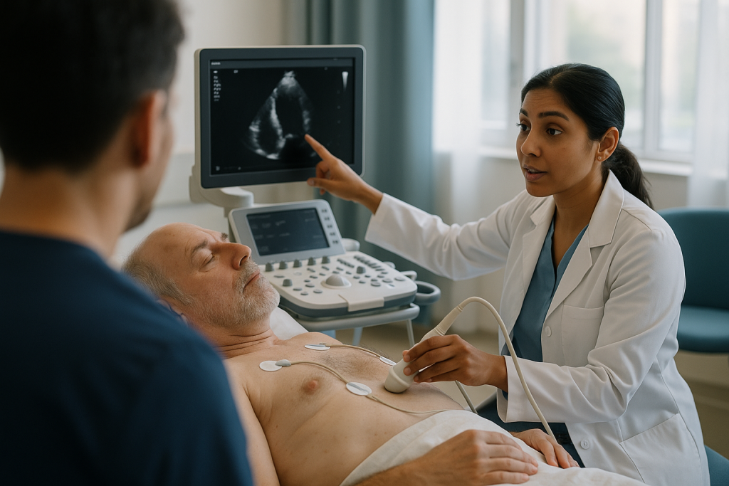
The headline number is the left ventricular ejection fraction, or LVEF: the percentage of blood the main pumping chamber pushes out with each beat. A healthy heart typically ejects somewhat more than half of what it holds. LVEF is the measurement that most monitoring decisions hinge on, because the definition of treatment-related heart damage in cardio-oncology guidance is built around a fall of ten or more percentage points from baseline to a level below the normal range (ESC guideline, PubMed).
Modern scanners also measure global longitudinal strain, or GLS, which tracks how much the heart muscle shortens along its length during each beat. Strain tends to change earlier than ejection fraction, so a relative fall of more than fifteen percent in GLS is treated as an early warning even when LVEF still looks normal (ESC guideline, PubMed). Think of LVEF as the volume of water leaving a pump and GLS as the flex of the pump housing: the housing often shows fatigue first.
The scan also looks at valves, the relaxing phase of the heartbeat, and the pressures inside the chambers. When ultrasound images are difficult to obtain, the team may use a cardiac MRI or a nuclear scan instead. Whichever tool is chosen, the principle is the same: measure carefully before, so that any later change is unmistakable.
Blood tests that hear the heart: troponin and natriuretic peptides
Troponin is the same protein emergency departments measure when someone arrives with chest pain. Heart muscle cells contain it, and when they are injured they leak it into the bloodstream, where a standard blood test can detect it within hours (MedlinePlus). In cancer care, a troponin drawn alongside routine blood counts turns each infusion visit into a quiet check on the heart.
A single raised value does not mean heart failure. It means injury is happening at a cellular level, which is often reversible and sometimes explained by something else entirely, such as kidney function or a recent infection. What the team watches is the pattern: a persistent rise across several visits, or a sudden jump, carries more weight than one borderline reading.
Natriuretic peptides, usually reported as BNP or NT-proBNP, tell a different story. The heart releases them when its walls stretch under pressure or volume load. A rising level suggests the heart is working harder to move the same blood, even before pumping strength falls. Fluid given with chemotherapy, kidney changes, and irregular rhythms can also raise these markers, so results are always read in context.
Where these tests earn their place is in immunotherapy. Immune-related myocarditis is rare, affecting roughly one in a hundred people or fewer on checkpoint inhibitors, but it tends to appear in the first few months and can progress quickly (ESC guideline, PubMed). Troponin can catch it days before symptoms would. For anthracyclines, a persistently raised troponin during treatment is one of the signals that prompts a cardio-oncology review.
Neither marker replaces the scan. Together with it, they form a three-legged stool: structure, injury, and strain.
Which cancer treatments carry heart risk, and what monitoring they usually trigger
Grouping treatments by mechanism makes the monitoring plan easier to understand. The table below summarizes the main classes discussed in cardio-oncology guidance; individual regimens vary, and the treating team decides what applies to a given patient (ESC guideline, PubMed; Cleveland Clinic).
| Treatment class | Main heart concern | Typical timing of effect | Usual monitoring emphasis |
|---|---|---|---|
| Anthracyclines | Weakened pumping (cardiomyopathy) | During treatment through years later; cumulative | Baseline and repeat echocardiogram; troponin |
| HER2-targeted therapy | Reversible fall in pumping strength | During treatment | Echocardiogram about every three months while on drug |
| Immune checkpoint inhibitors | Myocarditis (heart muscle inflammation) | Mostly first weeks to few months | Troponin and ECG early; urgent review if raised |
| Fluoropyrimidines | Coronary artery spasm | During or shortly after infusion | Symptom awareness; ECG if chest discomfort |
| VEGF pathway inhibitors | High blood pressure; clots | Days to weeks after starting | Home blood pressure monitoring |
| Chest radiation | Coronary disease, valve stiffening, pericardial scarring | Years to decades later | Long-term survivorship review |
| Certain kinase inhibitors for leukemia | Rhythm disturbance; artery disease | Variable | ECG; blood pressure; lipid checks |
Two patterns stand out. Damage from anthracyclines and radiation is slow and cumulative, which is why survivorship follow-up matters. Damage from HER2 drugs and immunotherapy is faster but frequently reversible if caught, which is why scans and blood tests are clustered during active treatment. Combining an anthracycline with HER2-targeted therapy, a common sequence in breast cancer care, raises risk beyond either alone and typically brings the closest monitoring of all.
Who is usually monitored closely, and who is usually asked to wait
Not everyone needs the full program, and guidance is explicit about this. Monitoring intensity is matched to a baseline risk assessment that combines the treatment planned with the person receiving it (ESC guideline, PubMed).
Closer monitoring is usual for people with existing heart disease of any kind, including prior heart failure, heart attack, significant valve disease, or a weak pumping fraction on the baseline scan. Age over roughly sixty-five, high blood pressure, diabetes, kidney disease, obesity, smoking, and previous chest radiation or previous anthracycline exposure each add to the risk score. Someone with several of these factors receiving an anthracycline followed by a HER2 drug will typically see the cardio-oncology team before the first infusion and have scans at every stage.
People at low baseline risk receiving a single heart-active drug generally have a baseline scan, one or two repeats, and clinical review in between. Those receiving treatments with no established heart effect may need no cardiac imaging at all beyond what their general health warrants.
Who is asked to wait? Occasionally, a baseline scan reveals an unexpected problem: a pumping fraction already below normal, or a valve that needs attention. In that situation the teams may pause briefly to optimize heart treatment, choose an alternative cancer regimen, or proceed with heightened surveillance. The wait is usually short and always weighed against how urgently the cancer needs treating. Equally, a borderline result during treatment may lead to a scan being repeated in a few weeks rather than acted on immediately, because a single measurement can fluctuate with fluid status, heart rate, and image quality.
The decision at every branch rests with the oncologist and cardiologist together, and it should be explained in plain terms.
What the following weeks and months usually look like
Imagine the timeline for a common scenario: a heart-active chemotherapy given in cycles over several months, followed by a targeted drug for a year. This is illustrative, not a promise, and intervals follow international guidance rather than a fixed rule (ESC guideline, PubMed).
Before cycle one, an echocardiogram and baseline blood tests are done, and a risk score is calculated. If risk is high, a cardio-oncology consultation is arranged, possibly with blood pressure or cholesterol treatment adjusted by the prescribing clinician before starting.
During the chemotherapy phase, troponin may be checked with routine bloods at each cycle. A repeat scan is often scheduled once a set cumulative exposure has been reached, and again within a few weeks of the last cycle. Symptoms are reviewed at every visit.
During the targeted-therapy phase, echocardiograms recur roughly every three months. Most people become practiced at the routine: an ultrasound appointment slotted into an infusion week, results discussed at the next clinic visit.
At the end of active treatment, a scan is typically repeated within the first year, with the guideline suggesting around twelve months after completion for those who received anthracyclines or high-risk combinations (ESC guideline, PubMed). Those with normal results and low risk may then return to ordinary primary care follow-up, with a request to report new cardiac symptoms promptly. Those with any abnormality, or with high cumulative exposure, enter longer-term surveillance.
The practical texture of these months is mostly ordinary. Scans are painless, blood draws are already part of the routine, and the mental load is usually the waiting between test and result. Asking the team how and when results will be communicated removes much of that uncertainty.
Cardiotoxicity from chemotherapy: what happens when a scan shows a change
Cardiotoxicity is the medical term for heart damage caused by a treatment. In cardio-oncology it is graded rather than treated as a single event, and the grade determines what happens next.
A mild change, such as a fall in strain without a fall in ejection fraction, or a modest troponin rise with a normal scan, usually leads to closer monitoring and often to starting heart-protective medicines whose choice and timing belong to the prescribing clinician. Cancer treatment typically continues.
A moderate change, defined in guidance as a fall of ten or more points in ejection fraction to below the normal range but not into the severely reduced zone, triggers a cardio-oncology review, heart treatment, and a discussion between teams about whether to continue, pause, or modify the cancer drug (ESC guideline, PubMed). For HER2-targeted therapy, a pause with re-scan after a few weeks is a common path, because recovery of pumping strength is frequent and the drug can often be restarted.
A severe change, meaning heart failure symptoms or a pumping fraction that has fallen sharply, generally leads to the cancer drug being held while the heart is treated urgently. Even then, the aim is to recover enough function to resume cancer treatment or switch to an alternative.
What people fear most is permanent damage. Reversibility depends on the mechanism. Damage from HER2 drugs recovers in most cases once the drug stops; anthracycline damage is less predictable, and early detection with prompt heart treatment appears to improve the odds of recovery (Cleveland Clinic). The single most important variable the patient controls is reporting symptoms early, because the earlier the grade is identified, the more options remain.
Protecting your heart between infusions: what the evidence actually supports
The internet is full of supplements that claim to shield the heart from chemotherapy. The honest summary is that none of them has convincing evidence in people, and several can interfere with cancer drugs or blood tests. Anything taken beyond prescribed medicines should be declared to the oncology pharmacist first.
What does have support is unglamorous. Blood pressure control matters, because hypertension amplifies the strain any cardiotoxic drug places on the heart; home monitoring and prompt reporting of readings outside the agreed range are among the most useful things a patient can do (Mayo Clinic). Staying physically active within the limits of fatigue is associated with better heart outcomes during and after cancer treatment, and structured exercise during chemotherapy is safe for most people when cleared by the team (Cleveland Clinic). Not smoking, limiting alcohol, and managing blood sugar carry the same weight they carry in general cardiology.
Heart-protective medicines are sometimes prescribed preventively. Two classes, ACE inhibitors and beta-blockers, have been studied for this purpose. The evidence suggests they may blunt the fall in pumping strength in higher-risk patients, though trial results are mixed and the benefit for low-risk people is uncertain (ESC guideline, PubMed). Whether to use them, and when, is a decision for the prescribing clinician who knows the full picture.
One further point deserves emphasis. Fluid retention and shortness of breath during chemotherapy have many causes, most of them not cardiac. Reporting them promptly is not overreacting; it allows the team to tell the difference with a scan or a blood test rather than guessing.
Heart monitoring after treatment ends: the survivorship years
The final infusion is a milestone, not a finish line for the heart. Two treatment types in particular leave a legacy that can take years to declare itself.
Anthracycline damage is cumulative and can surface long after exposure. Survivors of childhood cancers treated decades ago have taught cardiology much of what it knows here, and the pattern in adults is similar: a small proportion develop a weakened heart years later, with risk rising alongside total lifetime exposure and additional factors such as chest radiation or hypertension (Mayo Clinic).
Radiation to the chest, common in breast cancer, lymphoma, and lung cancer treatment, accelerates the ordinary aging of the arteries and valves within the treated field. Coronary narrowing, stiffened valves, and scarring of the sac around the heart typically appear five to fifteen years later or more, which is why survivorship guidance recommends periodic cardiovascular risk review even in people who feel entirely well (ESC guideline, PubMed).
What surveillance looks like depends on cumulative risk. For many, it means a scan around a year after finishing treatment, then ordinary cardiovascular prevention through primary care: blood pressure, cholesterol, blood sugar, and lifestyle. For higher-risk survivors, echocardiograms are repeated every few years and a cardio-oncology clinic may hold the relationship long term.
The practical advice is simple. Keep a written summary of the cancer treatments received, including drug classes, approximate cumulative exposure, and any radiation fields. Share it with every new doctor. Decades from now, a clinician meeting a survivor for the first time will make better decisions about chest pain or breathlessness if that history is one page rather than a memory.
What people often get wrong about heart monitoring during chemotherapy
“If they are checking my heart, they must think it is damaged.” Baseline scans are ordered for almost everyone starting a heart-active drug. They establish a reference point, and in most people every subsequent result stays close to it.
“A chemo heart problem means stopping chemo.” Most detected changes are mild or moderate and managed with heart treatment while cancer therapy continues or pauses briefly. Outright discontinuation for cardiac reasons is the exception, and even then alternatives are usually available (Cleveland Clinic).
“Only old chemotherapy hurts the heart.” Newer targeted drugs and immunotherapies have their own cardiac effects, some of which appear within weeks. Monitoring has expanded to match, not shrunk.
“Feeling fine means the heart is fine.” Early cardiotoxicity is silent. Strain changes and troponin rises precede symptoms, which is the whole rationale for scheduled tests rather than waiting for breathlessness.
“A supplement can protect me.” No over-the-counter product has demonstrated protection against chemotherapy heart damage in people, and some interact with treatment. Blood pressure control and physical activity have far better evidence.
“Radiation is over when the course ends.” Radiation-related heart disease is a late effect, often surfacing a decade or more later. Lifelong cardiovascular prevention is part of survivorship, not an optional extra.
“Cardio-oncology is only for people with heart disease.” The specialty exists mainly to keep hearts healthy through cancer treatment. Many of its patients have never had a cardiac diagnosis and never will.
Each of these misconceptions carries a cost: unnecessary fear, skipped scans, or a symptom left unreported. Correcting them is part of the treatment.
Questions to ask your care team about heart monitoring
Good questions turn a monitoring plan from something done to you into something done with you. These are worth bringing to the first oncology visit or the cardio-oncology consultation, ideally written down.
- Which of my planned treatments are known to affect the heart, and through what mechanism?
- What is my baseline risk level, and what factors contributed to it?
- Which tests will I have before starting, and how often will they be repeated?
- What results would prompt a change to my plan, and what would that change look like?
- Will a cardiologist be involved from the start, or only if something changes?
- Should I monitor my blood pressure at home, and what readings should I report?
- Which symptoms between visits should I report the same day, and to whom?
- Are there heart-protective medicines you would consider for me, and what is the evidence in my situation?
- How will I receive scan and blood test results, and how quickly?
- Is physical activity safe for me during treatment, and are there limits?
- What will follow-up look like after treatment ends, and for how long?
- Can I have a written summary of my cancer treatments to keep for future doctors?
Two further questions matter for people with existing heart conditions. First, whether any current heart medicines interact with the planned cancer drugs, so that the prescribing clinician can review them. Second, whether the cardiologist who already knows you will be looped in, or whether a cardio-oncology specialist will take over that role for the duration of treatment. Continuity across teams prevents the most common gap, which is a result seen by one clinic and not the other.
When to call your doctor: red-flag signs during and after chemotherapy
Scheduled tests catch silent changes. Symptoms catch the ones that arrive between appointments, and the team would far rather hear about a false alarm than miss a real one.
Call emergency services or go to an emergency department if you experience chest pain or pressure, especially during or shortly after an infusion; sudden severe breathlessness or breathlessness at rest; fainting or near-fainting; a very rapid, pounding, or chaotic heartbeat that does not settle within a few minutes; or new confusion, weakness on one side, or difficulty speaking, which may indicate a stroke.
Contact your oncology or cardio-oncology team the same day if you notice new or worsening breathlessness on exertion, such as stairs you managed easily last month; swelling of the ankles, legs, or abdomen; needing to sleep propped up on extra pillows or waking breathless at night; a persistent cough without a cold; unexplained rapid weight gain over a few days, which can signal fluid retention; home blood pressure readings above the range your team set; or a heartbeat that feels irregular, skipping, or unusually fast at rest.
People receiving immunotherapy should treat any new chest discomfort, breathlessness, palpitations, or unusual fatigue in the first few months as urgent, because myocarditis can progress quickly and is most treatable early (ESC guideline, PubMed).
After treatment has finished, the same symptoms remain worth reporting for years, alongside any new exertional chest tightness in people who received chest radiation. Mention your cancer treatment history to whoever assesses you; it changes how they interpret the findings.
None of this is a checklist for self-diagnosis. It is a list of reasons to pick up the phone and let the people with your scans decide.
Frequently asked questions
What happens to your heart during chemo, and does everyone need monitoring?
Most people’s hearts are unaffected, and monitoring depends on which drugs are used and the person’s baseline risk. Anthracyclines can gradually weaken heart muscle, HER2-targeted drugs can cause a usually reversible dip in pumping strength, immunotherapies can rarely inflame the heart, and some drugs raise blood pressure or cause artery spasm. People receiving treatments with no known heart effect may need no cardiac testing beyond routine care.
Why did my oncologist make a cardio-oncology referral before I even started treatment?
Because the goal is to protect the heart so cancer treatment can proceed as planned. Guidance recommends a specialist review before heart-active drugs for people with existing heart disease, high blood pressure, diabetes, prior chest radiation, or several other risk factors. The referral produces a shared plan between oncology and cardiology; it is planning, not a diagnosis, and it does not mean anything is wrong.
What does an echocardiogram before chemotherapy involve, and does it hurt?
It is a painless ultrasound taking roughly twenty to sixty minutes, with no radiation. You lie on your left side while a technician moves a gel-coated probe between your ribs to image the beating heart. It measures ejection fraction, the percentage of blood pumped out each beat, plus muscle strain, valve function, and chamber pressures. These baseline numbers become the reference for every later scan.
How often is heart monitoring during chemotherapy repeated?
Intervals follow the drug and the person’s risk. Anthracyclines usually prompt a repeat scan after a set cumulative exposure and at the end of treatment; HER2-targeted therapy about every three months while on the drug; immunotherapy emphasizes early troponin blood tests. A scan around twelve months after finishing high-risk treatment is commonly recommended. Your team sets the schedule, and it can change if results shift.
Is cardiotoxicity from chemotherapy permanent?
It depends on the mechanism. Damage from HER2-targeted drugs recovers in most people once the drug is paused. Anthracycline damage is less predictable, but early detection with prompt heart treatment appears to improve the chance of recovery. Immune-related myocarditis can be serious yet often responds when caught early. Reporting symptoms promptly and attending scheduled scans are the factors most within a patient’s control.
What happens if you stop chemo halfway through because of a heart problem?
Stopping outright is uncommon; most cardiac changes lead to a brief pause, heart-protective treatment, and a re-scan, after which the cancer drug is often resumed or switched to an alternative with a different heart profile. The oncologist and cardiologist weigh the cancer’s urgency against the heart’s condition together. Never stop or skip a cycle on your own; raise concerns with the team, who will explain the options.
Can a supplement protect my heart during chemotherapy?
No over-the-counter product has demonstrated protection against chemotherapy heart damage in people, and several can interact with cancer drugs or distort blood tests. The measures with real evidence are controlling blood pressure, staying physically active within your limits, not smoking, and managing blood sugar. Any supplement should be declared to the oncology pharmacist before you take it.
Can you have visitors during chemotherapy, and does that affect heart monitoring?
Visitors are generally welcome, subject to infusion unit rules and your infection risk, which the team will explain. Heart monitoring is unaffected by company, though a companion can be genuinely useful: a second set of ears for results, a driver home after a scan, and someone who notices breathlessness or swelling you might dismiss. Ask the unit about its current visitor policy.
How should I prepare my house for chemo with heart monitoring in mind?
Keep it simple. A validated home blood pressure cuff, if your team asks you to monitor, and a notebook or phone log for readings, weight, and symptoms are the most useful additions. Arrange seating so stairs are not the only route to essentials during fatigue, and store your treatment summary and clinic contact numbers somewhere visible for anyone who might need to call on your behalf.
Do I still need heart checks years after cancer treatment ended?
Often, yes, particularly after anthracyclines or chest radiation, whose effects can surface five to fifteen years later or more. For many survivors this means ordinary cardiovascular prevention through primary care with periodic review; higher-risk survivors may have echocardiograms every few years. Keep a written summary of your treatments and share it with every new doctor, since it changes how symptoms are interpreted.
References
This article is for general information only and is not a substitute for professional medical advice. Please consult a qualified doctor about your individual situation.

More from the Blog

Cardiothoracic Surgery in Older Adults: How Age and Other Conditions Are Weighed
Age alone rarely decides whether an older adult can have cardiothoracic surgery. Surgical teams weigh frailty, kidney and lung function, diabetes, memory, mobility and…

Why Does Congenital Heart Disease Need Lifelong Cardiology Follow-Up After Repair?
Repairing a congenital heart defect changes the heart's anatomy, but it rarely makes the heart identical to one that formed normally. Over decades, patches…

How Heart Bypass Surgery Reroutes Blood Around Blocked Arteries, Step by Step
Heart bypass surgery, also called coronary artery bypass grafting (CABG), treats blocked coronary arteries by sewing a healthy blood vessel taken from the chest…

When Cardiac Rehab Ends: Keeping the Exercise, Nutrition and Medication Habits Going
After cardiac rehab ends, most people are encouraged to continue the same pattern they practiced in the program: regular moderate aerobic activity on most…

Blood Sugar Spikes: What They Do to Energy, Hunger and Arteries, and How to Blunt Them
Blood sugar spikes are sharp rises in glucose after eating, usually peaking within an hour and settling within two to three hours in healthy…

Vinegar Before Meals and Food Order: Two Blood-Sugar Hacks Under the Evidence Lens
Eating vegetables and protein before the carbohydrate portion of a meal (food order) and taking a small amount of diluted vinegar before eating both…