

How a Stent Graft Is Placed Inside an Aortic Aneurysm Through the Groin Arteries
Key Takeaways
- The NHS defines an abdominal aortic aneurysm as a widening of the aorta to 3 cm or more, and repair is usually considered once it reaches 5.5 cm, grows quickly or causes symptoms.
- A stent graft is a fabric-lined tube that seals off a bulge from the inside, whereas a coronary stent is a bare mesh that props open a narrowing; they share a name and nothing else.
- The graft is delivered in folded modular pieces through the femoral arteries at the top of each thigh and assembled inside the aorta under live X-ray, a procedure MedlinePlus puts at roughly 2–4 hours.
- Hospital stays after endovascular repair are typically about 1–3 days according to MedlinePlus, compared with a week or more after open surgery, though fatigue commonly lasts a few weeks.
- Randomized trials found endovascular repair carries lower risk around the time of surgery but similar long-term survival to open repair, largely because stent grafts more often need later touch-up procedures.
- The device is designed to be permanent, but the seal between graft and artery can loosen silently, so imaging follow-up continues for life and missed scans are the main preventable failure.
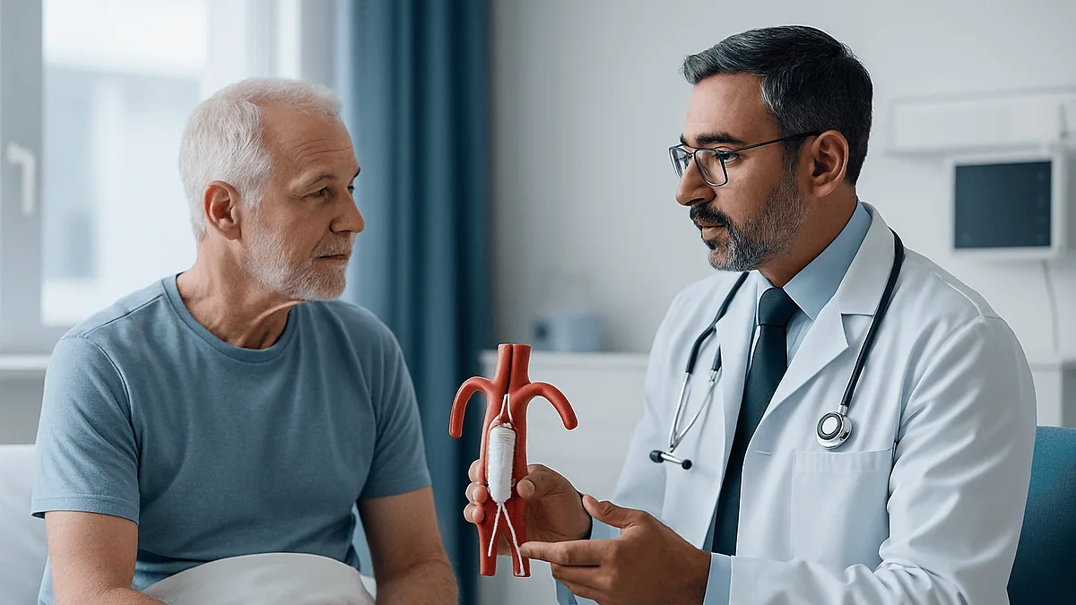
An aortic aneurysm stent graft is placed through small openings in the femoral arteries at the top of each thigh. Guided by live X-ray, the surgeon threads a folded fabric-covered metal tube up to the aneurysm and releases it so it seals against healthy artery above and below the bulge. Blood then flows through the graft instead of pressing on the weakened wall.
Ray is 71, a retired postal carrier with a bad knee and a good sense of humor, and until a routine ultrasound he had never heard of his aorta. Now a radiologist has circled a bulge on a screen and said the words “five point six centimeters.” The surgeon who follows explains that the repair can be done through the groin. Ray looks at his thigh, then at his belly, and asks the question half the waiting room is thinking: how does anything get from there to there?
That question, how aortic aneurysm stent graft is placed through two arteries you can feel with your fingertips, is the whole subject of this article. It is a genuinely clever piece of engineering, and it is also a real operation with real trade-offs, follow-up that lasts for life, and a set of myths that travel faster than the facts.
What follows is the version a seasoned vascular nurse would give you if she had an hour and no hurry.
What is an aortic aneurysm, and why does a stent graft help?
The aorta is the body’s main artery, a pipe roughly the width of a garden hose that runs from the heart down through the chest and abdomen before splitting into the two iliac arteries that feed the legs. An aneurysm is a segment of that pipe that has stretched and ballooned outward because its wall has weakened. The NHS defines an abdominal aortic aneurysm as a widening of 3 cm or more, compared with a normal adult aorta of about 2 cm.
Why does the width matter so much? Physics. The tension on the wall of a tube rises as the tube gets wider, so a larger aneurysm is under more strain at the very moment its wall is thinnest. That is the mechanism behind rupture, and it is the reason vascular teams watch small aneurysms and repair large ones rather than simply waiting.
A stent graft is a tube of woven fabric supported by a metal frame. “Endovascular” means working from inside the blood vessel, and the procedure is usually called EVAR, short for endovascular aneurysm repair. Nobody cuts the aneurysm out. Instead, the graft is placed inside the bulge and anchored to healthy artery above and below it, creating a new channel for blood to travel through. The weakened wall is still there, but it is no longer taking the pressure. Over months the sac around the graft often shrinks because it is no longer being inflated with every heartbeat.
Think of relining an old chimney with a new flue. The brickwork stays, the smoke goes through the liner, and the pressure the bricks used to bear is gone.
What is the difference between a stent and a stent graft?
People hear “stent” and picture the tiny mesh tube a cardiologist uses to prop open a blocked heart artery. That device is a bare or drug-coated metal scaffold a few millimeters across. Its job is to hold a narrowed vessel open so blood can pass. Blood touches the metal and flows through the gaps in the mesh.
A stent graft has a different job and a different design. It is lined, inside or outside, with a tightly woven synthetic fabric, most commonly polyester or a fluoropolymer, so nothing leaks through its wall. The metal frame is there to hold the fabric open and to grip the artery at each end. Blood cannot escape sideways into the aneurysm sac; it has to travel the length of the tube. The stent vs stent graft difference, in one sentence: a stent opens a narrowing, a stent graft seals off a widening.
Scale is the other difference. An aortic stent graft has roughly the diameter of the aorta itself, and it comes in modular pieces. The main body looks like a pair of trousers with one long leg and one very short leg. Once the main body is anchored below the arteries that feed the kidneys, a second component is fed up from the opposite groin and docked into the short leg to complete the second trouser leg into the other iliac artery. Extensions can be added if the fit needs adjusting.
This modular design is what makes groin access possible. Nothing that size could be pushed up an artery in one piece, but folded components delivered one at a time can.
Who is usually offered a stent graft, and who is asked to wait?
Size drives most decisions. The NHS describes three tiers for abdominal aortic aneurysms: small (3.0–4.4 cm), medium (4.5–5.4 cm) and large (5.5 cm or more). Small and medium aneurysms are usually monitored with repeat ultrasound rather than repaired, because the risk of the procedure would outweigh the risk of rupture at that size. Large aneurysms are usually offered repair. Rapid growth between scans, or an aneurysm that is causing pain or tenderness, can also move someone toward treatment earlier, according to Mayo Clinic.
Being large enough is only the first gate. A stent graft needs somewhere to grip. The surgeon looks for a “neck,” a stretch of reasonably straight, healthy aorta just below the kidney arteries, long enough for the top of the graft to seal. The iliac arteries in the pelvis need to be wide enough and not too twisted for the delivery system to pass. When the anatomy does not cooperate, options include more complex grafts with side openings for the kidney arteries, or open surgery through the abdomen.
Some people are asked to wait for reasons unrelated to the aneurysm: an uncontrolled infection, a recent heart event, kidney function that needs protecting before contrast dye is used, or a chest infection that makes anesthesia unwise for now. Others, particularly those who are very frail with other serious illnesses, may be advised with their families that continued surveillance is the more sensible path.
None of this is decided by a scan alone. Vascular surgeons, radiologists and anesthesiologists typically review the case together, and the person at the center of it has the final say about what they are willing to undergo.
Before the procedure: the CT scan that decides everything
Screening ultrasound finds aneurysms; CT plans their repair. A CT angiogram, which is a CT scan performed while an iodine-based contrast dye highlights the arteries, shows the aorta in three dimensions down to the millimeter. From it the team measures the neck below the kidney arteries, the diameter of the aorta at each sealing point, the angles the graft must turn, the length to each iliac artery and the width of the femoral arteries in the groin. Those numbers are used to select graft components sized slightly larger than the artery so they press outward and hold.
Because contrast dye is filtered by the kidneys, blood tests before the scan and again before the procedure check kidney function. People with reduced function may be given extra fluids or have the imaging protocol adjusted. Tell the team about any previous reaction to contrast.
Medicines are reviewed, not stopped on your own. Blood thinners, antiplatelet drugs, diabetes medicines and some blood pressure tablets may need timing changes around the operation, and the anesthesiologist or surgeon will give specific instructions. MedlinePlus advises asking your team which medicines to take on the morning of surgery rather than guessing.
Smoking is the one preparation step with clear evidence behind it. Tobacco weakens the aortic wall, slows healing and raises the chance of breathing problems under anesthesia. Even a few weeks without cigarettes before surgery helps, and vascular clinics can connect people with support.
Expect fasting instructions, a pre-assessment visit, and a conversation about anesthesia choices. Bring a list of questions. The scan may decide the technical plan, but the pre-assessment visit is where your priorities get written into it.
How an aortic aneurysm stent graft is placed, step by step
Here is the EVAR procedure step by step, as it usually unfolds in a hybrid operating room fitted with an X-ray imaging arm.
You lie flat on a narrow table. The groins are cleaned and covered with sterile drapes. Using ultrasound, the surgeon locates the femoral artery at the top of each thigh, the same vessel you can feel pulsing in the crease of your groin. Access is either a needle puncture through the skin or a small cut a few centimeters long to expose the artery directly.
A thin guidewire is passed into the artery and advanced under X-ray up through the iliac artery, through the aneurysm and into the healthy aorta above it. A short plastic tube called a sheath follows, giving the team a protected doorway into the vessel. Contrast dye injected through a catheter produces a live map of the aorta and the kidney arteries.
The main body of the graft arrives folded tightly inside a delivery catheter roughly the thickness of a pen. It is pushed up over the guidewire until its top edge sits just below the kidney arteries. The surgeon then withdraws the outer cover. The frame, made of a spring-like nickel-titanium alloy, expands on its own and presses against the aortic wall.
From the other groin, a second wire is steered into the short trouser leg of the main body, and the second limb is delivered and expanded inside it. A soft balloon is inflated briefly at each junction and sealing zone to mould the graft snugly against the artery. A final contrast injection confirms blood is flowing through the graft and not around it.
Wires and sheaths come out. The artery openings are closed with stitches or a purpose-made closure device. MedlinePlus puts the whole procedure at roughly 2–4 hours.
What you feel during the procedure: anesthesia and the room
Many people are surprised to learn that a repair of the body’s largest artery does not always require being fully asleep. MedlinePlus lists three approaches: general anesthesia, where you are unconscious throughout; regional anesthesia, an injection into the lower back that numbs everything below the waist while you drift under sedation; and local anesthetic at the groins combined with intravenous sedation. The choice depends on your lungs, your heart, how long the case is expected to run, and your own preference. Someone with severe lung disease may do better avoiding a breathing tube; someone anxious about lying still for hours may prefer to be asleep.
If you are awake or lightly sedated, a few sensations are common. The contrast dye can bring a brief wave of warmth through the pelvis and a metallic taste. The balloon moulding step can cause a dull pressure in the back for a few seconds. Lying flat on a firm table for two or more hours becomes uncomfortable, and the team will pad and reposition where they can.
The room itself is a hybrid of operating theatre and X-ray suite. A large C-shaped imaging arm swings over the table, screens show the live picture, and staff wear lead aprons because the procedure uses ionizing radiation. Your exposure is monitored and kept as low as the imaging allows; for a one-time procedure the dose is considered acceptable against the benefit, though it is one more reason teams avoid unnecessary repeat CT scans afterward.
Throughout, an anesthesiologist tracks blood pressure, oxygen and heart rhythm. A urinary catheter is often placed so the bladder can drain during a long case. It usually comes out within a day.
Stent graft (EVAR) vs open repair: how the two approaches compare
Open repair is the older operation. The surgeon makes a long incision in the abdomen, clamps the aorta above and below the aneurysm, opens the sac and sews a fabric graft directly into the vessel. It is durable and it is a bigger physiological hit. The table summarizes typical differences drawn from MedlinePlus and NHS descriptions; individual experiences vary.
| Feature | Stent graft (EVAR) | Open surgical repair |
|---|---|---|
| Incision | Small punctures or cuts at both groins | Long abdominal incision |
| Anesthesia | General, regional or local with sedation | General |
| Typical hospital stay | About 1–3 days (MedlinePlus) | Longer, often a week or more (MedlinePlus) |
| Return to usual activity | Typically a few weeks | Typically several weeks to months |
| Anatomical requirements | Needs a suitable neck and access arteries | Fewer anatomical restrictions |
| Lifelong imaging follow-up | Required | Usually less frequent |
| Chance of a later touch-up procedure | Higher | Lower |
What does the evidence say about outcomes? Large randomized trials comparing the two approaches, summarized by Mayo Clinic and the NHS, found that people having EVAR were less likely to die around the time of surgery, but that this early advantage narrowed over the following years, largely because stent grafts more often need additional procedures. Long-term survival ended up similar. That pattern shapes advice: EVAR spares the body a major operation up front, and in exchange asks for a lifetime of scans and a willingness to have small adjustments if the graft needs them.
Which trade you make is a conversation, not a formula, and it belongs with your surgical team.
The first hours and days after aortic stent graft placement
You wake, or surface from sedation, in a recovery area with a blood pressure cuff cycling on your arm and a nurse checking the pulses in both feet. Foot pulses matter because they confirm blood is reaching the legs through the new graft limbs. Expect them to be checked often in the first night.
The groins are the focus of early care. If closure devices or stitches were used, you will usually be asked to lie flat and keep the legs straight for several hours so the artery seals. A firm dressing and sometimes a small weighted pad help. Bruising that spreads across the upper thigh over the next few days is common and looks worse than it is; a firm, painful, rapidly enlarging lump is not, and should be reported.
Most people sit up the same evening and walk with assistance the next morning. Early walking reduces the chance of blood clots in the leg veins and helps the lungs re-expand. The urinary catheter, if one was placed, typically comes out within a day. Eating and drinking usually resume quickly because the bowel has not been handled, one of the practical differences from open surgery.
A mild fever, achiness and a washed-out feeling in the first few days are recognized after stent graft placement and are thought to reflect the body’s inflammatory response to the new material and the clotting inside the excluded sac. Your team will still check for infection if a fever appears, because the two can look alike.
MedlinePlus notes a typical hospital stay of 1–3 days. Before discharge you should have written wound care instructions, a plan for your usual medicines, and a date for your first follow-up scan.
How long does it take to recover after aortic stent graft repair?
Recovery after aortic stent graft repair is measured in weeks, not months, for most people, but it is rarely as quick as the small groin wounds suggest. The body has still been through a long procedure, contrast dye, anesthesia and a night or two of poor sleep. Fatigue that lingers for two or three weeks is ordinary and does not mean something has gone wrong.
The MedlinePlus discharge guidance for endovascular repair describes a gradual return: walking daily from the first week, increasing distance as comfort allows, and avoiding heavy lifting or straining for several weeks so the groin arteries heal fully. Desk-based work is often possible within a couple of weeks; heavier manual work waits longer. Your surgeon will give you a personal timeline, and it may differ from a neighbor’s.
Driving deserves a specific question to your team, because groin discomfort can slow an emergency brake reaction and because sedative painkillers impair judgement. Sexual activity can resume when the groins are comfortable; there is no graft-related reason to wait beyond that.
The groin wounds themselves need little: keep them clean and dry, shower rather than soak for the first week or so, and watch for redness, warmth or discharge. A pea-sized firm lump under the puncture site, which is scar tissue around the closure, can persist for weeks and usually softens.
By comparison, the NHS notes that recovery from open repair can take several months, reflecting the abdominal incision and the bowel handling it involves. That gap is the main practical reason people who are anatomically suited to EVAR often choose it. The gap in recovery does not, however, exempt anyone from the follow-up scans discussed next.
How long do aortic stent grafts last, and why scans continue for life
Stent grafts are built from materials intended to remain in the body permanently. The fabric does not dissolve and the metal frame does not wear out in any ordinary sense. So the honest answer to “how long do aortic stent grafts last” is that the device is designed to last a lifetime, while the seal between the device and a living, changing artery is what needs watching.
Four things can undermine that seal over the years. An endoleak, the term for blood finding its way back into the aneurysm sac around or through the graft, is the most common. Type I endoleaks occur at the top or bottom sealing zones. Type II endoleaks come from small side branches that bleed backward into the sac; many of these settle on their own. Type III leaks arise between the graft’s modular components, and type IV describes seepage through the fabric itself, which is rare with modern materials. The second problem is migration, the graft slipping downward as the aorta lengthens with age. Third, a graft limb can kink or narrow. Fourth, the aorta above the graft can itself widen over time.
Surveillance catches these before they matter. MedlinePlus describes imaging within the first months after repair and then at regular intervals for life, using CT or, increasingly, duplex ultrasound to spare the kidneys and reduce radiation. If the sac is shrinking or stable and no leak is seen, intervals may lengthen. If the sac grows, the team may add an extension, block a feeding branch or, rarely, convert to open repair.
Many people never need a second procedure. Some need a small one. Missing scans is how a manageable leak becomes an emergency.
Risks of stent graft repair, in plain clinical language
Every procedure that enters an artery carries risk, and a good consent conversation names them without drama. These are the ones vascular teams routinely discuss, drawn from MedlinePlus and Mayo Clinic descriptions.
At the groin: bleeding, bruising, a false aneurysm where blood pools beside the puncture, and damage to the femoral artery that occasionally needs a stitch repair or a small patch. Wound infection is uncommon but more likely in people with diabetes or a high body weight.
Inside the vessels: injury to the iliac arteries as the delivery system passes, dislodging debris that blocks a smaller artery in the leg or, rarely, the bowel or a kidney, and the endoleaks described above. Very rarely the graft cannot be positioned safely and the team converts to open surgery during the same anesthetic.
Kidney injury from contrast dye is a specific concern for people with pre-existing kidney disease, and hydration protocols are used to reduce it. Heart attack, stroke, pneumonia and blood clots are general risks of any major procedure in an older population, which is who most aneurysm patients are.
Infection of the graft itself is rare but serious. Spinal cord injury causing weakness in the legs is mainly a risk of grafts placed in the chest portion of the aorta, not the abdominal repair described here, but teams mention it where relevant.
Alternatives are part of the same conversation: open repair for those whose anatomy does not suit a graft or who prefer a single durable operation; more complex fenestrated or branched grafts when the neck is too short; and continued surveillance with risk-factor management when the aneurysm is not yet large or the person is too frail. Weighing these is the treating team’s job, done with you.
What people often get wrong about aortic stent grafts
“It’s the same stent my brother had in his heart.” It is not. A coronary stent is a bare scaffold that opens a blockage; an aortic stent graft is a fabric-lined tube that seals off a bulge. They share a word and little else.
“They removed my aneurysm.” The sac stays. It is bypassed from the inside, depressurized and, often, shrinks over time. Because it is still there, it still needs watching.
“Keyhole means it’s minor.” The wounds are small; the operation is not. It involves the main artery of the body, contrast dye, radiation and several hours of anesthesia or sedation. Recovery is faster than open surgery, not trivial.
“Once it’s fixed I’m done.” Follow-up imaging is part of the treatment, not an optional extra. Endoleaks are usually silent. The scan is the only way to know the seal is holding.
“I can never have an MRI again.” Most modern grafts are labelled as safe for MRI under specific conditions. You will be given an implant card; show it to any imaging department and let them check the exact device.
“The graft fixes the underlying problem.” It fixes the mechanical danger. The arterial disease that weakened the aorta, driven by smoking, high blood pressure and cholesterol, is still present in the rest of the body. Stopping smoking, controlling blood pressure and taking any medicines your clinician has prescribed for cardiovascular protection matter as much after repair as before.
“Airport scanners and microwaves are a problem.” They are not. The graft has no electronics. The implant card is for imaging departments, not security lines.
“I’ll feel the graft.” You will not. The aorta has no sensation of that kind, and people are unaware the device is there.
Questions to ask your care team before stent graft repair
The best consultations are the ones where you leave understanding not just what will happen but why this option was chosen for your particular aorta. Write questions down beforehand; the appointment goes faster than you expect. Bring someone to listen with you.
About the aneurysm and the decision:
- How large is my aneurysm now, and how fast has it grown between scans?
- Does my anatomy suit a standard stent graft, or would I need a more complex device or open surgery?
- If we did nothing for now and kept scanning, what would you expect to happen?
About the procedure:
- What kind of anesthesia are you recommending for me, and why?
- Will the groin access be by puncture or a small cut, and what does that mean for my recovery?
- What is your plan if the graft cannot be positioned as intended?
About medicines, without changing anything on your own:
- Which of my regular medicines should I take on the morning of the procedure, and which should I pause, and who will tell me when to restart?
- Will I be started on anything new afterward, and what is it for?
About afterward:
- When is my first follow-up scan, what kind will it be, and how often after that?
- What symptoms should make me call, and what should make me go straight to emergency care?
- When can I drive, lift, return to work and travel?
- Who do I contact with a question in the first two weeks, and how?
Ask, too, how the team will keep track of you over the years. Surveillance works only if someone is holding the diary. Knowing whether that is the vascular clinic, your primary care doctor or you yourself prevents the most common failure of all: a scan that quietly never gets booked.
When to call your doctor: red-flag signs after stent graft placement
Most recoveries are uneventful, and most of what you notice in the first fortnight, bruising, tiredness, a tender groin, is expected. A short list of symptoms is different in kind and should not wait for a scheduled appointment.
Call emergency services or go to the nearest emergency department if you have:
- Sudden severe pain in the abdomen, back or side, especially if it is tearing in quality or comes with sweating, faintness or collapse. This can signal a leak or rupture and is an emergency at any time after repair, whether one week or ten years.
- A leg that becomes cold, pale, numb, weak or intensely painful. This can mean a graft limb or leg artery has blocked and blood is not reaching the foot.
- Active bleeding from a groin wound that does not stop with firm pressure, or a groin lump that is rapidly growing and hard.
- Chest pain, sudden breathlessness, or fainting.
Contact your surgical team or primary care doctor the same day if you notice:
- A fever, shaking chills, or a groin wound that is increasingly red, warm, swollen or leaking fluid.
- Passing much less urine than usual, or none, in the first days after contrast dye.
- New or worsening pain in the back or abdomen that is not severe but is not improving.
- Persistent nausea, vomiting or inability to keep fluids down.
- Bloody or black stools.
Two habits protect you over the long term. Keep every surveillance scan, and reschedule promptly if one is missed. Tell any new doctor, dentist or imaging department that you have an aortic stent graft, and carry the implant card. Sudden severe abdominal or back pain in someone with a graft is treated as a possible aortic emergency until proven otherwise, and telling the emergency team about the graft in the first sentence saves time that matters.
Frequently asked questions
How long do aortic stent grafts last?
The graft itself is made from permanent materials and is intended to stay in place for the rest of your life. What can change is the seal between the device and the artery, since the aorta keeps aging around it. Endoleaks, slippage or a kinked limb can develop years later, usually without symptoms, which is why imaging follow-up is lifelong. Many people never need another procedure; some need a minor adjustment identified on a routine scan.
What is the success rate of aortic stent graft surgery?
No single percentage applies to everyone, and reputable sources avoid quoting one because outcomes depend heavily on age, other illnesses and anatomy. Large randomized trials summarized by Mayo Clinic and the NHS found that endovascular repair carried a lower risk of death around the time of surgery than open repair, but that long-term survival was similar, partly because stent grafts more often need follow-up procedures. Your surgeon can discuss how those findings apply to your situation.
How long does it take to recover from aortic graft surgery?
Recovery after endovascular stent graft repair usually takes a few weeks, with a hospital stay of about 1–3 days according to MedlinePlus. Walking is encouraged from the first day, heavy lifting is avoided for several weeks while the groin arteries heal, and tiredness commonly lingers for two or three weeks. Open surgical repair involves a long abdominal incision and the NHS notes recovery can take several months. Your team will give you a personal timeline.
What is the difference between a stent and a stent-graft?
A stent is a bare metal mesh tube, usually a few millimeters across, placed to hold a narrowed artery open; blood passes through the gaps in the mesh. A stent graft is a much larger metal frame lined with woven fabric so nothing can leak through its wall. It is used to seal off a widened, weakened artery rather than to open a blocked one, redirecting blood through the tube and taking pressure off the aneurysm sac.
What does the EVAR procedure step by step actually involve?
The surgeon accesses the femoral artery in each groin by needle puncture or a small cut, passes a guidewire up to the aorta under X-ray, and injects contrast dye to map the vessels. The folded main graft is delivered through a catheter, positioned just below the kidney arteries and released to expand. A second limb is fed from the other groin and docked into it. Balloons mould the seals, a final X-ray confirms no leak, and the groin openings are closed.
Is aortic stent graft placement done under general anesthesia?
Not always. MedlinePlus describes three options: general anesthesia, regional anesthesia through an injection in the lower back that numbs the lower body, or local anesthetic at the groins combined with sedation. The choice depends on your lung and heart health, the expected length of the procedure and your preference. People with significant lung disease are sometimes steered away from general anesthesia, while others prefer to be fully asleep. The anesthesiologist decides this with you at pre-assessment.
Can I have an MRI after an aortic stent graft?
In most cases yes, under specific conditions. Modern aortic stent grafts are generally labelled MRI-conditional, meaning scanning is permitted within defined settings. You will receive an implant card naming your exact device; show it to any imaging department before a scan so staff can check the manufacturer’s conditions. Airport security scanners and household appliances are not a concern, because the graft contains no electronics. When in doubt, ask your vascular team before booking.
Why do I need scans for life after stent graft repair?
Because the aneurysm sac is still there and problems with the seal are usually silent. Blood can find its way back into the sac around or through the graft, the graft can shift as the aorta lengthens with age, or a limb can narrow. None of these cause symptoms until they are serious. MedlinePlus describes imaging within the first months and then at regular intervals; ultrasound is increasingly used to limit radiation and contrast exposure.
What does recovery after aortic stent graft repair feel like at home?
Expect bruising spreading across the upper thighs, groin tenderness when climbing stairs, and a level of tiredness that surprises people given how small the wounds are. Daily walking, gradually lengthened, is the main rehabilitation. Heavy lifting and straining wait several weeks, and driving should be cleared by your team. A small firm lump at the puncture site is common and usually softens. Fever, a rapidly growing groin lump or a cold painful leg need prompt medical attention.
Who is not suitable for an aortic stent graft?
People whose aorta lacks a straight, healthy segment below the kidney arteries for the graft to seal against, or whose pelvic arteries are too narrow or twisted for the delivery system, may be offered open surgery or a more complex fenestrated graft instead. Those with small or medium aneurysms are usually monitored rather than repaired. Very frail people with serious other illnesses may, after discussion, choose continued surveillance. The treating vascular team makes this assessment from the CT scan.
References
- MedlinePlus: Aortic aneurysm repair – endovascular
- MedlinePlus: Aortic aneurysm repair – endovascular – discharge
- NHS: Abdominal aortic aneurysm
- NIH National Heart, Lung, and Blood Institute: Aortic Aneurysm
This article is for general information only and is not a substitute for professional medical advice. Please consult a qualified doctor about your individual situation.

More from the Blog

When Does Hypertension Need Medication? How Doctors Weigh Readings, Risk and Lifestyle
High blood pressure usually needs medication when confirmed readings stay at or above 140/90 mm Hg, or at 130/80 mm Hg or higher in…

TAVI (TAVR) or Open-Heart Valve Surgery: How the Heart Team Decides Who Is Suited to Which
TAVI, also called TAVR, and open-heart valve surgery both treat severe aortic stenosis, and a multidisciplinary heart team decides between them for each person.…

Who Is Considered for Mitral Valve Clip Repair When Open-Heart Surgery Is Not Advised
Mitral valve clip repair, known as transcatheter edge-to-edge repair, is usually considered for people with severe, symptomatic mitral regurgitation whose surgical risk a heart…

Does High Cholesterol Cause Symptoms? Tiredness, Headaches and the Myths
High cholesterol itself does not usually cause symptoms; most people feel entirely well until it has quietly damaged arteries. Tiredness, headaches and dizziness are…

Metformin for Longevity: What the Trials Show and Why Healthy People Are Not Prescribed It
Metformin is a prescription medicine for type 2 diabetes, and no randomized trial has yet shown that it extends life or slows aging in…

When Is Disease-Specific Therapy Started for Cardiac Amyloidosis? How Specialists Decide
Disease-specific therapy for cardiac amyloidosis is usually started once the amyloid type is confirmed, because light-chain (AL) and transthyretin (ATTR) disease are treated completely…