

How a Ureteral Stone Is Located Before Treatment: CT, Ultrasound and Kidney Function Tests

Key Takeaways
- The standard scan for a suspected ureteral stone is a non-contrast CT of the abdomen and pelvis; dye is left out because it would hide the stone rather than reveal it.
- Ultrasound is the preferred first test in pregnancy and childhood because it uses no radiation, but it often cannot see stones in the middle part of the ureter.
- Three report findings drive decisions: stone width in millimeters, its position in the proximal, mid or distal ureter, and whether the kidney above it is swollen.
- Pain intensity reflects ureteral spasm rather than stone size, so a tiny stone can be agonizing and a large one silent.
- Fever with a known obstructing stone is the single finding that most often changes the plan from watchful waiting to urgent drainage.
- Pain stopping does not prove the stone has passed; follow-up imaging is recommended when no stone has been caught in a strainer.
A ureteral stone is usually located with a non-contrast CT scan of the abdomen and pelvis, which shows the stone's size and exact position in the tube between kidney and bladder. Ultrasound is often used first in pregnancy and in children. Blood tests for creatinine and a urine test for blood or infection complete the picture before the care team decides on treatment.
The pain often arrives at an inconvenient hour: a deep ache under one rib that turns, over an hour or two, into waves that make sitting still impossible. In the emergency department the questions come quickly. Where exactly does it hurt? Any blood in the urine? Any fever? Then a cup, a needle, and a trip to the scanner.
What happens next is less dramatic than the pain suggests, and more interesting. A CT scan for ureteral stone is less about confirming that a stone exists than about answering three practical questions: how wide is it, where in the ureter is it sitting, and is urine backing up behind it. Those three answers shape nearly every decision that follows.
This explainer walks through the tests in the order they usually happen, what each one can and cannot tell the team, and why the same stone can be watched in one person and treated promptly in another.
What a ureteral stone is, and why its exact position matters
A kidney stone is a hard crystal that forms when minerals in urine, most often calcium combined with oxalate, become concentrated enough to clump together. A ureteral stone is simply a kidney stone that has left the kidney and entered the ureter, the narrow muscular tube that carries urine down to the bladder. Each ureter is roughly as long as a pencil and, at its tightest points, about as wide as a cooked spaghetti strand.
That narrowness is the whole story. Inside the kidney a stone can sit quietly for years. Once it enters the ureter it has to squeeze through three natural pinch points: where the ureter leaves the kidney, where it crosses the pelvic blood vessels, and where it tunnels into the bladder wall. A stone that lodges at any of these causes the ureter to spasm, and urine pools above the blockage. The kidney’s drainage system stretches, which is the source of the characteristic colicky pain described by the Mayo Clinic as coming in waves and shifting as the stone moves.
Position changes the plan. A stone at the top of the ureter has the longest journey ahead and is often approached differently from one already at the bladder entrance, which may be only a short distance from passing on its own. Imaging exists to pin that location down with precision, and to check the one complication the team cares about most: swelling of the kidney from trapped urine, known as hydronephrosis.
So when a clinician orders a scan, they are not asking whether you have a stone. The pain and a drop of blood in the urine usually make that likely already. They are asking the measurement questions that decide whether waiting is safe.
What are the first signs of a ureteral stone?
People often expect the first sign to be pain when passing urine. More commonly it starts higher and vaguer: an ache in the flank, the soft area between the lowest rib and the hip, on one side. Over minutes to hours the ache sharpens into cramping waves. According to the NHS, the pain can be severe and may spread toward the lower abdomen or groin as the stone descends, and it is often accompanied by nausea or vomiting.

The pattern tells the team something before any machine does. Pain that sits high and radiates around the side suggests a stone near the top of the ureter. Pain that moves toward the groin, the testicle or the labia, along with a frequent urge to urinate, points to a stone near the bladder. Blood in the urine, visible as pink or tea-colored or found only under the microscope, is common because the stone scrapes the ureter lining. Johns Hopkins notes that restlessness is typical: unlike people with appendicitis, who tend to lie very still, people with a ureteral stone pace and shift because no position relieves the spasm.
None of this is a self-diagnosis tool. Several conditions share the flank-pain-plus-nausea picture, including infection higher in the urinary tract, gallbladder problems and, in women, ovarian causes. That overlap is one reason imaging is used rather than relying on the story alone.
One sign changes the urgency immediately. Fever or shaking chills alongside the pain suggests infection trapped behind the stone, a situation the Cleveland Clinic lists among the reasons to seek emergency care. Imaging in that setting is done quickly, and the goal shifts from measuring the stone to relieving the blockage.
Why a non contrast CT kidney stone scan is the usual first test
CT stands for computed tomography: an x-ray tube rotates around the body while a computer stacks the readings into cross-sectional slices, like looking at a loaf one piece at a time. For suspected ureteral stones the standard version is a non-contrast CT of the abdomen and pelvis, meaning no iodine dye is injected. The Mayo Clinic describes CT as able to reveal even tiny stones, and the National Institute of Diabetes and Digestive and Kidney Diseases (part of the NIH) lists it among the main imaging tests used to diagnose stones.
Why skip the dye? Almost all stones are dense enough to appear bright white on CT without help, and contrast would pool in the ureter and hide them. Avoiding contrast also removes the small risks of allergic reaction and kidney strain that come with iodinated dye, which matters when one kidney is already under pressure.
The scan answers the location question in a single pass. Because it images the whole urinary tract from kidney to bladder, it finds stones at every pinch point, measures them in millimeters, and shows whether the kidney above is swollen. It also catches stones that older plain x-rays miss, including uric acid stones, which are invisible on ordinary films because they contain no calcium.
There is a second benefit that patients rarely hear about. The same images show the other organs in the neighborhood: the appendix, the gallbladder, the ovaries, the aorta. If the flank pain turns out not to be a stone, the CT frequently points to the real cause in the same sitting. That is a large part of why emergency departments reach for it first in adults with a classic presentation.
What actually happens during a CT scan for ureteral stone
The experience is shorter than most people expect. You lie on a padded table that slides into a wide ring; it is open at both ends, not a tunnel, and the imaging itself typically takes a matter of minutes according to MedlinePlus. There is no injection for a non-contrast study, so no cannula is needed for the scan itself, though you may already have one in place for pain relief or fluids.
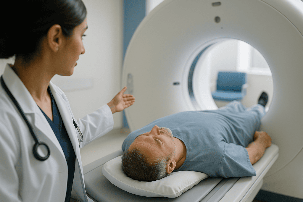
The technologist asks you to lie on your back with arms above your head, which keeps dense arm bones out of the picture. You will hear a hum as the tube spins and a recorded voice asking you to hold your breath for several seconds. Breath-holding freezes the kidneys, which otherwise move with each breath and blur the images. For someone in the middle of a colic wave, that few seconds is the hardest part, and staff will usually time it between spasms or offer pain relief first.
Many centers now use a low-dose protocol for stone searches. The scanner delivers less radiation than a standard abdominal CT by accepting a grainier image, which is acceptable because stones are so bright that they still stand out clearly. The Mayo Clinic notes that this low-dose approach is widely used for stone evaluation. If you are slimmer the dose can be reduced further; in people with larger body habitus the scanner compensates upward to keep the picture readable.
Afterward there are no restrictions. You can eat, drink and move normally. The images are reviewed by a radiologist, a doctor trained to interpret scans, who measures the stone, records its position in one of the three ureter segments and notes any swelling of the kidney. Those measurements, not the picture itself, are what the treating team acts on.
Ultrasound vs CT for kidney stones: when sound is the better first choice
Ultrasound builds an image from high-frequency sound waves bounced off tissue. It uses no radiation at all, which is why the NIDDK and Mayo Clinic both describe it as the preferred first test in pregnancy and in children, and why it is increasingly used for younger adults with a typical presentation.
Its strength is the kidney itself. Ultrasound shows hydronephrosis, the swelling of the kidney’s collecting system behind a blockage, very clearly, and it can spot stones sitting inside the kidney. Its weakness is the middle of the ureter. That part of the tube runs behind loops of bowel, and gas scatters sound waves, so a stone in the mid-ureter often simply cannot be seen. Small stones near the bladder may also hide. In practice, an ultrasound that shows a swollen kidney on the painful side strongly suggests an obstructing stone even when the stone itself is not visualized.
A short plain x-ray of the abdomen, sometimes called a KUB for kidney, ureter and bladder, is occasionally added. It shows calcium-containing stones as white specks and is useful later for tracking a known stone without repeat CT, but it misses uric acid stones and overlaps with bowel content and bone.
The honest summary from mainstream guidance is this: CT finds more stones and measures them more accurately; ultrasound avoids radiation and reliably shows whether the kidney is under pressure. Neither is wrong. In many emergency pathways an initial ultrasound is followed by CT only if the result is unclear or the pain does not settle, and the choice between them is a judgment about your age, your history and how typical your symptoms are.
Kidney function tests for kidney stones: what the blood and urine reveal
Imaging shows the stone; laboratory tests show what the stone is doing to the rest of you. Three groups of results arrive alongside the scan.
The first is a blood test for creatinine, a waste product muscles release at a steady rate that healthy kidneys filter out. A rise above your usual level suggests the obstructed kidney is struggling or that the other kidney is not compensating. From creatinine the laboratory calculates eGFR, the estimated glomerular filtration rate, which expresses overall kidney filtering capacity in a single number. The Mayo Clinic lists these blood tests among the standard work-up because they also reveal too much calcium or uric acid in the blood, which hints at why the stone formed.
The second is a urine test. A dipstick checks for blood, which is present in most people with a stone, and for white cells and nitrites, which point to infection. Infection behind an obstruction is the single finding that most reliably moves someone from watchful waiting to prompt drainage, so this cheap test carries disproportionate weight. A sample may also be sent for culture to identify the organism.
The third is a complete blood count. A raised white cell count, especially with fever, reinforces the infection concern.
Later, once the acute episode has settled, the NHS and Mayo Clinic describe two further steps: analyzing any stone you catch in a strainer to learn its chemistry, and in people with repeat stones, a 24-hour urine collection to measure the minerals that drive stone formation. Those tests do not change the first week’s decisions, but they shape the advice that lowers the chance of a next time.
Who gets scanned straight away, and who is usually asked to wait
Not everyone with flank pain walks straight into a CT scanner, and the triage logic is worth understanding because it explains why your experience may differ from a friend’s.
Prompt imaging, usually CT, is the norm for a first episode in an adult, because the diagnosis has not been confirmed before and other causes need excluding. It is also the norm whenever there is fever, a known single kidney, a kidney transplant, pregnancy with suspected obstruction (usually by ultrasound), pain that pain relief does not control, or vomiting that prevents keeping fluids down. In these situations the information changes management within hours.
A different group is often managed more conservatively. Someone who has had several documented stones, whose pain feels identical to past episodes, who has no fever, normal kidney blood tests and can drink, may be offered an ultrasound rather than a repeat CT, or may be observed with a plan to image if things do not settle. The intent is to avoid accumulating radiation in people who form stones repeatedly over decades.
Children and pregnant patients are asked to wait for CT in a specific sense: ultrasound comes first, and CT is reserved for when the ultrasound leaves a question the team cannot safely leave open. The NIDDK and Mayo Clinic both describe this ordering.
Then there is the follow-up scan. After a stone is diagnosed and a decision is made to let it pass, imaging is usually repeated after a few weeks if the stone has not been seen, to confirm it has gone. That check is frequently a plain x-ray or ultrasound rather than a second CT. Whichever path applies to you, the sequencing is a clinical judgment by the treating team, weighing your particular risks.
Reading the report: size, position and hydronephrosis side by side
A radiology report for a ureteral stone typically contains three measurements that drive the conversation that follows. Knowing what they mean lets you take part in that conversation rather than nod along.
| Report item | What it describes | Why the team cares |
|---|---|---|
| Stone size (mm) | Largest diameter, usually measured on the CT slice where the stone is widest | Smaller stones are more likely to pass unaided; larger ones more often need a procedure |
| Position | Proximal (near kidney), mid, or distal ureter including the junction with the bladder | Distal stones have less distance to travel; proximal stones may be approached differently |
| Hydronephrosis grade | Degree of swelling in the kidney’s collecting system, graded mild to severe | Severe or worsening swelling, especially with infection, raises urgency to relieve the blockage |
| Fat stranding or perinephric fluid | Inflammation or leaked urine around the kidney | Suggests high pressure; adds weight to earlier intervention |
| Other kidney | Appearance and any stones on the opposite side | Confirms the unobstructed kidney is healthy and working |
Two caveats keep these numbers honest. Size on CT is a measurement of a shadow, and the same stone can read a millimeter larger or smaller depending on the slice; clinicians treat it as a range rather than a decimal. Hydronephrosis grading is also partly subjective, and a kidney can look mildly swollen for a day or two even after a stone has passed.
What the report cannot tell you is how the stone will behave. A stone’s chemistry, the ureter’s own anatomy and how much it spasms all influence passage, and none of them appear on the scan. That is why the team combines the report with your symptoms, your labs and your history before recommending a course.
What size ureter stone requires surgery? The honest answer
This is the question people most want a number for, and the mainstream answer is a range rather than a threshold. The NHS states that most kidney stones are small enough to pass in the urine, and that larger stones may need a procedure to break them up or remove them. The Mayo Clinic describes small stones as often passing with fluids, pain relief and sometimes a medicine that relaxes the ureter, while stones too large to pass on their own are considered for shock wave therapy, ureteroscopy or, rarely, open-style surgery.
In everyday urology, stones measured at a few millimeters sitting low in the ureter are the ones most often given time. Stones wider than that, and especially those near the top of the ureter, pass less reliably, and the balance tips toward intervention as width increases. But size alone has never been the rule. The same mid-sized stone will be watched in a comfortable person with normal kidney tests and treated promptly in someone with fever, a single kidney, uncontrolled pain, or a job such as piloting where sudden incapacitating pain is a safety issue.
The medicine mentioned above belongs to a class called alpha blockers, which relax smooth muscle in the lower ureter and are sometimes prescribed to help a distal stone move. Whether it is suitable, and for how long, is a decision for the prescribing clinician; evidence suggests it helps most for larger distal stones and little for very small ones.
The procedures themselves are described in neutral terms by the NHS: shock wave lithotripsy focuses sound energy from outside the body to fragment the stone; ureteroscopy passes a thin telescope up through the bladder to break or retrieve it, often leaving a temporary internal tube called a stent; percutaneous surgery enters the kidney through the back for very large stones. Which, if any, applies to you is a joint decision with your urologist, informed by exactly the measurements the CT provides.
How long results take, and what the following days and weeks look like
Timing depends heavily on where the scan happens. In an emergency department the radiologist’s report is usually available within the same visit, often within an hour or two, because the result decides whether you go home or are admitted. For a scan booked as an outpatient, the report typically reaches the referring clinician within a few working days, and the clinic then contacts you. If you have not heard anything after about a week, it is reasonable to call rather than assume no news is good news.
Once a stone has been located and the plan is to let it pass, the following weeks have a recognizable shape. The NHS advises drinking enough water to keep urine pale, continuing pain relief as prescribed, and catching urine through a strainer so the stone can be analyzed. Pain tends to come in episodes separated by quiet stretches as the stone moves and sticks. Many people notice a final sharp twinge, then relief, then the stone in the strainer.
Follow-up imaging is standard if nothing has been caught. Guidance from the NIDDK and Mayo Clinic supports re-checking within a few weeks, commonly with ultrasound or a plain x-ray, to confirm the ureter is clear. A stone that stays put beyond that window, even if the pain has faded, is not considered safe to ignore because a silent blockage can quietly damage the kidney. That is the point at which the team often revisits the options.
If a procedure is chosen instead, the days after ureteroscopy frequently involve stent-related urgency and mild bleeding until the stent is removed, while shock wave therapy is commonly followed by passing gritty fragments for a period. Your team will give the specific timeline for your situation.
Is it worth getting a CT scan for kidney stones? Weighing radiation against information
Radiation is the only real cost of a CT scan to your body, and it deserves a straight answer rather than reassurance. A CT of the abdomen and pelvis delivers more radiation than a chest x-ray, and the risk, while small for any single scan, is cumulative over a lifetime. MedlinePlus notes that the benefits usually outweigh the risks when a scan answers an important clinical question, and that children are more sensitive than adults.
For a first episode of severe flank pain in an adult, most mainstream guidance treats CT as worth it. The scan confirms the diagnosis, excludes dangerous look-alikes such as a leaking aortic aneurysm, and provides the measurements that decide treatment. The low-dose stone protocol, now widespread, lowers the exposure substantially without losing the ability to see stones, which shifts the balance further in its favor.
The calculation changes for people who form stones repeatedly. Someone who has had five episodes in ten years and a CT each time has accumulated a meaningful dose. For them, clinicians increasingly reach for ultrasound first, and use CT selectively. Pregnancy and childhood sit at the far end of the spectrum, where ultrasound is the default and CT is a last resort.
Two practical points help you manage this yourself. Keep a record of the scans you have had, including where and when, so that a new team can review old images rather than repeat them. And ask whether a low-dose protocol is being used; in most modern departments it is, but asking costs nothing.
The question, then, is not whether CT is worth it in general. It is whether this scan, for this episode, answers a question that will change what happens next. When it does, the evidence supports it.
What people often get wrong about locating a ureteral stone
“If the scan shows a stone, I need an operation.” Most people who are scanned for a stone do not go on to have a procedure. The NHS and Mayo Clinic both describe spontaneous passage as the usual outcome for smaller stones. The scan is what makes watchful waiting safe, by confirming the stone is a size and position where waiting is reasonable.
“The pain stopped, so the stone must have passed.” Pain settles when the ureter stops spasming, which can happen while the stone is still lodged. A persistently blocked kidney can become quiet and still lose function over weeks. This is why follow-up imaging is recommended when no stone has been caught.
“A normal ultrasound rules out a stone.” Ultrasound often cannot see the mid-ureter, so a normal study does not exclude a stone there. It is reassuring if it also shows no kidney swelling, but it is not the final word.
“I should drink huge amounts of water to flush it out.” Staying well hydrated helps, but drinking to excess while vomiting or in severe pain can make you feel worse, and extreme volumes do not push a stuck stone through a narrowed ureter. Pale urine is the target the NHS describes, not a gallon challenge.
“Kidney stones show up on any x-ray.” Uric acid stones, which are not rare, are invisible on plain films. That is one reason CT replaced x-ray as the first-line test.
“Bigger pain means a bigger stone.” Pain reflects how tightly the ureter is clamping down, not stone size. Some of the most painful stones are tiny, and some large kidney stones cause nothing at all until they move.
“Contrast dye would show the stone better.” For stone detection it does the opposite; bright dye in the ureter can hide a bright stone. Contrast is reserved for other questions.
Questions to ask your care team after the scan
A CT report arrives full of measurements, and the ten minutes you get with the clinician afterward go further if you know what to ask. These questions are drawn from what patients most often wish they had raised.
- How large is the stone in millimeters, and where exactly in the ureter is it sitting?
- Is there any swelling of the kidney, and if so, how severe did the radiologist grade it?
- Do my blood tests show my kidneys are working normally, including the kidney on the other side?
- Is there any sign of infection in my urine or blood?
- Given the size and position, is waiting for it to pass a reasonable option for me, and for how long?
- What symptoms would mean the waiting plan is no longer safe?
- If a procedure becomes necessary, which approach would you consider and why?
- Was a low-dose CT protocol used, and can I get a copy of my images and report for future reference?
- Should I strain my urine, and what should I do with a stone I catch?
- When will follow-up imaging happen, and will it be ultrasound, x-ray or CT?
- Once this episode is over, what tests would help explain why I formed a stone?
Write the answers down, or ask someone to. People in pain, or recently given strong pain relief, remember remarkably little of what they are told. It is also fair to ask for a printed copy of the report; most health systems provide it on request, and having the numbers in hand makes any second conversation, including with a different clinician, far more productive.
None of these questions imply distrust. A good care team welcomes them, because a patient who understands the plan is the one most likely to return at the right moment rather than too late.
When to call your doctor: red-flag signs after a ureteral stone is found
Most people who are sent home with a known ureteral stone and a plan to let it pass do exactly that, uneventfully. A small number develop complications, and the difference between a good outcome and a serious one is often how quickly they return. The Cleveland Clinic, NHS and Mayo Clinic are consistent about the warning signs.
Seek urgent care, by emergency services if needed, for any of the following:
- Fever, shaking chills or feeling suddenly very unwell: these suggest infection trapped behind the blockage, which can progress rapidly.
- Pain that the prescribed pain relief no longer controls, or pain so severe you cannot find any position of comfort.
- Persistent vomiting that prevents you from keeping fluids or medicines down.
- Passing very little or no urine over several hours, which may mean both kidneys are affected or you have only one functioning kidney.
- Heavy or clotted blood in the urine, or urine that looks like red wine rather than faintly pink.
- Confusion, drowsiness, rapid breathing or a racing heartbeat alongside any of the above.
Call your doctor’s office within a day or two, rather than waiting for the scheduled follow-up, if pain has lasted longer than you were told to expect, if you were asked to strain your urine and nothing has appeared after several weeks, if a new ache has started on the opposite side, or if you simply feel the plan is not working. Rising creatinine on a repeat blood test, if one was arranged, is another reason the office may contact you.
None of these signs means something terrible has happened. Each means the balance between waiting and acting may have shifted, and that judgment belongs to the clinicians caring for you, armed with the latest information. Calling early gives them that chance.
Frequently asked questions
Is it worth getting a CT scan for kidney stones?
For a first episode of severe flank pain in an adult, mainstream guidance generally supports CT because it confirms the stone, measures it precisely and rules out dangerous look-alike conditions. Low-dose protocols reduce radiation considerably. For people with many previous stones, or in pregnancy and childhood, ultrasound is often tried first. The right choice depends on your history and is a decision for the treating team.
Why is a non contrast CT used for a kidney stone instead of one with dye?
Stones are already dense enough to appear bright white on CT, so dye adds nothing to their detection. Injected contrast collects in the ureter and shows up bright too, which can mask a stone sitting in it. Leaving the dye out also avoids the small risks of allergic reaction and kidney strain, which matters when one kidney is already under pressure from a blockage.
How long does it take to get results from a CT scan for kidney stones?
In an emergency department the report is usually available during the same visit, often within an hour or two, because it determines whether you are admitted or sent home. Outpatient scans are typically reported within a few working days and the referring clinic then contacts you. If roughly a week passes without word, it is reasonable to phone the clinic rather than assume all is well.
What are the first signs of a ureteral stone?
The earliest sign is usually a one-sided ache between the lowest rib and the hip that sharpens into cramping waves over minutes to hours. Nausea, vomiting, restlessness and pink or tea-colored urine commonly follow. As the stone descends, pain may shift toward the groin with an urge to urinate often. Fever alongside these symptoms suggests infection and warrants urgent assessment rather than waiting.
Ultrasound vs CT for kidney stones: which one finds more?
CT finds more stones and measures them more accurately, including small ones and those hidden in the mid-ureter behind bowel gas, where ultrasound often cannot see. Ultrasound reliably shows whether the kidney is swollen from a blockage and uses no radiation, which is why it is preferred first in pregnancy and children. Many pathways start with ultrasound and add CT only if a question remains.
What size ureter stone requires surgery?
There is no single cutoff. Smaller stones, especially those low in the ureter, are usually given time to pass; wider stones and those near the kidney pass less reliably and are more often treated with shock wave therapy or ureteroscopy. Fever, a single kidney, uncontrolled pain or failure to pass after several weeks can prompt intervention regardless of size. Your urologist weighs all of these together.
What kidney function tests are done for kidney stones?
A blood test measures creatinine and calculates eGFR, which together estimate how well the kidneys are filtering. A urine dipstick checks for blood and signs of infection, and a culture may follow. A complete blood count looks for a raised white cell count. After the acute episode, stone analysis and sometimes a 24-hour urine collection help explain why the stone formed.
Can a ureteral stone be missed on a CT scan?
It is uncommon but possible. Very small stones, stones that have just passed into the bladder, or stones in a person who cannot hold still can be hard to identify. Some rare stone types linked to certain medicines are less dense and harder to see. If symptoms persist after a scan reported as clear, the team may repeat imaging or look for other causes of the pain.
What does hydronephrosis from a ureteral stone mean?
Hydronephrosis is swelling of the kidney’s internal drainage system because urine cannot flow past the blockage. On CT or ultrasound it appears as a dilated collecting system and is graded mild to severe. Mild swelling is expected with any obstructing stone; severe or worsening swelling, particularly with infection or a rising creatinine, increases the urgency to relieve the blockage rather than wait.
Will I need another CT scan after the stone is found?
Usually not. If the stone is not caught in a strainer, follow-up imaging within a few weeks is recommended to confirm it has gone, but this is often a plain x-ray or ultrasound rather than a second CT, to limit radiation. A repeat CT is reserved for situations where those tests leave an important question unanswered or symptoms suggest a new problem.
References
- NHS: Kidney stones
- NIH NIDDK: Diagnosis of kidney stones
- Cleveland Clinic: Kidney stones
- MedlinePlus: CT scans
This article is for general information only and is not a substitute for professional medical advice. Please consult a qualified doctor about your individual situation.

More from the Blog

How Sling Surgery for Stress Incontinence Works: Anesthesia, the Procedure and Time in Hospital
A sling surgery procedure treats stress urinary incontinence by placing a narrow strip of synthetic mesh or the patient's own tissue under the urethra…

How Do a Bladder Diary and Urodynamic Testing Pinpoint Female Bladder Problems?
A bladder diary records, over roughly three days, what a woman drinks, when she passes urine, how much, and when leaks or urgency occur,…

Is Your Nephrostomy Tube Not Draining or Leaking? What Is Normal and When to Call the Team
A nephrostomy tube that stops draining is most often kinked, clamped, blocked by a clot or debris, or pulled out of position, and it…

Fluid Management With Heart or Kidney Disease: Why the Drinking Plan Is Individually Set
Fluid management in heart and kidney patients is individually set because the safe amount of fluid depends on how well the heart pumps, how…

Bowel and Urinary Preparation Before Minimally Invasive Urology: What the Instructions Mean
Bowel prep before urology surgery ranges from nothing beyond standard fasting to a full colonoscopy-style clean-out, depending on whether the surgeon will work near…

Inherited Kidney Disease Care: How Alport Syndrome and Similar Conditions Are Monitored
Hereditary kidney disease treatment for conditions such as Alport syndrome and polycystic kidney disease centers on lifelong monitoring rather than a single procedure: regular…