

How Brain Aneurysm Imaging Works: CT Angiography, MRA and the Catheter Angiogram

Key Takeaways
- CT angiography detects roughly 97 percent of brain aneurysms larger than about 3 mm, but its sensitivity falls substantially for smaller ones, according to the AHA unruptured aneurysm guideline.
- A plain head CT without contrast can show bleeding from a ruptured aneurysm but cannot reliably show an unruptured one; only CTA, MRA or a catheter angiogram images the vessel wall.
- The catheter angiogram remains the reference standard because direct arterial contrast and real-time X-ray reveal aneurysms of 1 to 2 mm and show how blood flows through them.
- Neurological complications from diagnostic cerebral angiography occur in well under 1 percent of procedures in large series, with permanent deficits rarer still, per AHA guidance.
- Time-of-flight MRA needs no contrast or radiation, which is why it is favored for screening high-risk families and for long-term surveillance of small untreated or coiled aneurysms.
- The AHA guideline suggests a first follow-up scan around six to twelve months after an untreated small aneurysm is found, then yearly or every two years, usually with noninvasive imaging.
CT angiography (CTA) is usually the first test for a suspected brain aneurysm because it is fast, widely available and detects most aneurysms larger than about 3 mm. Catheter angiography remains the reference standard, showing small aneurysms and neck anatomy in finer detail, but it is invasive and carries a small stroke risk. MRA avoids radiation and suits screening and follow-up. The care team chooses based on urgency, aneurysm size and planned treatment.
The radiologist’s report arrives as a single line in a patient portal: “3 mm outpouching of the right middle cerebral artery, likely saccular aneurysm, recommend further characterization.” You were scanned for migraines. Now you are searching “cta vs angiogram brain aneurysm” at midnight, trying to work out whether the next test involves a needle in your groin and whether the first one could have been wrong.
That uncertainty is the ordinary experience of aneurysm imaging. Three tests, CT angiography, magnetic resonance angiography and the catheter angiogram, look at the same blood vessels in three different ways, and each has a blind spot the others cover. The order in which they are used is not arbitrary. It follows how urgent the situation is, how small the suspected aneurysm is, and whether anyone is already planning to treat it.
This explainer walks through what each scan actually does, what the evidence says about how much each one misses, and what the day and the following weeks feel like from the patient’s side.
What is the difference between a CTA and an angiogram for a brain aneurysm?
A brain aneurysm is a weak, bulging spot in the wall of an artery inside the skull. Finding one means seeing the artery itself, not the brain tissue around it, and that is what separates these tests from an ordinary head scan.
A CT angiogram (CTA) is a CT scan timed to catch iodine contrast dye as it fills the arteries. The dye goes in through an ordinary arm vein. The scanner spins around the head for a few seconds and a computer reconstructs the vessels in three dimensions. Nothing enters an artery.
A catheter angiogram, also called cerebral angiography or digital subtraction angiography (DSA), is different in kind. A doctor threads a thin tube from an artery in the groin or wrist up to the neck vessels and injects contrast directly into them while X-ray images are taken. Because the dye arrives exactly where it is needed and images are captured as it flows, the pictures are sharper and show blood moving in real time. The trade-off is that a catheter is inside your arteries.
Magnetic resonance angiography (MRA) sits between them: no radiation, often no contrast at all, but slower and more sensitive to movement.
When people compare CTA vs angiogram for a brain aneurysm, they are really asking two separate questions. First, which test is most likely to find an aneurysm that exists? Second, which one is worth the extra risk? The American Heart Association’s guideline on unruptured aneurysms answers the first with numbers and the second with judgment, and the rest of this article follows that structure.
How each aneurysm scan actually works, in plain language
Picture a garden hose with a bubble in it. You can spot the bubble by shining a light through the hose from outside, or by putting a dye in the water and watching where it pools. The three tests do versions of both.
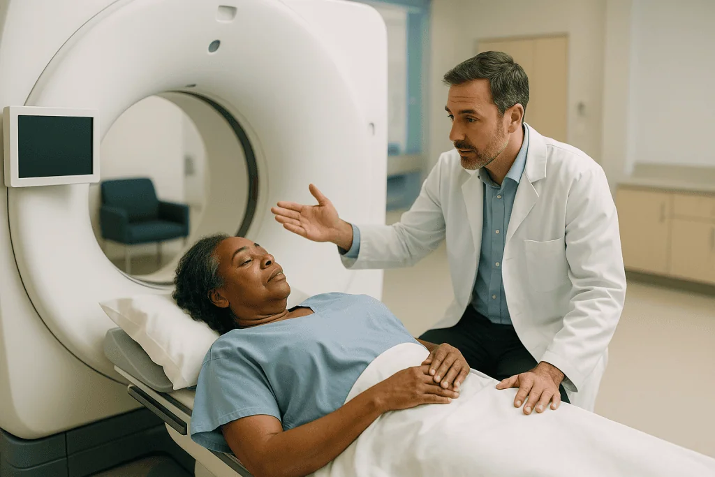
For a CTA, you lie on a narrow table that slides into a ring-shaped scanner. A technologist places an intravenous line, usually in the arm, and a pump injects contrast over a few seconds. The scanner is programmed to fire as the dye reaches the brain arteries, so timing matters more than it does for a plain head CT. The scan itself is over in under a minute; most of the appointment is preparation. Afterwards, software peels away bone and displays the arteries as a 3D model that can be rotated on screen.
MRA uses a strong magnet and radio waves. One common technique, time-of-flight MRA, needs no dye at all: it exploits the fact that moving blood looks different from stationary tissue. You lie still inside a tube for roughly ten to twenty minutes while the machine knocks and hums. Any movement blurs the vessels, which is why MRA is harder for people who are restless, claustrophobic or acutely unwell.
The catheter angiogram takes place in an angiography suite rather than a scanner. After local anesthetic numbs the skin, an interventional neuroradiologist or neurosurgeon guides a catheter through the arterial tree using live X-ray. Each injection of dye produces a short movie of blood filling and emptying the vessels. MedlinePlus describes the whole procedure as typically lasting one to three hours, most of it positioning and imaging rather than the puncture itself.
Can a CTA detect an aneurysm? Where it excels and where it misses
Yes, and for most aneurysms it does so reliably. The AHA guideline on unruptured intracranial aneurysms summarizes pooled studies showing CTA sensitivity of roughly 97 percent and specificity close to 98 percent when compared against catheter angiography. In plain terms, if an aneurysm is present, CTA finds it about 97 times in 100, and when it reports one, it is almost always right.
The catch lives in the small print about size. Those figures are driven by aneurysms larger than 3 mm. For aneurysms under 3 mm, the same guideline notes that reported CTA sensitivity drops substantially, with wide variation between studies. A 2 mm bulge on a curving vessel next to the skull base can be lost in bone artifact or mistaken for a normal branch point.
Location matters too. CTA performs well for aneurysms of the middle cerebral and anterior communicating arteries, which sit away from dense bone. It struggles more around the cavernous segment of the internal carotid artery and the posterior circulation, where bone and venous structures crowd the picture.
Newer multidetector scanners and post-processing have narrowed the gap, and research into software that flags possible aneurysms for a radiologist’s review is active. Those tools are decision support, not diagnosis, and none of them changes the physics of a tiny lesion beside bone.
So the honest answer to whether a CTA can detect an aneurysm is: almost always for the ones most likely to matter, less dependably for the smallest ones, and best when a specialist reads it knowing what the clinical question is.
MRA vs CTA for aneurysm: when doctors choose magnets over X-rays
MRA and CTA answer similar questions with different costs to the body. MRA involves no ionizing radiation and, in its time-of-flight form, no contrast. That makes it the natural choice when the same person will be scanned repeatedly over years.

The AHA unruptured aneurysm guideline reports MRA sensitivity in the mid-90s percent range for aneurysms above 3 mm, comparable to CTA, with the same fall-off for smaller lesions. Where MRA lags is speed and robustness. A CTA of the head takes seconds; an MRA takes many minutes of holding still. In an emergency department with a possible ruptured aneurysm, that difference is decisive, and CT-based imaging wins.
Several situations tilt toward MRA:
- Screening people with a strong family history, where the AHA guideline suggests considering imaging when two or more first-degree relatives have had aneurysms, and where lifetime radiation exposure adds up.
- Follow-up of known small aneurysms that are being watched rather than treated.
- People with reduced kidney function or a documented reaction to iodinated contrast.
- Pregnancy, where avoiding radiation is preferred when clinically reasonable.
MRA has its own exclusions. Certain older pacemakers, cochlear implants, metal fragments in the eye and some aneurysm clips are not compatible with the magnet, and every MRI department screens for these before anyone enters the room. Claustrophobia and inability to lie still degrade the images.
There is no universal winner in the MRA vs CTA for aneurysm debate. The pattern in practice is CTA for speed and acute situations, MRA for repetition and radiation-sparing, and either one as the gatekeeper that decides whether a catheter angiogram is worth doing at all.
Why the catheter angiogram is still called the reference standard
Every accuracy figure quoted for CTA and MRA is measured against something, and that something is the catheter angiogram. It earns the title “reference standard” for three reasons rooted in physics rather than habit.
Resolution comes first. Contrast injected directly into the carotid or vertebral artery is dense and undiluted, and the X-ray detector captures it at a spatial resolution the noninvasive scans cannot match. Aneurysms of 1 to 2 mm, tiny daughter sacs on the dome, and small branches arising from the aneurysm neck become visible.
Time is second. Because DSA records a sequence of frames as dye passes through, it shows flow: how quickly the aneurysm fills, whether it empties slowly, whether a nearby vessel is being fed from an unexpected direction. A CTA is a single frozen moment; an angiogram is a short film.
Three-dimensional rotational angiography, in which the X-ray arm sweeps around the head during one injection, adds a rotatable model that helps the team measure the neck of the aneurysm and plan whether a coil, a stent or a surgical clip would fit.
The AHA guideline recommends catheter angiography when noninvasive imaging is equivocal, when a subarachnoid hemorrhage has occurred and CTA has not shown a cause, and when detailed anatomy is needed before endovascular treatment. It is not recommended as a routine first test for every incidental finding.
The reference standard is not flawless. Very small aneurysms can hide behind overlapping vessels on a given projection, and the test depends on the operator choosing the right angles. Its authority comes from being the best available comparison, not from being perfect.
CTA vs angiogram for brain aneurysm: a side-by-side summary
The table below pulls together what the preceding sections describe. Accuracy figures are those summarized in the AHA guideline on unruptured intracranial aneurysms and apply to aneurysms above roughly 3 mm; all three tests perform less well below that size.
| Feature | CT angiography (CTA) | MR angiography (MRA) | Catheter angiogram (DSA) |
|---|---|---|---|
| How dye is given | Arm vein | Often none; sometimes arm vein | Directly into neck arteries via catheter |
| Radiation | Yes | No | Yes, usually more than CTA |
| Scan time | Seconds | 10–20 minutes | 1–3 hours including preparation |
| Sensitivity, aneurysms over 3 mm | About 97% | Mid-90s percent | Reference standard |
| Shows blood flow over time | No | Limited | Yes |
| Main risks | Contrast reaction, kidney strain | Metal incompatibility, claustrophobia | Puncture-site bleeding, small stroke risk |
| Typical role | First-line, emergencies | Screening, long-term follow-up | Equivocal cases, treatment planning |
Two patterns stand out. The noninvasive tests are close to each other in accuracy and differ mainly in speed and radiation. The catheter angiogram stands apart in what it can show and in what it asks of the patient.
That asymmetry is why the sequence usually runs CTA or MRA first, angiogram only if the answer is unclear or treatment is being planned. A negative CTA in a low-risk person often ends the workup. A 2 mm question mark near the skull base in someone whose sibling had a rupture may not.
Who is usually offered which test, and who is asked to wait
Three groups arrive at aneurysm imaging by different doors, and the door shapes the test.
The first group is in an emergency department with a sudden, severe headache or a collapse. Here the concern is a ruptured aneurysm bleeding into the space around the brain, called subarachnoid hemorrhage. A plain CT comes first to look for blood. The AHA guideline on aneurysmal subarachnoid hemorrhage supports CTA as the immediate next step to find the source, with catheter angiography if CTA is negative or treatment is planned. Nobody in this group is asked to wait.
The second group has an incidental finding: an aneurysm spotted on a scan done for something else. The Mayo Clinic notes this is increasingly common as brain imaging becomes routine. For a small, smooth aneurysm in a low-risk location, the treating team may propose surveillance with MRA or CTA rather than an angiogram, because the AHA guideline recognizes that many small unruptured aneurysms carry a low yearly rupture risk and that treatment has its own risks. “Wait” here means watchful imaging on a schedule, not neglect.
The third group is being screened without symptoms because of family history or a condition such as autosomal dominant polycystic kidney disease. The AHA guideline suggests considering noninvasive imaging for people with two or more affected first-degree relatives; a single affected relative is a discussion, not an automatic scan.
Who is asked to hold off entirely? People whose only symptom is a common headache pattern with no red flags, for whom guidelines do not support aneurysm imaging, and anyone whose kidney function or contrast history makes a particular test unsafe until it can be adjusted. Those decisions belong to the clinician who knows the whole picture.
Which is safer, an angiogram or a CT scan? Cerebral angiogram risks weighed honestly
Safer for whom, and against what alternative? Framed properly, the comparison is less lopsided than it first appears, though the catheter angiogram does carry risks the scans do not.
CTA’s hazards are those of any contrast CT. Iodinated contrast can trigger allergic-type reactions, most mild, a few serious. It can strain kidneys that are already impaired, which is why kidney function is checked in people at risk. The radiation dose from a head CTA is modest for a single scan; the concern is cumulative exposure in someone scanned yearly for decades, which is one reason follow-up often shifts to MRA.
MRA’s risks are almost entirely about metal and confinement. Gadolinium contrast, when used, has a very low rate of serious reaction.
The catheter angiogram adds two categories. At the puncture site, bruising is common and a larger collection of blood, a hematoma, or damage to the artery is uncommon. Inside the head, the catheter or dislodged plaque can block a small vessel. MedlinePlus and Johns Hopkins both list stroke as a recognized complication. The AHA subarachnoid hemorrhage guideline describes neurological complications from diagnostic cerebral angiography as occurring in well under 1 percent of procedures in large series, with permanent deficits far rarer still, and notes that risk is higher in older patients and those with atherosclerosis.
So the honest answer: a CT scan is safer than a catheter angiogram for the same person on the same day. The angiogram is justified when it will change what happens next, because a missed aneurysm or an imprecisely planned treatment carries risks of its own. That trade is the treating team’s to weigh with you, not a fixed rule.
Is a brain angiogram painful? What the day actually feels like
Most people describe a cerebral angiogram as strange rather than painful, and the strangeness is worth knowing about in advance.
You will be asked not to eat for several hours beforehand and to tell the team about every medicine you take, especially blood thinners and diabetes medicines, since these may need adjustment under the prescribing clinician’s direction. Do not stop anything on your own.
In the angiography suite you lie on a hard table with your head cradled. The groin or wrist is cleaned and numbed with local anesthetic; the injection stings for a few seconds, after which pressure is felt but not sharp pain. Many people receive light sedation and remain awake, because the team may ask you to hold your breath or keep still at specific moments. MedlinePlus notes that some centers use deeper sedation or general anesthesia, particularly for children or people who cannot lie still.
You will not feel the catheter moving through your arteries; arteries have no pain sensors for this. What people do feel is the contrast injection: a sudden warm flush across the face and behind the eyes, sometimes a metallic taste, occasionally brief flashes of light or a fleeting sensation of heat in the jaw. Johns Hopkins describes this as normal and short-lived. Some people feel a headache during injections.
When the imaging is done, the catheter comes out and the puncture is sealed either by firm manual pressure or a small closure device. The soreness afterwards is at the puncture site, usually described as a bruise-like ache for a day or two. The part patients most often complain about is not pain but the hours of lying flat afterwards.
What the following days and weeks usually look like
The three tests diverge sharply once you leave the room.
After a CTA or MRA, most people go straight home or back to the ward. Drinking fluids over the following day helps the kidneys clear contrast. There are no activity limits. Results are typically read by a radiologist within hours in an emergency setting and within days for outpatient scans; the referring clinician, not the scanner, delivers them.
After a catheter angiogram, recovery follows a predictable script. MedlinePlus describes lying flat for several hours so the arterial puncture can seal, with staff checking the site, pulses in the foot or hand, and neurological signs at intervals. Wrist access generally allows earlier sitting up than groin access. Many people go home the same day with a companion; some stay overnight, particularly if the angiogram was part of a hospital admission.
The first days at home are about protecting the puncture. Johns Hopkins and MedlinePlus advise avoiding heavy lifting, strenuous exercise and driving for a short period, typically a day or two, and keeping the site clean and dry. A small bruise is expected; a rapidly expanding, tense lump is not.
The weeks that follow depend entirely on what the imaging showed. If no aneurysm was found, the pathway may end. If a small aneurysm is being watched, the AHA unruptured aneurysm guideline suggests a first follow-up scan at around six to twelve months, then yearly or every two years, with MRA or CTA rather than repeated angiograms. If treatment is planned, the angiogram often becomes the map for the procedure, and the timeline is set by the treating team based on rupture status and anatomy.
Imaging after an aneurysm has been treated: coils, clips and follow-up scans
Treatment does not end the imaging; it changes what the imaging is looking for. Two main methods are used to treat aneurysms. Endovascular coiling packs the sac with soft platinum coils delivered through a catheter, sometimes alongside a stent that reshapes the vessel. Surgical clipping places a tiny metal clip across the neck of the aneurysm during an open operation. Each leaves metal behind, and metal is what complicates follow-up scans.
The question after treatment is whether the aneurysm is fully closed off or whether blood is finding its way back in, which is called recurrence or recanalization. Coiled aneurysms can compact over time and reopen at the neck, so guidelines from the AHA recommend periodic follow-up imaging after coiling. Clipped aneurysms recur far less often but are not immune.
Here the three tests trade places. CTA is degraded by coils, which produce bright streaks across the image and hide the neck. MRA copes better with coils, and time-of-flight MRA has become a common noninvasive follow-up tool after coiling, though it is checked for compatibility with any stent used. Clips, by contrast, scatter the MRA signal and can be assessed more clearly on CTA, provided the clip material is confirmed MRI-safe if MRA is ever needed.
The catheter angiogram remains the most precise judge of whether a treated aneurysm is truly closed, and many teams perform one at a set interval after coiling before relaxing to noninvasive surveillance. How often, and for how long, is individualized to the aneurysm’s size, the completeness of the initial treatment and the person’s age. Those intervals are set by the treating team, not by a standard calendar.
What people often get wrong about aneurysm scans
Aneurysm imaging attracts a particular set of misunderstandings, partly because the stakes feel high and partly because the tests have confusingly similar names.
“I had a normal head CT, so I don’t have an aneurysm.” A plain CT without contrast looks at brain tissue and blood, not vessel walls. It can show bleeding from a ruptured aneurysm; it cannot reliably show an unruptured one. Only angiographic imaging does that.
“The angiogram is the best test, so I should insist on it.” More detail is not the same as more benefit. For a low-risk incidental finding, the AHA guideline supports noninvasive follow-up, and the small but real stroke risk of catheter angiography is not justified when the result would not change management.
“A negative CTA means I’m in the clear forever.” It means no aneurysm above the scan’s detection threshold was seen on that day. Aneurysms can form later in life, which is why screening in high-risk families is periodic rather than one-off.
“Finding an aneurysm means it will burst.” The National Institute of Neurological Disorders and Stroke estimates that roughly one in fifty adults has an unruptured aneurysm, the great majority of which never rupture. Size, location, shape and personal risk factors drive the estimate, and the treating team weighs them individually.
“Contrast dye is dangerous.” Serious contrast reactions are uncommon, and screening for kidney function and allergy history is routine. Refusing contrast without discussing it can mean a less accurate scan.
Each of these errors pushes in one of two directions: too little imaging, or too much. The evidence sits in the middle.
Questions to ask your care team before aneurysm imaging
A good consultation about aneurysm imaging is a conversation about thresholds: what the scan needs to show to change the plan. These questions tend to surface that reasoning.
- What exactly are we looking for, and what result would change what we do next?
- Why this test rather than one of the others? If a catheter angiogram is proposed, what would a CTA or MRA have missed?
- How large and where is the suspected aneurysm, and how does that affect the accuracy of the scan you are ordering?
- What are the specific risks for me, given my kidney function, allergies, age and any blood thinners I take?
- Should any of my regular medicines be adjusted before the test, and who will tell me how?
- Will I be sedated, and do I need someone to drive me home?
- How long will I need to lie flat afterwards, and how long before I can return to work and exercise?
- Who reads the scan, and how and when will I receive the result?
- If the finding is small, what would surveillance look like, and which test would you use for it?
- If the aneurysm is treated, what imaging will I need afterwards, and for how long?
- Is there a reason to involve a second specialty, such as neurosurgery or interventional neuroradiology, before deciding?
Write the answers down or bring someone who will. The details that matter most, the size in millimeters, the artery involved and the planned interval to the next scan, are exactly the ones that slip away in a short appointment. None of these questions second-guesses the team; they help you understand the decision that is being made with you.
When to call your doctor
Two sets of warning signs matter here: those that suggest an aneurysm may have ruptured, and those that suggest a complication after a catheter angiogram.
Call emergency services immediately, do not drive yourself, if you or someone with you experiences a sudden, explosive headache unlike any before, often described as the worst headache of their life, particularly with a stiff neck, vomiting, sensitivity to light, a seizure, a drooping eyelid, double vision, confusion or loss of consciousness. The NHS and Mayo Clinic both describe this pattern as the hallmark of subarachnoid hemorrhage, and minutes matter.
After a catheter angiogram, seek urgent care for any of the following:
- Bleeding from the puncture site that does not stop with firm pressure, or a lump that is growing, tense or increasingly painful.
- A leg or hand below the puncture that becomes cold, pale, numb, weak or changes color.
- New weakness or numbness on one side of the body, difficulty speaking or understanding speech, facial drooping, or sudden vision loss. These are signs of stroke and are emergencies.
- Fever, spreading redness or discharge at the puncture site in the days afterwards.
- Hives, swelling of the face or throat, or difficulty breathing, which may indicate a delayed contrast reaction.
Contact the team that performed the test, rather than waiting for a scheduled follow-up, if you notice a persistent headache that is worsening, dizziness on standing, or markedly reduced urine output, which can signal kidney strain from contrast.
None of these lists replaces clinical judgment. If something feels wrong and is not on this page, call anyway. The people who ordered and performed your imaging expect those calls and would rather answer an unnecessary one than miss a necessary one.
Frequently asked questions
What is the most accurate test for diagnosing a brain aneurysm?
Catheter angiography, also called digital subtraction angiography, is the most accurate test and the reference standard against which the others are measured. It shows aneurysms as small as 1 to 2 mm and reveals blood flow in real time. Because it is invasive, guidelines reserve it for cases where CTA or MRA is unclear, where a bleed has occurred without an obvious source, or where treatment is being planned. For most aneurysms above 3 mm, CTA and MRA are accurate enough to guide decisions.
Can a CTA detect an aneurysm?
Yes. CT angiography detects about 97 percent of brain aneurysms larger than roughly 3 mm, with specificity close to 98 percent, according to the AHA guideline on unruptured aneurysms. Its weakness is very small aneurysms, particularly those close to dense bone at the skull base, where sensitivity drops and results vary between studies. A negative CTA is reassuring for the aneurysms most likely to matter, but a specialist may still recommend further imaging if clinical suspicion is high.
Which is safer, an angiogram or a CT scan?
A CT angiogram is safer than a catheter angiogram for the same person on the same day. CTA risks are limited to contrast reactions, kidney strain and a modest radiation dose. The catheter angiogram adds puncture-site bleeding and a small risk of stroke, described in AHA guidance as well under 1 percent for neurological complications in large series. The angiogram is justified when its extra detail will change treatment; otherwise noninvasive imaging is preferred.
Is a brain angiogram painful?
Most people find a brain angiogram uncomfortable rather than painful. The local anesthetic injection at the groin or wrist stings briefly. You will not feel the catheter moving through the arteries. Contrast injections cause a sudden warm flush across the face, sometimes a metallic taste or brief flashes of light, which pass within seconds. Afterwards the puncture site aches like a bruise for a day or two. The part patients most often dislike is lying flat for several hours afterwards.
How is MRA vs CTA for aneurysm decided?
The choice usually comes down to speed and radiation. CTA takes seconds and is preferred in emergencies and when a rapid answer is needed. MRA takes ten to twenty minutes, uses no radiation and often no contrast, so it suits screening of high-risk families and repeated follow-up over years. Both have similar accuracy for aneurysms above 3 mm. Kidney function, contrast allergy, pregnancy, implanted metal and the ability to lie still all influence which one a clinician orders.
What are the main cerebral angiogram risks?
The main risks are at the puncture site and inside the head. At the groin or wrist, bruising is common and a larger blood collection or artery injury is uncommon. Inside the arteries, the catheter or dislodged plaque can block a vessel and cause a stroke; AHA guidance places neurological complications well under 1 percent in large series. Contrast reactions and kidney strain are also possible. Risk rises with age and atherosclerosis, which is why the test is used selectively.
Why did my CTA find an aneurysm when I had no symptoms?
Most unruptured aneurysms cause no symptoms, and they are increasingly found by chance on scans done for other reasons such as headaches, dizziness or head injury. The National Institute of Neurological Disorders and Stroke estimates that about one in fifty adults has an unruptured aneurysm, the majority of which never rupture. An incidental finding starts a discussion about size, location, shape and personal risk factors, and often leads to surveillance imaging rather than immediate treatment.
How long do I have to lie flat after a cerebral angiogram?
MedlinePlus describes lying flat for several hours after a groin puncture so the artery can seal, with staff checking the site, foot pulses and neurological signs at intervals. Wrist access generally allows earlier sitting up. Many people go home the same day with a companion, while some stay overnight if the angiogram was part of a hospital admission. Your team will give you a specific time based on the access site and whether a closure device was used.
Do I need imaging after a brain aneurysm is coiled or clipped?
Yes, follow-up imaging is standard, particularly after coiling, because coils can compact and allow blood back into the aneurysm neck. MRA is often used after coiling since coils distort CTA images, while clipped aneurysms are frequently assessed with CTA. Many teams perform a catheter angiogram at a set interval after coiling before switching to noninvasive surveillance. The schedule is individualized to the aneurysm’s size, how completely it was closed, and the type of device used.
Can a normal head CT rule out a brain aneurysm?
No. A plain head CT without contrast shows brain tissue and blood, so it can detect bleeding from a ruptured aneurysm, but it does not image the artery walls where an unruptured aneurysm sits. Detecting an intact aneurysm requires angiographic imaging: CTA, MRA or a catheter angiogram. If you were told a head CT was normal and are concerned about aneurysm risk because of family history or symptoms, ask your clinician whether vessel imaging is appropriate.
References
- Brain aneurysm: Diagnosis (NHS)
- Cerebral angiography (MedlinePlus)
- Cerebral Aneurysms (NIH National Institute of Neurological Disorders and Stroke)
This article is for general information only and is not a substitute for professional medical advice. Please consult a qualified doctor about your individual situation.

More from the Blog

Life After a Craniotomy: Hair Regrowth, Scars, Driving and Long-Term Neurosurgery Follow-Up
Life after craniotomy usually means a hospital stay of several days, a scar hidden under regrowing hair within a few months, fatigue that eases…

How Congenital Hydrocephalus Is Treated and Why Relieving Pressure Early Protects Development
Congenital hydrocephalus is treated with surgery that redirects or reroutes the excess cerebrospinal fluid pressing on a baby's brain, most often a ventriculoperitoneal shunt…

ALS vs Other Motor Neuron Diseases: Why the Diagnosis Changes the Care Plan
ALS is the most common motor neuron disease, but the two terms are not the same thing. Motor neuron disease is an umbrella for…

How Does Recovery Differ After Brain Aneurysm Coiling and Clipping? Timelines Side by Side
After an unruptured brain aneurysm is treated, coiling recovery is usually shorter: most people go home within one to two days and ease back…
What Happens During Gamma Knife Radiosurgery: Frame or Mask, Planning and the Session Itself
During Gamma Knife radiosurgery, a lightweight head frame or a custom face mask holds the head still, detailed MRI or CT images are taken,…

Who Is a Candidate for TMS? Why Implants, Metal in the Head and Seizure History Matter
TMS is usually considered for adults with depression that has not improved after standard treatment, and for a few other approved conditions. Candidacy depends…