

How Digital Planning Maps Tooth Movement Before Clear Aligners Are Made

Key Takeaways
- A clear aligner digital plan is built from a surface scan of the crowns; roots and bone are visible only on X-rays, which is why imaging is part of the diagnosis rather than an optional extra.
- In the most cited prospective measurement, summarized in a systematic review, teeth achieved a mean of about 41 percent of their planned movement, with extrusion lowest at roughly 30 percent.
- Each tray typically moves teeth by a fraction of a millimeter, and the studies in that review changed trays about every two weeks, a schedule clinicians now vary by movement type.
- Attachments, enamel polishing between teeth (IPR) and deliberate overcorrection are planned, on-screen features of a treatment plan, not signs that something has gone wrong.
- The NHS gives a typical orthodontic treatment window of around 18 to 24 months depending on complexity, and any aligner estimate assumes near-full-time wear and typical biology.
- Retainers, often worn indefinitely, are the only established way to hold a result after the last tray, because teeth drift back toward their old positions for years.
Clear aligner digital planning starts with a 3D scan of the teeth, often alongside X-rays, from which software separates each tooth and lets the clinician set a proposed final position. The plan is divided into small staged movements, each manufactured as one aligner. Research shows achieved movement often falls short of what is planned, so the on-screen result is a clinical target that the treating team reviews and adjusts.
The screen tips toward the chair. A dentist nudges a slider with one finger, and a set of teeth that have overlapped since adolescence slide, turn and settle into a tidy arc in about eight seconds. The patient laughs, then asks the only question that matters: is that real?
It is real in the sense that every tooth on that screen is hers, measured to a fraction of a millimeter. It is not real in the sense of being finished. Clear aligner digital planning sits in the gap between those two meanings. It is the process by which a scan of a mouth becomes a proposed end point, a route to get there and a stack of trays built to walk the teeth along it.
Knowing how that route is drawn, and how often teeth decline to follow it exactly, turns a persuasive animation into something more useful: a plan you can question before you agree to it.
What is clear aligner digital planning, in plain language?
Clear aligner digital planning is the work that happens between the moment your teeth are scanned and the day the first tray is handed over. It is a computer model of your mouth in which every tooth has been separated from its neighbors, placed in a proposed final position, and then walked backward, stage by stage, to where it sits today. Each stage becomes one aligner.
The idea borrows from an old orthodontic habit. For decades, clinicians built a diagnostic set-up: plaster teeth were cut from a stone model, rearranged into the planned result and glued down in wax. Digital planning does the same thing with pixels instead of plaster, and it adds something plaster never could, an exact, measurable path for every tooth expressed in millimeters and degrees.
Three ingredients go into that path. The first is geometry, meaning the shape and current position of each crown captured by the scanner. The second is a set of clinical decisions made by the orthodontist or dentist: which teeth should move, which should stay put as anchors, whether any space needs to be created, how the bite should close at the end. The third is a set of biological limits, because a tooth cannot move faster than the bone around it can remodel.
What the software produces is a proposal, not an instruction. A well-run plan is reviewed and often edited several times by the clinician before anything is manufactured, and the version a patient sees on screen is the intended route rather than a guaranteed destination. Published reviews make clear that achieved movement regularly falls short of planned movement for certain tooth types, which is exactly why human judgment stays in the loop (Rossini et al., Angle Orthodontist).
How a 3D dental scan for aligners captures your teeth
Most plans begin with an intraoral scanner, a handheld wand about the size of an electric toothbrush that projects patterned light onto the teeth and photographs it thousands of times a second. Software stitches those frames into a single three-dimensional surface. Within a few minutes the clinician has a digital model of both arches and a record of how they meet when you bite together.
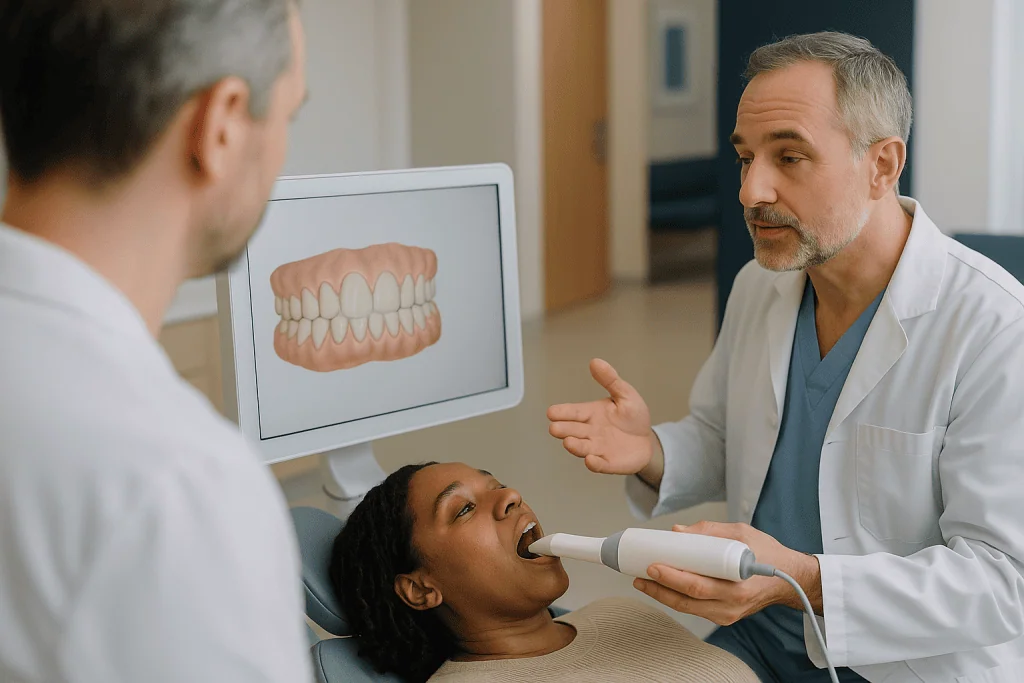
Patients who remember the tray of cold, minty putty from an earlier visit often ask whether that step is gone. Not always. Conventional impressions can still be taken and then scanned on a laboratory desktop scanner, and some clinicians prefer them for specific situations. Either route ends in the same place: a surface file the planning software can read.
A few practical points matter during the scan. Teeth should be clean and dry, because saliva and plaque scatter the light and leave holes in the model that must be recaptured. The scanner records only what it can see, so the gum line, the biting surfaces and the tight contacts between neighboring teeth need deliberate passes. The bite record, where you close gently in your usual position, is one of the most consequential moments of the appointment. If it is captured with the jaw pushed forward or clenched to one side, every later decision about how the bite should finish is built on a false starting point.
What the scan cannot show deserves equal honesty. It records the crown, the part of the tooth above the gum, and nothing below it. Root length, root angle and the thickness of the bone holding the tooth are invisible to it. Those come from X-rays, which is why the next step exists.
Do you need X-rays before clear aligner planning?
Dental X-rays use low-level radiation to show the parts of the tooth and jaw that no scanner can reach: roots, the bone around them, impacted or missing teeth, cysts and the condition of fillings and crowns (MedlinePlus, Dental x-rays). For orthodontic planning the two most common views are a panoramic film, which sweeps around the whole jaw in one image, and a lateral cephalometric film, a side-on view of the skull used to measure how the jaws relate to each other and to the face.
Why does a plan that moves crowns need pictures of roots? Because the crown is the handle and the root is the part doing the moving. A tooth with a short or curved root behaves differently from one with a long straight root. Teeth already tilted into a thin plate of bone cannot be pushed much further in that direction without the root emerging from bone, and a plan drawn from crowns alone would never know.
Some clinicians add a cone-beam CT, abbreviated CBCT, which is a three-dimensional X-ray of the jaws showing each root inside its bone. It is not routine for every case. The extra radiation has to be justified by a specific question, such as an impacted canine, a suspected root problem or a plan that pushes teeth outward near the limits of the bone. When the CBCT and the surface scan are merged, the software can display the root as it moves, not just the crown.
Patients sometimes ask to skip imaging to save time. The honest answer is that the X-ray is part of the diagnosis rather than an optional extra, and the decision about which views are needed, and how often they are repeated, belongs to the clinician responsible for treatment, who weighs the question being asked against the radiation involved.
How the software separates and moves each tooth on screen
Once the scan is loaded, the first job the software performs is segmentation, which simply means drawing a boundary around each tooth so it can be moved independently of its neighbors and of the gum. Early software needed a technician to trace every outline by hand; current tools do most of it automatically, but a human still checks the margins, because a boundary that cuts through enamel or includes a sliver of gum will mis-shape every aligner that follows.
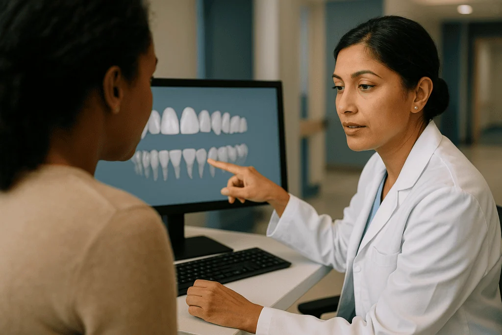
Each segmented tooth is then given a center point and three axes, so that movement can be described precisely. Orthodontists talk about six kinds of movement: tipping (the crown leans while the root stays nearer its place), bodily translation (crown and root slide together), rotation (the tooth turns on its long axis), intrusion (pushing a tooth deeper into bone), extrusion (drawing it further out) and torque (tilting the root while the crown stays). The software displays each as a number, and those numbers become the plan’s vocabulary.
Then comes the part patients see. The clinician, or a technician working from the clinician’s instructions, drags the teeth into a proposed end position. Crowded teeth are spread, rotated teeth are turned, the arch form is smoothed and the upper and lower teeth are checked for how they will meet. Collision detection flags any point where two virtual teeth would occupy the same space, and the program reports how many millimeters of room are missing or spare.
The animation of the whole sequence, where teeth glide into line in a few seconds, is the most persuasive and the most misleading part of the process. It is a true record of the intended geometry and a poor record of the time and biological uncertainty it represents. Reading the planned movement for each tooth tells a patient far more than watching the loop.
How aligners move teeth: the biology the plan has to respect
A tooth does not sit welded into the jaw. It hangs in its socket by the periodontal ligament, a thin cushion of collagen fibers, blood vessels and nerves roughly the thickness of a few sheets of paper. When an aligner presses on a crown, the ligament is squeezed on one side and stretched on the other. Cells respond to that strain: on the squeezed side, bone-removing cells called osteoclasts dissolve a little socket wall; on the stretched side, bone-forming cells called osteoblasts lay new bone down. The socket migrates and the tooth goes with it.
This is slow by design. Remodeling takes days to begin and weeks to produce measurable change, and it works best under light, continuous pressure. Heavy force does not speed it up; it can crush the ligament’s blood supply and stall movement, and it raises the risk of root shortening, a recognized complication of orthodontic treatment of any kind. The pace of a plan is therefore set by the ligament, not by the software.
Aligners deliver force differently from braces. A fixed appliance pulls a tooth toward a wire; an aligner pushes it from a tray manufactured already in the next position, so the tray is slightly misfitting on the day it goes in and the tooth moves to relieve that misfit. That is why the first day or two of each new tray feels tight. It is also why the tray must stay in for most of the day: force applied for a few hours and then removed does not sustain the cellular signal.
Several movements are hard for a push-based plastic shell to deliver. Pulling a tooth outward and rotating round-crowned teeth such as premolars give the plastic little to grip, and the research reflects that. Good digital planning anticipates these weak spots rather than pretending the tray can do everything equally well.
How a clear aligner treatment plan is split into stages
With the start and end positions fixed, the software divides the journey into stages. Each stage is a small, simultaneous shift of every tooth scheduled to move, and each stage is manufactured as one aligner. The prospective study that remains the most cited measurement of aligner accuracy programmed roughly a quarter of a millimeter of movement, or a couple of degrees of rotation, per tray, and changed trays every two weeks (summarized in Rossini et al., Angle Orthodontist).
Staging is where clinical judgment shows most clearly. Not every tooth should move at once. If an upper canine has to rotate through 30 degrees and the incisors only need to shuffle sideways, the plan may hold the incisors still for several stages while the canine turns, then release them. Some teeth are deliberately left untouched as anchors, so their roots resist the reaction force generated when neighbors move. Moving everything simultaneously feels efficient but can let teeth drift in unwanted directions.
Sequencing also decides where space comes from. A crowded arch needs room, and the plan can create it by tipping front teeth forward, by expanding the arch outward, by polishing a fraction of enamel from between teeth, or by removing a tooth. Each option has limits set by bone and gum, and the choice is clinical, not computational.
The number of stages gives a rough sense of treatment length, but only rough. Forty trays changed every ten days point to a little over a year; the same forty trays at two weeks each run closer to eighteen months. The NHS notes that orthodontic treatment generally lasts around 18 to 24 months depending on complexity (NHS, Orthodontics), and aligner plans, once refinement rounds are included, tend to fall within a similar broad window. Any figure quoted at the planning stage assumes full-time wear and typical biology.
Attachments, interproximal reduction and overcorrection: the plan's fine print
Three features in a clear aligner treatment plan surprise patients who expected nothing but smooth plastic.
Attachments are small bumps of tooth-colored filling material bonded to selected teeth before the first tray goes in. The aligner is made with a matching pocket, so the bump gives the plastic a ledge to push against. Without one, a smooth round tooth can simply slide inside the tray instead of rotating. The plan shows exactly where each attachment sits and which movement it serves, and a patient can reasonably ask why each one is there.
Interproximal reduction, usually shortened to IPR, means polishing a fine layer of enamel from the contact point between two teeth to create a sliver of space. The amounts are tiny, a fraction of a millimeter per contact, and the software tells the clinician which contacts, how much and at which stage. It is done with a thin abrasive strip or disc and stays within the outer enamel. It is also irreversible, which is why the planned total should be reviewed and agreed before treatment starts.
Overcorrection is a quieter concept. Because achieved movement tends to lag behind planned movement, a clinician may deliberately program a rotation or an extrusion to go slightly past the target, expecting the tooth to settle short of it. The on-screen final position may therefore look a little too corrected. That is intentional and should be explained rather than hidden.
Elastics, the small rubber bands hooked between upper and lower trays or onto bonded buttons, may also appear when the bite needs to shift forward or back. All of these are planned, visible on screen and open to discussion. None of them is a sign that something has gone wrong.
How accurate is clear aligner digital planning? What the evidence shows
The question patients most want answered is whether the teeth end up where the screen said they would. The evidence is clearer, and more sobering, than marketing suggests.
A systematic review in the Angle Orthodontist gathered the clinical studies that compared planned movement with the movement actually measured at the end of treatment (Rossini et al.). Its central finding, drawn from a prospective study of adult patients, was a mean accuracy of about 41 percent: on average, teeth achieved a little under half of the movement programmed for them. Accuracy varied sharply by movement type. Narrowing the front teeth inward was most predictable. Extrusion, drawing a tooth outward from the bone, was least predictable at roughly 30 percent. Rotations of rounded teeth such as canines and premolars, and movements meant to open or deepen the bite, also lagged.
The review concluded that aligners could reliably manage mild to moderate crowding and spacing in patients who had finished growing, with less predictable results for bodily movement of back teeth, large rotations, bite correction and extrusion. Later studies, using newer materials and attachment designs, report somewhat higher figures, but no published study has shown planned and achieved movement matching one for one.
What does this mean for the plan on screen? It means the end-of-treatment animation is the clinician’s target, not a measurement. It means some teeth will probably need a second round, discussed below. And it means the honest form of the question is not whether your teeth will look like the video, but which of your planned movements are the predictable kind and which are the kind that often needs extra help. A clinician who has read the research can answer that tooth by tooth.
Who is clear aligner treatment usually for, and who is asked to wait?
Digital planning can model almost any mouth. Whether aligners are the right way to treat what the model shows is a separate decision.
The strongest candidates, according to the published evidence, are adults and older teenagers with mild to moderate crowding or spacing, whose jaws have stopped growing and whose bite is broadly correct (Rossini et al.). Many kinds of malocclusion, the general term for teeth and jaws that do not line up, fall into this group: a few overlapping lower incisors, a gap between front teeth, mild relapse after earlier braces (MedlinePlus, Malocclusion of teeth).
People are commonly asked to wait, or offered a different appliance, in several situations. Children still growing may do better with treatment timed to growth, and aligner wear demands a daily discipline not every young patient can sustain. Large discrepancies between the upper and lower jaws usually need fixed appliances, sometimes with surgery, because the problem is skeletal rather than dental. Severe rotations, teeth that must be drawn out a long way, or molars that need to move bodily backward are the exact movements the research marks as least predictable. Active gum disease, untreated decay or an unstable bite from heavy grinding need resolving first, since moving a tooth in inflamed bone can accelerate its loss.
A few people wait for reasons unrelated to the teeth. Someone about to have extensive restorations, implants or jaw surgery will usually have the sequence planned around those procedures. Anyone unsure they can keep a tray in for most of the day will be told plainly that a plan built on near-constant wear, the pattern assumed in the published studies, cannot work on occasional wear.
In every case the clinician who examines you decides, and the reasoning should be shared in words you can repeat back.
Clear aligners vs fixed braces: how the planning differs
Fixed braces and aligners can treat many of the same problems, and the biology underneath is identical. What differs is where the planning decisions are made, how force is delivered, and how easily the plan can be revised once treatment is under way.
| Planning aspect | Clear aligners | Fixed braces |
|---|---|---|
| Starting record | Digital scan or scanned impression plus X-rays | Models or scan plus X-rays |
| When movements are decided | Almost entirely before manufacture, in the digital set-up | Broadly at the start, refined at each visit by wire changes and bends |
| Source of force | Misfit between the tooth and a tray made in the next position | Wire returning to shape, pulling brackets with it |
| Movements with strongest evidence | Mild to moderate crowding and spacing; tipping; alignment in non-growing patients | Wide range including bodily movement, extrusion, large rotations and bite correction |
| Changing course mid-treatment | Requires a new scan and a refinement set of trays | Adjusted chairside at a routine visit |
| Daily responsibility | Patient controls hours of wear; progress depends on it | Appliance works continuously regardless of patient effort |
| Hygiene | Trays out for brushing; risk from drinks consumed with trays in | Brushing around brackets is harder; enamel marking risk |
| Monitoring | Progress scans compared with the digital target | Visual checks, occasional X-rays |
The table is not a scorecard. A plan that depends on near-full-time wear is a poor fit for someone who knows they will remove trays often, however neat the animation looks. A plan involving movements the research grades as unpredictable may be better served by brackets, or by a combination: a period of fixed appliances followed by aligners, or aligners with a few bonded buttons and elastics. Many clinicians treat the two as tools in the same kit rather than rival philosophies. The evidence base for aligners is younger and narrower than for fixed appliances (Rossini et al.), which is a reason for honest conversation, not for ruling either one out.
What the first days and weeks with aligners usually look like
The first appointment after the plan is approved usually involves bonding any attachments, performing any first-stage IPR and fitting the first tray. Trays are changed on a schedule set by the clinician; the studies in the published review used around two weeks per tray, and many clinicians now use shorter or longer intervals depending on the movement planned (Rossini et al.).
Expect pressure rather than sharp pain in the first two or three days of each new tray, concentrated on whichever teeth are scheduled to move at that stage. Speech may lisp slightly for the first week until the tongue adapts. The plastic edge can rub the gum or tongue until it is smoothed. Many people find a new tray easiest to seat before bed so the first hours of tightness pass in sleep.
Daily life organizes itself around the tray. It comes out to eat and to drink anything other than water, and the teeth are brushed before it goes back in; sugary or acidic drinks sipped with a tray in sit against the enamel for hours. Clinicians often provide small foam cylinders to bite on for a few minutes, because a tray that is not fully seated delivers the wrong force.
Check-ups usually fall every several weeks, more often early on. The clinician looks for tracking, which means whether each tooth is actually sitting where the tray expects it. A visible gap between the plastic edge and the biting surface of a tooth is the classic sign of a tooth lagging behind the plan. At the end of the planned trays a new scan is taken and compared with the digital target. If some teeth have fallen short, a refinement round is planned: a fresh, shorter sequence of trays built from the new scan. Refinements are common enough to count as part of normal treatment rather than a failure, and the number needed cannot be promised in advance.
What people often get wrong about digital aligner planning
“The computer designs the plan, so the clinician just approves it.” The software proposes; the clinician disposes. The clinical questions, which teeth anchor, where space comes from, how the bite should finish, whether a root is safe to move, are not decided by an algorithm. Two clinicians given the same scan can produce quite different plans.
“The video shows my final result.” It shows the target. The accuracy figures cited above mean some teeth will typically arrive short of it, and refinement trays exist for that reason.
“A scan means I don’t need X-rays.” The scan sees crowns only. Roots, bone and anything hidden under the gum come from radiographs, which are part of the diagnosis (MedlinePlus, Dental x-rays).
“More trays means a more thorough plan.” Tray count reflects how far teeth must travel and how finely the movement is staged. A long plan for a simple problem may mean over-staging; a short plan for a complex one may be unrealistic. Neither number is a quality score.
“Aligners are gentler, so the risks are lower.” The biology is the same. Root shortening, gum recession where teeth are moved outward through thin bone, and decay around trays worn over unbrushed teeth are all possible with any orthodontic appliance.
“Once the trays finish, the teeth stay put.” They do not. Teeth drift back toward their old positions for years, and retainers, worn as advised and often indefinitely, are the only established way to hold the result (NHS, Orthodontics).
“If the software accepts the plan, it can be done.” Software will happily animate a movement that the bone cannot support. Acceptance by a program is not a clinical judgment, and a plan nobody has argued with is a plan nobody has checked.
Questions to ask your care team before approving the plan
A plan reviewed on a screen can feel like a product demonstration. Treat it as a consultation instead, and bring questions. These are the ones that tend to change decisions.
- Which of my planned movements fall into the predictable category in the research, and which are the kind that often needs refinement or extra help?
- Which teeth are anchors, and what happens to them if the moving teeth lag?
- Where is the space for my crowded teeth coming from: tipping forward, widening the arch, enamel reduction or extraction?
- How much total interproximal reduction is planned, on which contacts, and at what stages?
- Why is each attachment there, and which movement does it serve?
- What X-rays were used, and did the plan consider root position and bone thickness where teeth move outward?
- Has the final position been overcorrected anywhere, and why?
- How many hours a day of wear does this plan assume, and what happens to the timeline if I fall short?
- How will you check that my teeth are tracking, and what would make you change the plan mid-course?
- What retainer is planned afterward, and for how long?
Two further questions are worth asking out loud even when the answers seem obvious. First, who will actually review and modify the plan: the clinician examining you, or a technician working remotely from the scan? Both arrangements exist, and you are entitled to know. Second, what is the alternative? Most malocclusions that aligners can treat can also be treated with fixed appliances, and some can be left alone without harm to health (MedlinePlus, Orthodontia). A clinician who can describe the other options without disparaging them is offering a genuine choice. Write the answers down; a plan is easier to live with when you understand why it was built the way it was.
When to call your doctor or dentist
Most of what aligners produce is ordinary: pressure for a few days, a mild lisp, a rubbed spot on the tongue. A few things are not, and they warrant a call to your dentist or orthodontist rather than waiting for the next scheduled visit.
Call the same day if a tooth becomes noticeably loose, far beyond the slight give its neighbors share, or if a tooth changes color, which can signal that its nerve is in trouble. Call if pain is severe, throbbing or worsening after the first few days of a new tray rather than easing, or if it is focused on one tooth and wakes you at night. Swelling of the gum or face, bleeding that does not settle with cleaning, a bad taste or discharge around a tooth, or fever alongside any of these, need prompt assessment, because moving teeth through infected bone is unsafe and the plan may need to pause.
Also make contact if a tray suddenly will not seat, if the bite feels dramatically different from one day to the next, if a tray cracks and leaves an edge that cuts, or if you have lost or broken several trays and are tempted to skip ahead. Moving to a later tray before the teeth are ready applies force to teeth that have not caught up and can undo progress.
Seek emergency care, not a dental appointment, for swelling of the lips, tongue or throat, difficulty breathing or swallowing, or a widespread rash after inserting a tray, which may indicate an allergic reaction to the material, and for facial swelling spreading toward the eye or neck. Numbness or tingling of the lip or chin that does not resolve should also be reported promptly.
None of these situations is something to manage alone with a search engine. Your treating team holds the scan, the X-rays and the plan, and every decision about pausing, adjusting or stopping treatment sits with them.
Frequently asked questions
What is a clear aligner treatment plan and who actually creates it?
A clear aligner treatment plan is the digital set-up showing each tooth’s current position, its proposed final position and the staged path between them. Software and technicians prepare a draft, but the clinical decisions about anchorage, space, bite and root safety belong to the orthodontist or dentist, who reviews and edits the proposal before any trays are manufactured.
How long does a 3D dental scan for aligners take?
An intraoral scan of both arches and the bite typically takes a few minutes of actual scanning, though the appointment is longer because teeth must be clean and dry and some areas may be re-captured. The scan records only the crowns above the gum, so X-rays are still needed to see roots and bone before a plan is finalized.
Do I still need X-rays if my teeth have been scanned digitally?
Yes, in almost every case. The scanner cannot see below the gum, while dental X-rays show roots, the surrounding bone, impacted or missing teeth and other hidden findings (MedlinePlus, Dental x-rays). Which views are taken, and whether a three-dimensional CBCT is justified, is a decision for the treating clinician, who weighs the information needed against radiation exposure.
How do aligners move teeth if the plastic is so thin?
Each tray is manufactured in the next planned position, so it fits slightly imperfectly on the day it goes in and presses on specific teeth. That light, continuous pressure strains the periodontal ligament around the root, prompting bone cells to remove bone on one side of the socket and add it on the other. The socket migrates and the tooth follows.
Why does the last position on my screen look slightly over-straightened?
That is usually deliberate overcorrection. Research shows teeth often achieve less than the programmed movement, especially for rotations and extrusion, so a clinician may plan a tooth to go a little past the target expecting it to settle short. Ask your care team to point out where overcorrection has been built in and why.
What are aligner refinement scans and will I need one?
A refinement scan is a new 3D scan taken when the planned trays are finished, compared against the digital target to see which teeth have fallen short. If some have, a fresh, shorter set of trays is made from that scan. Refinements are common enough to be considered part of normal treatment, and nobody can promise in advance how many a given mouth will need.
Why does my plan include small bumps bonded to some teeth?
Those are attachments, small shapes of tooth-colored filling material that give the aligner a ledge to push against. Without one, a smooth round tooth can slide inside the tray instead of rotating or moving as intended. Their position and shape are chosen in the digital plan to match specific movements, and they are removed when treatment ends.
Is interproximal reduction safe for my enamel?
Interproximal reduction, or IPR, removes a very thin layer of outer enamel between two teeth to create space, usually a fraction of a millimeter per contact. Kept within planned limits it stays in the enamel’s outer layer, but it is irreversible, so the total amount and the contacts involved should be reviewed and agreed with your clinician before treatment starts.
How many hours a day do the studies assume aligners are worn?
The clinical studies gathered in the published systematic review used protocols of near-full-time wear, with trays removed essentially only for eating and cleaning, and in the main accuracy study each tray was worn for about two weeks (Rossini et al.). Because the biology needs continuous light force, a plan built on that assumption cannot be expected to track on part-time wear.
Can digital planning tell me exactly how long my treatment will take?
No. The plan gives an estimate from the number of trays and the change interval, but it assumes full-time wear, typical biology and no refinement rounds. The NHS describes orthodontic treatment generally as lasting around 18 to 24 months depending on complexity, and aligner timelines tend to fall within a similar broad range once refinements are counted.
References
- NHS: Orthodontics
- MedlinePlus: Malocclusion of teeth
- MedlinePlus: Dental x-rays
- MedlinePlus: Orthodontia
This article is for general information only and is not a substitute for professional medical advice. Please consult a qualified doctor about your individual situation.

Planning This Treatment in Turkey
Our full guide for international patients: techniques, hospital stay, recovery and cost.
More from the Blog
How Long Does Sensitivity Last After Restorative Dental Treatment? Numbness and Temporaries
Mild sensitivity after restorative dental work, such as a filling or crown, is common and usually eases over a few days to a few…

Flying After a Tooth Extraction: How Long to Wait and How to Protect the Clot
Most dental teams prefer that you avoid flying for at least 24 hours after a simple tooth extraction, and many suggest 48 to 72…
Wisdom Teeth Removal Myths: Age Limits, Face Shape and Other Claims Worth Correcting
Most wisdom teeth removal myths do not hold up to the evidence. There is no upper age limit for extraction, although healing tends to…

What Happens in Orofacial Myology Sessions: Posture Checks, Exercises and Progress Reviews
Myofunctional therapy sessions are short, structured appointments with a trained therapist who checks how your tongue, lips and jaw rest and move, teaches targeted…
How a Dental Anatomy Evaluation Guides Your Plan: Tooth Charts, Bite Checks and Imaging
A dental anatomy evaluation is a structured, tooth-by-tooth assessment in which a dentist records each tooth on a numbered chart, checks how your upper…

Tooth Decay Myths: Sugar-Free Means Safe, Brushing Harder Helps and Other Errors
Most tooth decay myths fall apart under evidence. Sugar-free does not mean safe, because acidic drinks and starchy snacks still feed decay or erode…