

How Does Rehabilitation After Acute Flaccid Myelitis Rebuild Strength and Mobility?

Key Takeaways
- Acute flaccid myelitis injures the motor neurons in the spinal cord's gray matter, so weakness is limp rather than stiff and behaves differently from stroke weakness.
- Strength returns through three routes: recovery of inflamed neurons, slow collateral sprouting from surviving neurons, and trained compensation by other muscles and equipment.
- Resistance exercise cannot strengthen a muscle with no nerve supply; until signal returns, therapy protects joints, prevents contractures, and works in gravity-eliminated positions.
- The CDC reports that most people with AFM had a fever or respiratory illness beforehand, that most cases are in children, and that no specific treatment for the condition exists.
- Nerve transfer surgery reroutes a spare working nerve to a paralyzed muscle, and its timing is a clinical judgment balancing spontaneous recovery against the muscle's fading ability to accept new nerve.
- Breathing difficulty, a weak cough, trouble swallowing, or new spreading weakness during recovery are emergencies or same-day calls, not signs to wait and watch.
Rehabilitation after acute flaccid myelitis rebuilds strength by protecting joints and limp muscles while surviving spinal motor neurons recover or sprout new connections, then progressively loading muscles as signal returns. Physical and occupational therapy, bracing, breathing support, and sometimes nerve or tendon transfer surgery are combined over months, with the pace and plan set by the treating team.
The first thing a parent usually notices is an arm that will not lift. A child who was climbing the sofa on Tuesday cannot raise a spoon by Friday, and the limb hangs soft, not stiff. In the neurology ward the word that follows is unfamiliar and frightening, and the question that follows it is always the same: what happens now, and will the strength come back?
Acute flaccid myelitis rehabilitation is the honest, unglamorous answer to that question. It does not start with a machine or a medicine. It starts with a therapist adjusting a pillow so a shoulder does not stretch out of its socket overnight, and with a family learning how to move a limb that cannot yet move itself.
What follows is a plain account of how that work rebuilds movement, what the evidence supports, where it is thin, and which decisions belong with the care team.
What acute flaccid myelitis actually does to the spinal cord
Acute flaccid myelitis, usually shortened to AFM, is a rare condition in which inflammation damages the gray matter of the spinal cord, the central column that houses motor neurons. Motor neurons are the nerve cells that carry movement commands from the cord out to the muscles. When they are injured, the muscles they serve lose their signal and become weak or paralyzed within hours to days.
The word flaccid matters. It means limp and low in tone, the opposite of the stiff, tight weakness people often picture after a stroke. That distinction shapes every later decision in rehabilitation, because a limp muscle that has lost its nerve supply behaves very differently from a stiff muscle whose nerve supply is intact but poorly controlled.
The Centers for Disease Control and Prevention describes a typical pattern: most cases occur in children, weakness often begins in one arm or leg and can be uneven from side to side, and the great majority of patients had a fever or a mild respiratory illness in the days before the weakness appeared. Enteroviruses, particularly one called EV-D68, have been linked to outbreaks, although a cause is not confirmed in every case.
Sensation is frequently spared, which means a child may feel a touch on a hand they cannot move. Some patients develop weakness of the neck, face, or the muscles used for breathing and swallowing, and those cases need intensive monitoring early on.
Recovery is variable. The National Institute of Neurological Disorders and Stroke notes that some people regain much of their function over months, while others are left with lasting weakness. Rehabilitation exists to shape whatever recovery the nervous system can offer into the most usable movement possible.
How acute flaccid myelitis rehabilitation works, in plain language
Strength returns after AFM through three routes, and therapy is built around all of them.
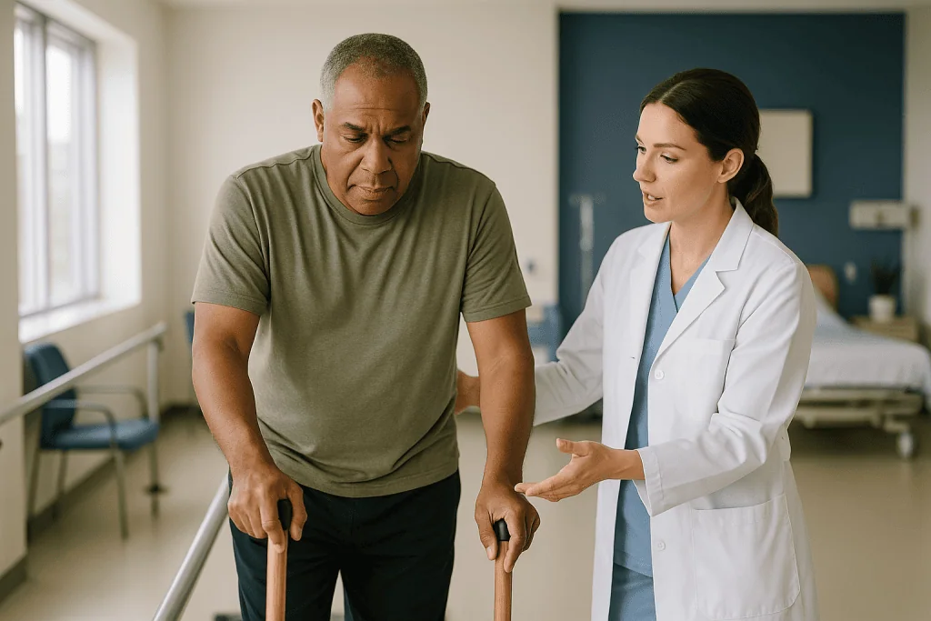
The first route is recovery of neurons that were inflamed but not destroyed. Swelling settles, signal transmission improves, and a muscle that produced only a flicker begins to contract. This tends to happen earliest and explains why some gains appear in the first weeks.
The second route is called collateral sprouting. Surviving motor neurons grow new branches that reach out and adopt muscle fibers whose original nerve has died. One neuron ends up controlling a larger territory than it did before. This is slow, measured in months, and it is the main biological reason therapists keep denervated muscle supple and protected: a muscle that has shortened or been overstretched cannot make good use of a new connection when it finally arrives.
The third route is compensation. Other muscles are trained to take over a task, equipment supplies what the body cannot, and daily activities are relearned in new ways. Compensation is not a consolation prize. For many families it is the difference between dependence and independence.
What therapy cannot do is force a muscle with no working nerve to grow stronger through exercise. Strength training only works once at least some signal is reaching the muscle. Before that point, the job is range of motion, positioning, and protection. After it, the job shifts to progressive loading and task practice. Knowing which phase a particular muscle is in, muscle by muscle, is the core skill of the team, which typically includes a rehabilitation physician, physical and occupational therapists, speech and respiratory therapists, an orthotist, and psychological support.
Who rehabilitation is for, and who is asked to wait on certain steps
Every person diagnosed with AFM is a candidate for rehabilitation. The question is never whether, but how intensively and in what setting. MedlinePlus describes rehabilitation as care that can take place in a hospital, a dedicated inpatient unit, an outpatient clinic, or at home, and people with AFM commonly move through several of these settings as they recover.
In the earliest days, someone who needs breathing support in intensive care still receives rehabilitation, but it looks like careful positioning, gentle movement of joints by a therapist, and attention to skin and lungs rather than active exercise. Medical stability comes first, and the team decides when the balance tips toward more active work.
Certain elements are deliberately delayed. Resistance training for a muscle that shows no voluntary contraction waits until reinnervation signs appear, because loading a muscle with no nerve supply risks stretching it without benefit. Formal evaluation for nerve transfer surgery typically waits a number of months so that spontaneous recovery has a chance to show itself, although surgical teams also want to see patients before the window for reinnervating a muscle narrows. Return to contact sports or high-impact activity waits until strength, balance, and bone health have been reviewed.
Fatigue deserves a specific mention. Because AFM injures the same motor neurons that polio did, therapists often borrow a lesson from post-polio care and watch for overwork weakness, where a limb becomes weaker after exercise rather than stronger. A muscle that is persistently more tired the day after a session is telling the team something, and the response is usually to change the load, not to push through.
What the first days and weeks of acute flaccid myelitis rehabilitation look like
The opening phase happens in hospital, often while the diagnosis is still being confirmed with spinal cord imaging and tests of spinal fluid. Rehabilitation begins alongside that medical work rather than after it.
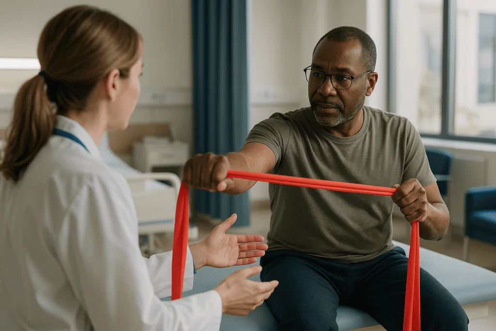
A therapist will assess which muscles move against gravity, which move only when gravity is removed, and which show nothing at all. That map becomes the baseline against which every later change is measured. Joints are moved through their full range at least daily to prevent contractures, which are permanent shortenings of muscle or joint tissue that can lock a limb in one position. A weak shoulder is supported so the weight of the arm does not gradually pull the joint out of alignment, and hands are often rested in light splints to keep the fingers in a usable position.
Breathing is watched closely. If the muscles of the chest wall or diaphragm are affected, respiratory therapists teach techniques to keep the lungs clear and may use devices that assist coughing. Swallowing is checked before food and drink are reintroduced if the neck or face is involved.
Pain is common and comes in two forms: aching from joints and muscles that are not moving normally, and burning or tingling nerve pain from the injured cord. Both are managed by the treating team, with positioning and movement doing a surprising share of the work.
Families are trained from the start. Learning how to move a child’s arm safely, how to position them in bed, and what to watch for at night gives parents a role in the recovery rather than a seat beside it. When the person is medically stable, the team decides whether inpatient rehabilitation, daily outpatient therapy, or a home program is the right next step.
Phases of recovery: a summary table
Recovery after AFM does not follow a fixed calendar. The National Institute of Neurological Disorders and Stroke describes improvement that can continue over months, with wide variation between individuals. The phases below describe what therapy focuses on at each stage, not how long each stage lasts for a particular person.
| Phase | Main goal | What therapy commonly involves |
|---|---|---|
| Acute (hospital) | Protect and stabilize | Positioning, passive range of motion, splints, breathing and swallowing support, pain control by the medical team |
| Early recovery | Find and nurture returning signal | Gravity-eliminated movement, assisted movement, functional electrical stimulation in some programs, seating and mobility equipment |
| Reinnervation | Build strength where nerve supply returns | Progressive resistance in muscles that can contract, task-specific practice, gait training with orthoses, fatigue monitoring |
| Surgical window (if considered) | Restore function to muscles that remain paralyzed | Evaluation for nerve transfer or tendon transfer, followed by a dedicated post-surgical therapy program |
| Long-term | Maintain gains and adapt with growth | Home program, periodic reassessment, orthotic adjustments, spine and hip surveillance in children, school and activity planning |
Two features of this table are worth underlining. Phases overlap, because different muscles in the same person are often at different stages: a shoulder may be in the protection phase while the hand is already strengthening. And the long-term phase is not an ending. Children grow, braces stop fitting, and a technique that worked at age four may need rethinking at age nine.
What are the 5 stages of rehabilitation, and do they apply here?
People searching for a roadmap often land on the five stages of rehabilitation, a framework that comes from sports and orthopedic medicine. The stages are usually listed as: control pain and protect the injured tissue; restore range of motion; rebuild strength; restore coordination and balance; and return to full activity. It is a sensible sequence for a torn ligament or a fractured ankle.
Applied to AFM, the framework is partly useful and partly misleading. The first two stages map well. Protecting joints and keeping them mobile is exactly what the acute phase demands, and it is where the model earns its keep by reminding families that early rehabilitation is about preventing loss, not chasing gains.
The third stage is where the model breaks. In an orthopedic injury, the muscle is healthy and simply needs loading. In AFM, the muscle may be healthy but disconnected. Strength cannot be rebuilt until nerve supply returns, and no volume of exercise changes that timeline. Therapists instead work in what they call gravity-eliminated positions, where a tiny returning signal can produce visible movement, and progress to loading only when the muscle can contract on its own.
The fourth stage, coordination and balance, becomes especially important because weakness in AFM is often uneven. A child with one strong leg and one weak leg has to learn a new way of walking, and the trunk muscles that keep them upright may themselves be affected.
The fifth stage, return to activity, is real but redefined. For some it means running again. For others it means playing a sport in a wheelchair, or managing a school day without exhaustion. A good team writes that goal with the family rather than assuming it.
Flaccid arm exercises: what therapists actually use for a limp arm
The arm is the most common site of lasting weakness after AFM, and the shoulder is the joint most at risk, so this is the question families ask most. What follows describes approaches therapists commonly use, not a program to start at home without guidance. Which techniques suit a particular arm depends on which muscles have signal, and only an assessment can tell.
Positioning comes first. A weak shoulder is supported in sitting and lying so the arm’s own weight does not stretch the joint capsule. Slings, lap trays, and pillows do this quietly all day long.
Passive and assisted range of motion keeps every joint moving through its full arc. A therapist or trained caregiver moves the limb, or the person uses the stronger arm to guide the weaker one.
Gravity-eliminated movement lets a faint signal show itself. Sliding the forearm across a smooth tabletop, moving the arm in a warm pool, or working with the limb suspended in a sling allows a muscle that cannot lift against gravity to practice contracting.
Task practice ties movement to purpose. Reaching for a cup, pushing a toy car, or turning a page recruits several muscles at once and is far more engaging for a child than isolated exercise.
Electrical stimulation, in which small electrical pulses make a muscle contract through pads on the skin, is used in some programs to maintain muscle bulk while nerve supply is absent. The evidence that it improves eventual recovery in AFM specifically is limited, and the NHS describes it as one of several tools physiotherapists may use rather than a treatment on its own.
The honest summary: no single exercise wakes a flaccid arm. Protection, patience, and matching the load to the returning signal are what the evidence supports.
Legs, trunk and breathing: the paraplegia-pattern rehabilitation protocol
When AFM affects both legs and the trunk, the pattern resembles a spinal cord injury, and rehabilitation borrows heavily from that field. People often search for a recommended protocol for paraplegia and hope for a single document. There is no universal protocol, but there is a consistent set of priorities that guideline-level care follows.
Skin and pressure come first. Someone who cannot shift their weight is at risk of pressure injuries, so seating, mattress choice, and regular repositioning are taught early and reinforced often.
Trunk control is the foundation of everything above it. Sitting unsupported, reaching without toppling, and transferring from bed to chair all depend on core muscles that may themselves be weak. Therapists work on this before expecting much from the legs.
Standing, even fully supported in a standing frame, is used in many programs to load the bones and hips, stretch the hip flexors, and help with bowel and bladder function. Weight-bearing matters particularly for children whose bones are still developing.
Walking is reintroduced when it is realistic, usually with orthoses that stabilize the ankle or the whole leg, and sometimes with a body-weight-supported treadmill. For those who will use a wheelchair long term, wheelchair skills are a rehabilitation goal in their own right, not a fallback.
Breathing rehabilitation runs alongside all of this when chest muscles are weak. Techniques to deepen breaths, assisted coughing, and monitoring of how well the lungs clear are part of the routine, because respiratory complications are among the most serious risks the CDC associates with severe AFM.
Bowel and bladder management, where affected, is handled by the medical team, and any medicines involved are prescribed and adjusted by that team alone.
Bracing, splints and equipment: what they do and why they change
Equipment in AFM rehabilitation is not a sign that therapy has failed. It is how a weak body borrows structure while the nervous system repairs itself, and it is how a child keeps moving, playing, and growing in the meantime.
An orthosis is a brace worn on the body to support or align a joint. An ankle-foot orthosis holds the foot at a right angle so it does not drag; a knee-ankle-foot orthosis extends up the thigh to stabilize a knee that buckles. A resting hand splint keeps the fingers and thumb in a functional position overnight so that a returning signal has a useful hand to work with. A shoulder support reduces the downward pull on a weak joint.
Wheelchairs and adapted seating are chosen for posture as much as mobility. A chair that lets a child sit upright with the trunk supported frees the arms for eating, writing, and play.
The orthotist, the professional who designs and fits braces, becomes a long-term member of the team, because equipment that fits perfectly in the spring may not fit by autumn. Growth changes limb length; recovery changes which muscles need support; and a brace that was essential for standing may become optional for walking.
Children with uneven trunk weakness are watched for scoliosis, a sideways curvature of the spine that can develop when muscles pull unequally during growth, and for hip problems when the muscles around the joint are weak. Regular review by the rehabilitation physician, with imaging when indicated, is how these are caught early. Decisions about bracing for the spine, or about surgery if a curve progresses, sit with the treating team and are made case by case.
Nerve transfer for AFM: where surgery fits into rehabilitation
For a muscle that shows no sign of recovery, surgery can sometimes supply a new nerve. In a nerve transfer, a surgeon takes a branch of a working nerve that the body can spare and connects it to the nerve of a paralyzed muscle, so that the healthy nerve fibers grow along the old pathway and eventually drive the muscle. The National Institute of Neurological Disorders and Stroke notes that this approach has been used in some people with AFM, most often to restore shoulder or elbow movement.
Timing is the crux. A muscle that has lost its nerve supply slowly loses its ability to accept a new one, so surgical teams generally want to evaluate patients while that capacity remains, yet not so early that spontaneous recovery is cut short. Where exactly that balance falls for a given muscle is a clinical judgment made by the surgeon and the rehabilitation team together, informed by examination and often by electrical tests of nerve and muscle. No figure in this article should be read as a deadline.
Tendon transfers are a different operation, performed later, in which a working muscle’s tendon is rerouted to perform a lost function. They rely on strength that already exists rather than growing new nerve, and are considered once recovery has plateaued.
Surgery does not replace rehabilitation; it restarts it. After a nerve transfer, months pass before the new fibers reach the muscle, and the person then has to learn to fire the transferred nerve in a new way, a process therapists call motor re-education. The evidence base in AFM is small and consists largely of case series, so honest teams describe possible benefits and risks without promising results. Whether surgery is appropriate at all is a decision for the treating team, never a default.
Medicines during recovery: what they do and what they do not
There is no medicine proven to reverse the nerve damage of AFM. The CDC states plainly that there is no specific treatment for the condition, and that care focuses on supporting the body and rehabilitating function. That fact disappoints families, and it deserves to be said clearly rather than softened.
In the acute phase, clinicians sometimes use treatments aimed at the immune system. Corticosteroids reduce inflammation; intravenous immunoglobulin supplies pooled antibodies; plasma exchange filters antibodies out of the blood. Whether any of these changes the course of AFM is uncertain, and their use is decided individually by the neurology team, weighing possible benefit against known risks.
During rehabilitation, medicines address symptoms rather than the underlying injury. Nerve pain, the burning or electric sensation from an irritated spinal cord, may be treated with medicines originally developed for seizures or depression that dampen abnormal nerve signaling. Muscle cramps and aches are more often managed with positioning, heat, and movement than with tablets. Where bladder or bowel function is affected, medicines that relax or stimulate those muscles may be part of the plan.
Timelines for these symptom treatments are set by the prescriber, who will explain what to expect and when to review. Nothing here should prompt anyone to start, stop, or adjust a prescribed medicine; that conversation belongs with the clinician who knows the case.
Vaccination is a related question families raise. There is no vaccine for the enteroviruses most often linked to AFM. Staying current with routine immunizations, including polio, remains important because poliovirus causes a nearly identical illness, and the CDC confirms that AFM cases in recent outbreaks have tested negative for polio.
How long does flaccidity last after a stroke, and why AFM is different
Families often arrive with knowledge from a relative’s stroke recovery, and the comparison is worth making carefully because it explains why AFM rehabilitation follows different rules.
After a stroke, the injury is in the brain, above the motor neurons of the spinal cord. The affected limb is often flaccid at first, and then, over days to weeks, tone typically increases and the muscles become stiff or spastic as the spinal cord loses the brain’s inhibiting influence. Some people remain flaccid for longer, and a minority never develop increased tone, but stiffness is the common trajectory. Stroke therapists spend much of their time managing that stiffness while retraining brain pathways.
In AFM, the injury is in the motor neurons themselves. There is no upstream signal being poorly controlled; there is no signal at all until neurons recover or sprout. Flaccidity is therefore not a phase that gives way to spasticity. It persists as long as the muscle lacks nerve supply, which may be weeks, months, or permanently for particular muscles. Muscle wasting, the visible thinning of a limb, follows because muscle fibers without nerve input shrink.
That difference changes practice in three ways. Stretching in AFM aims to prevent shortening rather than to fight tightness. Strengthening waits for reinnervation rather than starting as soon as a person can participate. And the surgical option of nerve transfer, rarely relevant after stroke, becomes a serious consideration.
The two conditions do share one truth. The Mayo Clinic’s stroke recovery guidance emphasizes that rehabilitation gains depend on repetition, task practice, and starting early, and every one of those principles carries over. The nervous system, wherever it is injured, learns from what it is asked to do.
What people often get wrong about AFM recovery
The first misunderstanding is that more exercise means more recovery. For a muscle that can contract, progressive loading does help. For a muscle with no nerve supply, extra repetitions build nothing and may overstretch tissue or exhaust the muscles that are compensating. Matching effort to what the nerve can currently deliver is the skill, and it is why therapists test muscle by muscle rather than prescribing a blanket routine.
The second is that a plateau means the end. Recovery through collateral sprouting is slow and uneven, and function can improve months after strength scores stop changing, because a person learns to use the strength they have more efficiently. Equally, honesty requires saying that some muscles do not recover, and planning for that possibility is not giving up.
The third is that AFM is polio. It is not, and the CDC confirms that outbreak cases have tested negative for poliovirus. The two conditions injure the same cells and are managed with similar rehabilitation principles, which is why lessons from polio care are relevant, but they are different infections.
The fourth is that AFM is spread from person to person. The viruses associated with it are common and contagious, but AFM itself is a rare complication. Most children who catch the same virus develop nothing more than a cold.
The fifth is that using a wheelchair or brace will stop a child from walking. Equipment preserves energy, protects joints, and keeps a child participating while recovery unfolds. Therapists see it as a bridge, not a verdict.
The last is that the psychological side can wait. A sudden loss of movement in a healthy child is a trauma for the whole family. Psychological support is part of rehabilitation, not an optional extra.
Questions to ask your care team about AFM physical therapy and beyond
A rehabilitation plan is easier to trust when you understand it. These questions are ones therapists and rehabilitation physicians generally welcome, and the answers will be specific to the person in front of them.
- Which muscles currently show signal, which show none, and how will you track changes over time?
- What are the goals for the next few weeks, and how will we know whether we are reaching them?
- What should we do at home between sessions, and what should we avoid doing?
- How will you watch for overwork weakness or fatigue, and what should we report?
- Which joints are most at risk of contracture or subluxation, and how are we protecting them?
- Is breathing or swallowing affected, and what signs would tell us something has changed?
- What equipment do you expect to be needed, and how often will it be reviewed as growth or recovery changes things?
- At what point would a referral for surgical evaluation be considered, and what would that assessment involve?
- How is pain being managed, and who do we contact if it changes?
- What support is available for school, for siblings, and for the emotional side of this for the whole family?
- Who coordinates the team, and who is our single point of contact with questions?
Writing down the answers, and bringing the list to each review, turns a long and uncertain process into a series of understood steps. The NHS notes that physiotherapy works best when the person and family are active participants in setting goals, and that principle holds firmly here.
When to call your doctor: red-flag signs during AFM recovery
Most of AFM rehabilitation is steady and unhurried, but a handful of changes need a same-day call to the treating team, and a few need emergency care.
Seek emergency help immediately for any new difficulty breathing, shallow or labored breaths, a weak or ineffective cough, choking on food or saliva, blue or gray lips, or unusual drowsiness or confusion. The CDC identifies respiratory failure as the most serious complication of AFM, and it can develop quickly when the muscles of the chest or neck are involved.
Call the care team the same day for new or spreading weakness in a limb that had been stable or improving, new difficulty swallowing, a change in voice or facial movement, new numbness or a change in bowel or bladder control, or a fever alongside any of these. New weakness after the acute illness is unusual and should not be assumed to be part of the process.
Also contact the team promptly for a red, warm, or swollen area on the skin or over a joint, which may indicate a pressure injury or infection; for a limb that has become noticeably more painful, stiffer, or harder to move through its usual range; for a brace or splint that is rubbing, leaving marks, or no longer fitting; or for a child who is more tired the day after therapy on a consistent basis, since this may signal overwork.
Emotional red flags matter too. Persistent low mood, withdrawal, sleep disturbance, or expressions of hopelessness in a child or a caregiver deserve the same prompt attention as a physical symptom.
When in doubt, call. Rehabilitation teams expect these questions, and an early conversation is always simpler than a late one.
Frequently asked questions
Can acute flaccid myelitis rehabilitation reverse paralysis?
Rehabilitation cannot repair destroyed motor neurons, but it can maximize whatever recovery the nervous system produces. Inflamed neurons may recover, surviving neurons can sprout to adopt orphaned muscle fibers, and other muscles can be trained to compensate. The National Institute of Neurological Disorders and Stroke notes that improvement varies widely and can continue over months, while some weakness may be permanent. The team’s role is to protect, then progressively load, each muscle as signal returns.
What are good flaccid arm exercises after AFM?
The most useful approaches for a limp arm are protective and assisted rather than forceful: supporting the shoulder to prevent it stretching out of alignment, moving every joint through its full range daily, and letting the arm move in gravity-eliminated positions such as sliding across a tabletop or in warm water. Task practice, like reaching for a cup, adds purpose. Which techniques suit a particular arm depends on which muscles have signal, so a therapist’s assessment should guide any home routine.
What are the 5 stages of rehabilitation?
The five stages commonly described are protection and pain control, restoring range of motion, rebuilding strength, restoring coordination and balance, and returning to activity. The model comes from sports and orthopedic medicine. It fits AFM for the first two stages, but strength cannot be rebuilt until nerve supply returns, so the third stage waits for reinnervation, and the final stage is redefined around each person’s realistic goals rather than a return to a previous sport.
How long does flaccidity last after a stroke, and is AFM different?
After a stroke, a limb is often flaccid for days to weeks before tone increases and muscles become stiff, although some people stay flaccid longer. In AFM the injury is to the motor neurons themselves, so there is no upstream signal to become spastic. Flaccidity persists as long as the muscle lacks nerve supply, which may be weeks, months, or permanently for particular muscles. This is why AFM rehabilitation waits for reinnervation before strengthening.
What is the recommended rehabilitation protocol for paraplegia after AFM?
There is no single universal protocol, but care follows consistent priorities borrowed from spinal cord injury rehabilitation: preventing pressure injuries through seating and repositioning, building trunk control before expecting leg function, supported standing to load bones and hips, gait training with orthoses where realistic, wheelchair skills as a goal in their own right, and breathing support when chest muscles are weak. The specific plan is written by the treating team for the individual.
What is nerve transfer for AFM and when is it considered?
A nerve transfer connects a branch of a working, expendable nerve to the nerve of a paralyzed muscle so that healthy fibers grow along the old pathway and eventually drive it. Surgeons generally evaluate patients after enough time has passed to judge spontaneous recovery but before the muscle loses its ability to accept new nerve. The evidence in AFM is limited to small case series, and the decision belongs with the surgical and rehabilitation team.
How often is AFM physical therapy needed?
Frequency depends on the phase of recovery and the setting. In hospital and inpatient rehabilitation, therapy often happens daily. Outpatient programs commonly run several times a week early on and taper as a home program takes over, with periodic reassessment thereafter. MedlinePlus describes rehabilitation as care that moves across hospital, outpatient, and home settings as needs change, and the treating team adjusts the schedule based on progress, fatigue, and family circumstances.
Are AFM recovery exercises safe to do at home?
Yes, when they are taught and reviewed by the rehabilitation team. Home programs typically include positioning, range-of-motion movements, and functional tasks matched to the muscles that currently have signal. The main risks of unsupervised exercise are overstretching a joint that lacks muscle protection and overworking compensating muscles, which can leave a limb weaker the next day. Reporting persistent post-exercise fatigue or new pain lets the team adjust the plan.
Is acute flaccid myelitis contagious?
AFM itself is not spread from person to person. The viruses associated with it, particularly enteroviruses such as EV-D68, are common and contagious, but AFM is a rare complication of infection. Most children who catch the same virus have only a mild cold. The CDC recommends ordinary measures such as handwashing and staying up to date with routine vaccinations, including polio, since poliovirus causes a nearly identical illness.
Will my child need a wheelchair after AFM?
Some children do, at least for a period, and some do not; it depends on which muscles are affected and how much recovery occurs. A wheelchair or brace during recovery preserves energy, protects joints, and keeps a child participating in school and play while the nervous system repairs itself. Therapists regard equipment as a bridge rather than a verdict, and needs are reviewed regularly as recovery and growth change what is required.
References
- CDC: About Acute Flaccid Myelitis
- NIH National Institute of Neurological Disorders and Stroke: Acute Flaccid Myelitis
- MedlinePlus: Acute Flaccid Myelitis
- MedlinePlus: Rehabilitation
This article is for general information only and is not a substitute for professional medical advice. Please consult a qualified doctor about your individual situation.

More from the Blog

Confirming HSP: How Genetic Testing, MRI and the Walking Assessment Fit Together
Hereditary spastic paraplegia (HSP) is diagnosed by combining three pieces of evidence rather than by a single test. A neurologist examines walking and leg…

Supporting a Family Member Through Brain Tumor Treatment: Appointments, Rest and Recovery
Supporting someone with a brain tumor means helping with three practical things: getting to and understanding appointments, protecting rest and sleep during treatment, and…

Why Neurologists Ask for Videos of Abnormal Movements Before a Movement Disorder Evaluation
Neurologists ask for videos because many abnormal movements are intermittent, change with stress or medication timing, or quiet down in a clinic room, so…

What Does Autonomic Testing Involve? Tilt Table, Sweat and Heart Rate Tests Explained
Autonomic testing is a set of noninvasive measurements, usually completed in one visit of roughly one to two hours, that records how your heart…

Caring for Someone After a Traumatic Brain Injury: What Families Can Do in the First Weeks
In the first weeks after a traumatic brain injury, families help most by keeping the environment calm and predictable, protecting sleep, speaking slowly in…

When Is It Time to See a Neurologist for Migraine? How Frequency and Disability Guide It
Consider seeing a neurologist for migraine when attacks occur on about five or more days a month, when they regularly stop you from working…