

How Donor Bone Is Screened, Prepared and Placed During Allograft Bone Grafting
Key Takeaways
- An allograft is a type of bone graft, not an alternative to one; the term simply means the bone came from another human donor.
- Donor bone contains no living cells after processing, which is why no tissue matching or anti-rejection medicine is needed.
- Screening layers donor history, blood testing for HIV, hepatitis B and C and syphilis, tissue cultures and sterilization, and a graft's unique identifier lets it be traced back to its donor.
- The more aggressively donor bone is cleaned, irradiated or demineralized, the safer it becomes and the less biological activity it retains.
- Dense cortical grafts bear load immediately but incorporate slowly; porous cancellous chips incorporate faster but offer no structural support.
- MedlinePlus places typical bone graft recovery between two weeks and three months, with the graft itself taking three months or longer to heal, and spinal fusion taking several months according to Mayo Clinic.
Allograft bone grafting uses bone from a deceased human donor to fill or bridge a defect that a person's own bone cannot close on its own. Donors are screened through medical history review and infectious-disease testing, the bone is cleaned, processed and usually sterilized, then shaped and packed into the surgical site, where it acts as a scaffold that the recipient's own cells gradually replace with living bone.
The consent form says it in two words that stop most people mid-signature: donor bone. A man with a spine that needs fusing, a woman whose jaw has thinned too much to hold an implant, a teenager whose fracture refused to knit; each of them tends to look up at the same moment and ask the same question. Whose bone? And how do you know it is safe?
They are fair questions, and the answers are more concrete than most surgeons have time to give in a fifteen-minute visit. Understanding how allograft bone grafting works means following a piece of tissue from a donor’s family conversation, through a tissue bank’s laboratory, into a freezer, and finally into a surgical wound where it slowly stops being donor bone at all.
This article follows that path step by step, with the evidence attached and the uncertainties left in.
What is an allograft bone graft, and how does it differ from other bone grafts?
A bone graft is any material a surgeon places into a gap in bone so that new bone can grow across it. The word describes the job, not the source. An allograft is one specific source: bone donated by another human being, almost always a person who has died and whose family agreed to tissue donation. So when someone asks about the difference between a bone graft and an allograft, the honest answer is that an allograft is a type of bone graft, in the way that a sedan is a type of car.
The other sources sit alongside it. An autograft is bone taken from the patient’s own body, often the pelvic rim or a nearby part of the same bone. A xenograft comes from an animal, usually cow or pig bone stripped of its living material. Synthetic substitutes are manufactured from calcium-based ceramics or glass. Each brings a different mix of strength, biology and convenience, and surgeons frequently combine them.
Understanding how allograft bone grafting works starts with a single idea: bone heals by rebuilding itself along a framework. Donor bone supplies that framework without asking the patient to give up bone from a second site. The donor material contains no living cells by the time it reaches the operating room; what remains is mineral, collagen and, in some preparations, a small amount of the natural protein signaling that nudges cells toward bone formation.
Allografts appear across many specialties. Spine surgeons pack them around vertebrae to encourage fusion. Trauma teams use them to fill defects after fractures with bone loss. Oral surgeons place them into a jaw socket to preserve height before a dental implant. The material is the same in principle; only the shape and the setting change.
How allograft bone grafting works in plain language
Think of a trellis in a garden. The plant does the growing; the trellis just gives it somewhere to climb. Donor bone behaves the same way inside the body.
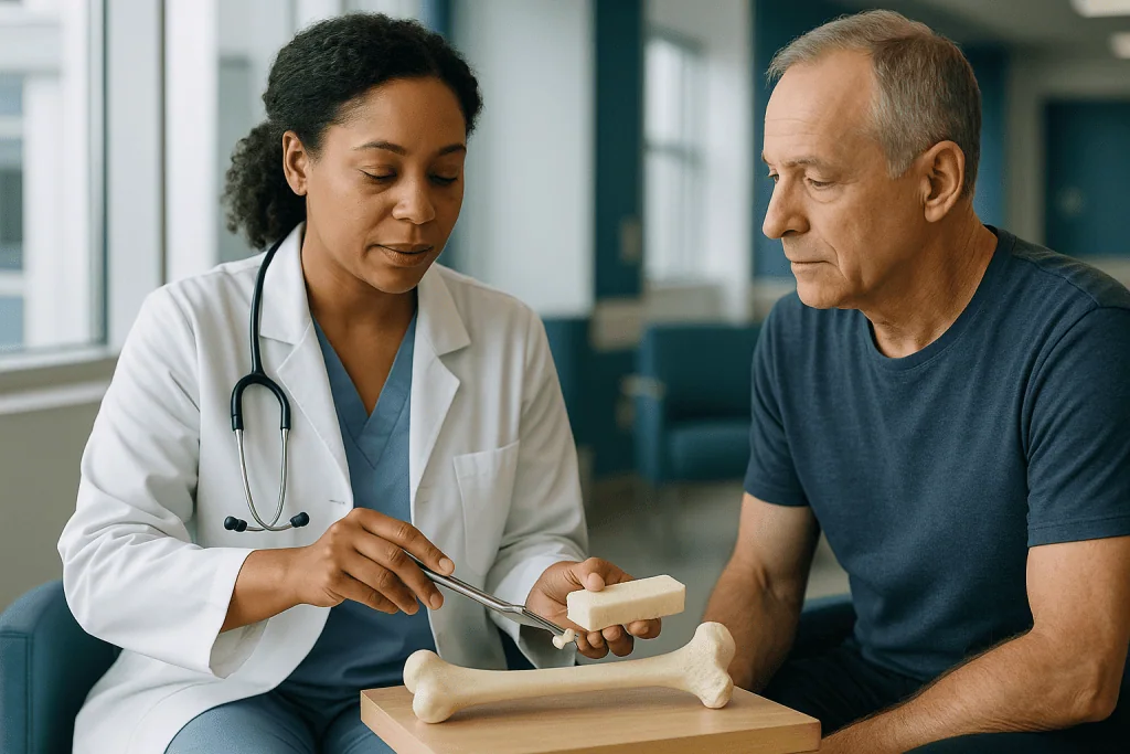
Bone specialists describe three properties a graft can have. Osteoconduction means the graft acts as a scaffold that the patient’s own cells can crawl along. Osteoinduction means the graft carries proteins that recruit nearby stem-like cells and prompt them to become bone-forming cells. Osteogenesis means the graft itself contains living cells that build bone. Allografts are strongly osteoconductive, modestly osteoinductive in some processed forms, and not osteogenic at all, because processing removes living cells (Kumar and colleagues, NIH PMC review).
Once placed, the graft sits in a bed of bleeding bone. Within days, tiny blood vessels begin to sprout into the porous spaces. Cells arrive with them. One type, the osteoclast, dissolves old donor mineral; another, the osteoblast, lays down fresh bone in its place. Over months the donor framework is chewed away and rebuilt, a process pathologists call creeping substitution. The name is apt: it creeps, working inward from the edges where living bone touches graft.
This is why a surgeon’s preparation of the site matters as much as the material. Graft dropped into a pocket with poor blood supply has no cells to borrow. Graft pressed against roughened, bleeding bone has everything it needs.
It also explains why there is no immune matching. Unlike a kidney or a heart, donor bone carries no living cells to provoke rejection. The body may mount a mild inflammatory response to foreign protein, but anti-rejection medicines are not part of bone grafting.
Where does donor bone actually come from?
Most allograft bone comes from deceased donors. When a person dies in circumstances that allow tissue recovery, a trained team may approach the family about donating tissue, a category that includes bone, tendon, skin and heart valves. Consent is documented before anything is recovered. A single donor can provide bone for many recipients, because the long bones, pelvis and spine yield a large volume of usable material.
A smaller stream comes from living donors. During a hip replacement, the ball of the thigh bone is removed and would otherwise be discarded. With the patient’s consent and the same screening applied to deceased donors, that femoral head can be processed and stored for someone else. It is a quiet form of recycling that many recipients never hear about.
Both streams pass through tissue banks, organizations that recover, test, process, store and distribute human tissue under national regulation. In the United States these banks operate under federal oversight and, in many cases, additional voluntary accreditation. The bank, not the operating surgeon, carries responsibility for confirming that the donor was suitable and the tissue safe to release.
Recovery itself is time-sensitive. Tissue is taken in a clean or sterile setting, generally within a day of death, and cooled promptly. Each piece receives a unique identifier that follows it through every later step, so that a graft placed in one patient can be traced back to its donor and forward to every other recipient from the same donor should a concern ever surface.
That traceability is the backbone of allograft safety. Screening removes unsuitable donors before processing begins; tracking lets the system respond if something is learned afterward.
How donors are screened before any bone is accepted
Screening happens in layers, and a donor is excluded if any layer raises doubt.
The first layer is history. Recovery staff interview the next of kin using a structured questionnaire that covers the donor’s medical conditions, cancers, infections, travel, tattoos, drug use and any behavior linked to higher infection risk. Medical records and the autopsy report, if one was performed, are reviewed. A physical examination of the body looks for signs of infection, injection marks or unexplained skin lesions.
The second layer is laboratory testing. A blood sample from the donor is tested for human immunodeficiency virus, hepatitis B, hepatitis C and syphilis, and in many programs for human T-lymphotropic virus as well. Modern panels combine antibody tests with nucleic acid tests, which detect the virus’s genetic material directly and shorten the window in which a very recent infection could be missed. Because blood collected after death can be diluted by fluids given during resuscitation, banks apply rules on sample timing and volume to keep results interpretable.
The third layer is the tissue itself. Swabs or fragments from the recovered bone are cultured for bacteria and fungi. Positive cultures may lead to discarding that tissue or routing it to a sterilization process validated against the organism found.
Exclusions are deliberately broad. Donors with most active cancers, systemic infections, certain neurological diseases or an unclear cause of death are typically declined, as are donors whose history cannot be adequately established.
None of this makes the risk zero, and honest surgeons say so. What screening does is stack independent barriers, so that a problem would have to slip past history, serology, culture and processing all at once for transmission to occur.
How donor bone is prepared: cleaning, preservation and sterilization
Once accepted, bone is cleaned before it is stored. Marrow, fat and blood are removed with mechanical washing, pressurized fluids and detergents or solvents. This step does two things at once: it lowers the load of any bacteria or viruses that may be present, and it strips out the cellular material most likely to provoke an immune response.
Preservation comes next, and the method shapes what the graft can later do. Fresh-frozen bone is stored at deep-freeze temperatures and keeps more of its mechanical strength and natural proteins. Freeze-dried bone, also called lyophilized, has its water removed under vacuum so it can be kept at room temperature; it is lighter and easier to store but more brittle until rehydrated. Demineralized bone matrix is made by soaking bone in dilute acid to dissolve the mineral, leaving collagen and growth proteins behind in a putty or powder that can be pressed into irregular spaces (Kumar and colleagues, NIH PMC review).
Sterilization is the final safeguard. Many banks expose bone to gamma irradiation or to validated chemical processes. Irradiation is effective against microbes but, at higher doses, weakens the collagen framework and reduces the graft’s ability to stimulate new bone. Banks therefore balance dose against intended use: a structural strut needs strength, a filler needs biology.
Every batch is tested again after processing, and each finished graft carries its identifier, expiration date and storage instructions. In the operating room, frozen grafts are thawed and freeze-dried grafts are soaked in sterile fluid according to the bank’s directions before the surgeon ever brings them to the wound.
The trade-off is unavoidable and worth saying plainly: the more aggressively bone is processed, the safer it becomes and the less biologically active it remains.
Forms of donor bone: chips, blocks, putty and demineralized matrix
Donor bone reaches the surgeon in several forms, and the choice is driven by one question: does this site need to bear weight, or does it simply need to be filled? Dense outer bone, called cortical bone, provides strength. Spongy inner bone, called cancellous bone, offers a porous honeycomb that blood vessels colonize quickly. Most operations use one or the other, and many use both.
| Form | What it is | Where it is often used | Main strength | Main limit |
|---|---|---|---|---|
| Cortical block or strut | Dense outer bone cut to shape | Spinal spacers, large defects, joint revision | Bears load from day one | Slow to incorporate; can crack |
| Cancellous chips | Spongy inner bone, crushed or cubed | Filling cavities, packing around fusion sites | Porous, incorporates relatively quickly | No structural support |
| Demineralized bone matrix | Acid-treated bone leaving collagen and proteins, as putty or gel | Dental sockets, small gaps, mixed with other graft | Some osteoinductive signal; molds to any space | No strength; potency varies between batches |
| Machined shapes | Cortical bone milled into wedges, dowels or rings | Spinal and foot-and-ankle fusion | Precise fit without metal | Limited sizes; same slow remodeling as block |
A surgeon rebuilding a collapsed vertebral space will reach for a machined cortical wedge, because the graft must hold the spine’s height while fusion happens behind it. The same surgeon filling the gap around a fracture plate will pour in cancellous chips, sometimes moistened with the patient’s own bone marrow drawn from the pelvis, to borrow living cells the allograft lacks.
Dental grafts sit at the small end of this spectrum. A tooth socket needs volume preserved, not weight carried, so particulate allograft or demineralized matrix is typical there.
Allograft vs autograft bone graft: how surgeons decide between them
For decades the patient’s own bone has been the reference material, and the reason is biological. An autograft arrives with living cells, its own growth proteins and a perfect immune match. It scores on all three properties: osteoconduction, osteoinduction and osteogenesis.
The price is a second wound. Harvesting from the pelvic rim adds operating time, blood loss and a separate site that can ache for weeks; some people describe the harvest site as more troublesome than the primary surgery. There is also a ceiling on supply. A large defect may simply need more bone than a person can spare (Cleveland Clinic).
Allograft flips that ledger. Supply is effectively unlimited, there is no donor-site pain and the operation is shorter. In exchange, the graft brings no living cells, incorporates more slowly, and carries a very small residual risk of disease transmission and a somewhat higher chance of failing to knit compared with autograft in demanding settings.
Surgeons therefore ask a series of practical questions. How large is the gap? Does it need to carry load? How good is the local blood supply? How much of the patient’s own bone is realistically available, and what would harvesting it cost in pain and time? Does the person smoke, have diabetes or take medicines that slow bone healing?
The answer is frequently both. A spine fusion might use a structural allograft wedge in front and the patient’s own bone shavings, collected during the operation, packed behind it. A revision hip replacement might use crushed allograft to fill a cavity and the patient’s marrow to seed it.
Neither option is universally superior. The right choice is the one matched to the defect and the person, and that judgment sits with the treating team.
What happens in the operating room when donor bone is placed
Grafting is rarely an operation on its own. It is a step inside a larger procedure: a fusion, a fracture fixation, a joint revision, a jaw reconstruction. Anesthesia is general for most orthopedic cases and local, sometimes with sedation, for many dental ones (MedlinePlus).
Before the graft comes out of its packaging, the surgeon prepares the bed. Scar tissue and dead bone are removed. The surface of the living bone is scraped or drilled until it bleeds, a step called decortication, because bleeding bone is where the recipient’s cells and vessels will come from. A graft placed against smooth, dry bone behaves like a seed on concrete.
The circulating nurse then checks the graft’s identifier against the patient record, confirms the expiration date and, if the graft is frozen or freeze-dried, thaws or rehydrates it in sterile fluid according to the tissue bank’s instructions. The surgeon shapes it: a block is trimmed to fit snugly, chips are packed until the cavity is full, putty is pressed into the corners. Snug matters. Gaps between graft and host bone are gaps that healing must bridge.
Because donor bone cannot hold itself in place, hardware usually does. Plates, screws, rods or cages immobilize the segment so the graft is not sheared or crushed while it incorporates. In dental sites, a thin collagen membrane may be laid over the particles to keep gum tissue from growing into the space before bone does.
Closure follows in layers, and an X-ray in the operating room or recovery area confirms position. From the patient’s point of view the graft is invisible; what they notice afterward is the incision and the restrictions on movement, both of which belong to the larger operation rather than to the graft itself.
Who allograft bone grafting is usually for, and who is usually asked to wait
Donor bone earns its place whenever a gap is too large, too poorly positioned or too slow-healing for the body to close alone. Common situations include spinal fusion, where vertebrae are joined to stop painful movement; fractures that have lost bone or failed to heal after months, called nonunion; revision joint replacement, where the old implant has left a hollow in the bone; benign tumor or cyst cavities after the lesion is scooped out; and jaw sockets or ridges that have thinned before a dental implant (Cleveland Clinic; Mayo Clinic).
Allograft is especially attractive when a patient has little spare bone to harvest, when a second incision would add meaningful risk, or when the volume needed is large.
Some people are asked to wait, and the reasons are usually about giving the graft a fair chance rather than about the graft itself. An active infection at or near the site is the clearest example; bacteria colonize dead bone readily, and a graft placed into infection tends to fail. Uncontrolled blood sugar slows every stage of bone healing. Nicotine, from cigarettes or vaping, constricts the small vessels the graft depends on, and many surgeons ask for a period of abstinence before and after surgery. Active gum disease is a reason to delay a dental graft. A history of radiation to the area, or of certain medicines that alter bone turnover, prompts a closer look and sometimes a different plan.
Children and adolescents are considered individually, because growth plates and rapid remodeling change the picture. Older adults are not excluded by age alone; bone quality, nutrition and overall health matter more than the number.
None of these is an automatic no. They are prompts for a conversation about timing, and that timing is the surgeon’s call with the patient.
How long does allograft bone take to heal? What the following weeks usually look like
The honest answer has two parts, because two different things are healing. The incision and soft tissue recover on a timescale of weeks. The graft itself incorporates on a timescale of months, and the distinction explains most of the confusion patients feel when they are told they look fine but still cannot lift, twist or bite down.
MedlinePlus puts overall recovery from a bone graft at roughly two weeks to three months depending on the site and the size of the defect, and notes that the graft may take up to three months or longer to heal fully. For spinal fusion specifically, Mayo Clinic describes fusion as taking several months to complete.
The first week is about the wound: swelling, bruising and pain managed by the surgical team, with early gentle movement of uninvolved joints to reduce clot risk. In the second to sixth weeks, the incision matures and most people are back to desk work or light daily activity, still under whatever weight-bearing or bite restrictions the surgeon set. During this window blood vessels are invading the graft and the first new bone is forming at its edges, none of which is visible on an X-ray yet.
Between roughly six weeks and three months, imaging begins to show blurring of the line between graft and host, the radiologic sign that incorporation is under way. Restrictions are usually eased in steps, guided by those images and by how the patient feels. Beyond three months, remodeling continues quietly; a large cortical allograft may go on being replaced for a year or more (Kumar and colleagues, NIH PMC review).
These are typical ranges, not promises. Smoking, diabetes, poor nutrition, infection and the size of the defect all stretch them, and the surgeon’s imaging, not the calendar, decides when it is safe to do more.
Cadaver bone graft risks and the real disadvantages of an allograft
People searching for cadaver bone graft risks usually mean disease. That fear deserves a straight answer: transmission of infection from screened, processed allograft bone is extremely rare, and modern testing and sterilization have made it rarer still, but it cannot be called impossible (Kumar and colleagues, NIH PMC review). Surgeons include it in consent because honesty requires it, not because it is a common event.
The disadvantages that actually change outcomes are more mundane. The first is slower, less certain incorporation. Because the graft brings no living cells and its natural proteins are diluted by processing, it relies entirely on the recipient’s biology, and in a poorly vascularized or infected bed it can fail to knit. The second is resorption: sometimes the body dissolves the graft faster than it rebuilds, leaving less volume than planned. The third, specific to large structural pieces, is fracture of the graft itself, particularly in the window when old bone has been removed but new bone has not yet fully replaced it.
Surgical-site infection is a risk of any operation, and dead bone is an inviting surface for bacteria, which is why site preparation and sterile handling receive so much attention. A mild inflammatory reaction to residual donor protein can occur and typically settles.
Some disadvantages are not medical. A few people hold religious or personal objections to receiving human tissue, and those views should be raised early so alternatives can be discussed. Others simply feel uneasy, which is a legitimate reason to ask more questions.
Set against these, allograft avoids the donor-site pain, extra operating time and supply limits of autograft. The decision weighs one set of risks against another, and it belongs to the patient and the treating team together.
What people often get wrong about how allograft bone grafting works
The most common misconception is that donor bone stays in the body as a permanent foreign object. In most cases it does not. Creeping substitution replaces it, piece by piece, with the patient’s own living bone. Years later a pathologist would struggle to find the boundary.
A close second is the belief that recipients need tissue matching or anti-rejection medicines, as organ transplant patients do. They do not. The living cells that trigger rejection are removed in processing, and the mineral and collagen that remain are nearly identical from one human to another.
Many people assume cadaver bone is inherently risky and their own bone inherently safe. The screening and sterilization chain described above makes allograft one of the more scrutinized materials in surgery. Autograft avoids donor risk entirely but introduces a second wound with its own infection and pain risks. Neither is risk-free.
Some believe allograft is always weaker. A machined cortical strut can bear more load on day one than a handful of the patient’s own cancellous chips; strength depends on form, not source. The reverse belief, that donor bone is just as biologically active as autograft, is also wrong. Processing removes cells and blunts growth signals.
Dental patients sometimes think their graft is a different technology from an orthopedic one. It is the same material and the same biology, scaled down.
Finally, people expect a single success rate. There is no such number. Outcomes depend on the site, the size of the defect, the form of graft, whether it was combined with the patient’s own bone or marrow, and the recipient’s smoking status, blood sugar and blood supply. Any figure quoted without those qualifiers is not describing your operation.
Questions to ask your care team before an allograft bone graft
A good consultation leaves you knowing not just that donor bone will be used, but why this form, why here, and what would change the plan. These questions tend to open that conversation.
- Why do you recommend donor bone for my situation rather than my own bone, a synthetic material, or a combination?
- Which form of allograft will you use: structural block, chips, demineralized matrix or a machined shape? What does that choice mean for how soon I can bear weight or bite?
- Where does the tissue come from, and what screening and sterilization does the tissue bank perform?
- Will you mix the donor bone with any of my own bone or bone marrow during the operation?
- What hardware will hold the graft in place, and does any of it need to come out later?
- What restrictions should I expect in the first two weeks, at six weeks and at three months, and how will you decide when to lift them?
- How will you check that the graft is incorporating, and what would a problem look like on imaging?
- What are the specific signs of infection or graft failure I should watch for at home?
- Do I smoke, take medicines or have conditions that could slow healing, and is there anything I should address before surgery?
- What would you do if the graft fails to incorporate? Is a second attempt possible?
- Is there anything about my religious, ethical or personal views on donated tissue that I should tell you now?
Write the answers down or bring someone who will. The details that matter most, such as weight-bearing limits and warning signs, are easy to lose in the relief of a plan being made.
When to call your doctor after allograft bone grafting
Most recovery days are uneventful: soreness that eases, a wound that dries and closes, stiffness that yields to the exercises you were given. The signs below are the exceptions, and they should prompt a call to the surgical team the same day rather than a wait for the next scheduled visit.
- Fever or shaking chills, particularly after the first few days when a low-grade temperature might otherwise be expected to have settled.
- Increasing redness, warmth, swelling or thick drainage at the incision, or a wound edge that opens.
- Pain that was improving and then worsens, or pain that is not controlled by the plan your team gave you.
- New numbness, tingling or weakness in a limb, or after spinal surgery any change in bladder or bowel control, which needs emergency evaluation.
- A sudden inability to bear weight or move the operated part after a fall or twist.
- Calf pain, swelling or tenderness in one leg, which can signal a blood clot.
- After a dental graft, persistent bleeding, a foul taste or odor, or a large shedding of graft particles that does not slow after the first days.
Go to an emergency department or call emergency services immediately for chest pain, sudden shortness of breath, coughing up blood, fainting, or one-sided weakness or facial droop. These can indicate a clot that has traveled to the lungs or a stroke, and minutes matter.
The surgical team wants to hear from you when you are unsure. Infection caught early can often be managed while preserving the graft; caught late it may cost the graft and require another operation. Whatever the finding, the decision about what happens next rests with the surgeon who knows your imaging, your hardware and your history.
Frequently asked questions
What is the success rate of allograft bone grafts?
There is no single success rate, because outcomes vary widely by site, defect size, graft form and patient health. A small dental socket graft and a large structural spinal strut are different operations with different odds. Smoking, diabetes, infection and poor blood supply lower the chance of incorporation for any graft. Ask your surgeon what published guideline or review data apply to your specific procedure rather than relying on a general percentage.
What is the difference between a bone graft and an allograft?
An allograft is one kind of bone graft. Bone graft is the general term for any material placed into a bone gap to help it heal, whether from the patient’s own body (autograft), a human donor (allograft), an animal (xenograft) or a laboratory (synthetic). When a surgeon says allograft, they are specifying the source, not describing a different procedure.
How does a cadaver bone graft work?
It works as a scaffold. The donor bone is placed against bleeding, roughened living bone, and within days the recipient’s blood vessels and cells grow into its porous structure. Over months, cells dissolve the donor mineral and lay down new bone in its place, a process called creeping substitution. Eventually most or all of the donor material is replaced by the patient’s own living bone.
What are the disadvantages of an allograft?
The main disadvantages are biological. Donor bone carries no living cells and reduced growth signals, so it incorporates more slowly and less reliably than the patient’s own bone, particularly in poorly supplied or infected sites. It can resorb faster than it rebuilds, and large structural pieces can fracture during remodeling. Disease transmission is extremely rare after screening and sterilization but is not zero. Some people also hold personal or religious objections.
How long does allograft bone take to heal?
Soft tissue heals in weeks, but the graft incorporates over months. MedlinePlus describes typical bone graft recovery as two weeks to three months, with the graft taking three months or longer to heal, and Mayo Clinic notes spinal fusion takes several months. Large cortical grafts may keep remodeling for a year or more. Your surgeon will use imaging, not the calendar, to decide when restrictions ease.
Can your body reject donor bone?
Not in the way it can reject a transplanted organ. Rejection is driven by living donor cells, and those are removed during processing, leaving mineral and collagen that are nearly identical between people. A mild inflammatory response to residual protein can occur and usually settles. Graft failure does happen, but it is generally caused by poor blood supply, infection, instability or smoking rather than immune rejection.
Is cadaver bone safe, and what are the infection risks?
Screened and processed donor bone is one of the more scrutinized materials in surgery. Donors are excluded for a broad range of conditions, blood is tested for HIV, hepatitis B and C and syphilis, tissue is cultured, and most grafts are sterilized. Transmission of infection is extremely rare but cannot be called impossible. The more realistic infection risk is an ordinary surgical-site infection, which applies to any operation.
What is an allograft bone graft used for in dental surgery?
In dentistry, donor bone is most often used to preserve a tooth socket after extraction, to rebuild a thinned jaw ridge before an implant, or to lift the sinus floor to make room for implants in the upper jaw. Particulate allograft or demineralized bone matrix is typical because these sites need volume preserved rather than weight carried. The biology is the same as in orthopedic grafting, at smaller scale.
Allograft vs autograft bone graft: which is better?
Neither is universally better. Autograft brings living cells and a perfect match but requires a second wound, adds operating time and is limited in quantity. Allograft avoids donor-site pain and supply limits but incorporates more slowly and depends entirely on the recipient’s biology. Many operations combine the two. The right choice depends on the defect, the site and the patient, and it rests with the treating team.
What can I do to help a donor bone graft heal?
The factors most within a patient’s control are avoiding nicotine in every form, keeping blood sugar well managed if you have diabetes, eating adequate protein and calories, following weight-bearing or bite restrictions exactly, and keeping the incision clean and dry as instructed. Take any prescribed medicines only as directed and raise questions with the prescribing clinician rather than adjusting on your own. Report fever, drainage or worsening pain promptly.
References
- MedlinePlus Medical Encyclopedia: Bone graft
- NIH PMC: Bone grafts in dentistry (Kumar P, Vinitha B, Fathima G)
This article is for general information only and is not a substitute for professional medical advice. Please consult a qualified doctor about your individual situation.

More from the Blog
Hip Arthroscopy: What Keyhole Hip Surgery Treats and How It Heals
Hip arthroscopy is keyhole surgery in which a surgeon uses a pencil-thin camera and small instruments, inserted through incisions about a centimeter long, to…
Cemented or Cementless Knee Replacement: How Implants Are Fixed to Bone and Why It Matters
Cemented knee replacement anchors the implant with a fast-setting acrylic grout that fills the gap between metal and bone, while cementless (uncemented) designs rely…

Yoga for Back Pain: What Trials Show and Which Poses to Avoid
Yoga for back pain has moderate-certainty evidence, mostly from randomized trials in chronic non-specific low back pain, showing small improvements in pain and daily…
What to Bring and How to Dress Your Baby for a Pavlik Harness Fitting Appointment
For a Pavlik harness fitting appointment, dress your baby in a short-sleeved bodysuit and a fresh diaper, skip pants and tight sleepers, and bring…

Hip Replacement Abroad: Costs, Safety Checks and Aftercare Planning
A total hip replacement abroad typically falls within a guide range of €9,100–16,900 for international patients, often well below typical UK (£11,000–17,000) or US…
Does Cracking Your Knuckles Cause Arthritis?
No, the available evidence does not show that cracking your knuckles causes arthritis. Studies comparing habitual knuckle crackers with people who never crack have…