

How Is Neurosarcoidosis Confirmed? MRI, Lumbar Puncture, PET-CT and Biopsy Explained

Key Takeaways
- Commonly quoted estimates place nervous system involvement at roughly 5% to 15% of people with sarcoidosis, and in some it is the first sign of the disease.
- Consensus criteria grade neurosarcoidosis as definite, probable or possible according to whether nervous system tissue, other tissue, or no tissue has shown granulomas.
- No blood test, including serum ACE, confirms or excludes sarcoidosis; ACE can be normal in active disease and raised in unrelated conditions.
- Contrast MRI is the most sensitive test for locating neurosarcoidosis, but its appearances overlap with infection, lymphoma and multiple sclerosis.
- Spinal fluid often shows raised lymphocytes and protein, yet its most decisive role is excluding infection and cancer cells.
- Most diagnoses rest on biopsy of a safer site such as a chest lymph node or skin, located with PET-CT, rather than on brain tissue.
Neurosarcoidosis is confirmed by combining a neurological examination, MRI of the brain or spine with contrast, analysis of spinal fluid from a lumbar puncture, imaging of the chest and body (often PET-CT) to find sarcoidosis elsewhere, and, wherever safely possible, a biopsy showing non-caseating granulomas. Consensus criteria grade the result as definite, probable or possible depending on whether nervous system or other tissue has been sampled and alternative causes excluded.
The neurologist has been talking for several minutes before the word finally lands: sarcoidosis. You came in because one side of your face stopped moving properly and your vision blurred for a week. You expected a stroke work-up, perhaps a conversation about multiple sclerosis. Instead you are hearing about a disease you associate, if at all, with lungs.
That gap between where the symptoms are and where the disease usually lives is exactly why how neurosarcoidosis is diagnosed deserves a slow, honest explanation. The condition rarely announces itself. It borrows the appearance of infections, tumors, lymphoma and inflammatory disorders, and no single scan or blood test can settle the question on its own.
What follows is the work-up as clinicians actually assemble it: a stepwise case built from MRI, spinal fluid, whole-body imaging and, when it can be done safely, a small piece of tissue under a microscope.
What is neurosarcoidosis, and why is it so hard to confirm?
Sarcoidosis is an inflammatory disease in which clusters of immune cells, called granulomas, form in tissues for reasons that are still not fully understood. Most people who develop it have granulomas in the lungs or the lymph nodes in the chest. Neurosarcoidosis simply means those granulomas have formed in the nervous system: the brain, the spinal cord, the thin membranes that cover them (the meninges), the cranial nerves that run to the face and eyes, or the peripheral nerves in the limbs.
Estimates of how often that happens vary, but commonly quoted figures from the National Institute of Neurological Disorders and Stroke and the Cleveland Clinic place nervous system involvement somewhere between about 5% and 15% of people with sarcoidosis. In a meaningful minority, the neurological problem is the first sign of the disease, before anyone has found anything in the lungs.
Three things make confirmation difficult. First, granulomas in the nervous system look, on scans, very much like other inflammatory or cancerous processes. Second, the nervous system is a poor place to take tissue samples; a brain biopsy carries real risk, so clinicians often have to prove the disease somewhere safer and infer the connection. Third, there is no blood test that reliably rules sarcoidosis in or out.
The practical result is that diagnosis is built as a case rather than delivered as a verdict. Each test adds a layer of probability. The treating team weighs the pattern of symptoms, the MRI appearance, the spinal fluid picture and the presence of granulomas elsewhere, and then, crucially, works to exclude the conditions that could mimic the whole picture. That exclusion step is not an afterthought; it is half of the diagnosis.
How neurosarcoidosis is diagnosed: the definite, probable and possible framework
Because certainty is hard to reach, neurologists use a tiered set of neurosarcoidosis diagnostic criteria rather than a yes-or-no label. The most widely used framework, developed by an international consortium of neurosarcoidosis specialists and reflected in patient information from the Cleveland Clinic and NINDS, sorts cases into three levels.

- Definite neurosarcoidosis requires a biopsy of nervous system tissue showing the characteristic granulomas, along with a clinical picture and work-up consistent with the disease and other causes excluded.
- Probable neurosarcoidosis applies when the neurological presentation and tests (typically MRI and spinal fluid) point to inflammation consistent with sarcoidosis, and a biopsy from outside the nervous system, such as a lymph node, lung or skin, confirms systemic sarcoidosis.
- Possible neurosarcoidosis is used when the clinical and imaging picture suggests the disease and alternatives have been reasonably excluded, but no tissue of any kind has been obtained.
Notice what the framework does. It separates the strength of the evidence from the decision about care. A person labelled “probable” is not being told the team is unsure whether something is wrong; they are being told exactly how the conclusion was reached and where the remaining uncertainty lies. Many people are treated at the probable level because the risks of obtaining brain or spinal cord tissue outweigh the value of upgrading the label.
The framework also builds in a non-negotiable requirement at every level: other explanations must be excluded. Infections such as tuberculosis, which also produces granulomas, lymphoma, which can shrink temporarily with steroids just as sarcoidosis does, and demyelinating diseases such as multiple sclerosis are the usual suspects. A diagnosis of neurosarcoidosis is, in a real sense, the diagnosis left standing after those have been investigated.
What actually happens during the work-up, step by step
Most people arrive at the question through a symptom, not a suspicion. The commonest neurological presentation is weakness of a cranial nerve, particularly the facial nerve, which can produce a drooping face that initially looks like Bell’s palsy. Others present with headaches, visual change, seizures, unexplained thirst and urination from pituitary involvement, numbness or weakness in the legs from spinal cord disease, or a slow change in thinking and mood.
The first step is a detailed neurological examination and history. The neurologist is listening for clues beyond the nervous system: a dry cough, breathlessness, skin lumps, painful red shins, eye inflammation, or joint pain, any of which hints that an inflammatory process is affecting several organs at once.
Imaging usually follows quickly. MRI of the brain, and often the spine, is performed with a gadolinium-based contrast agent, a dye injected into a vein that highlights areas where the protective barrier around blood vessels has been disrupted by inflammation. Blood tests are drawn at the same visit.
If the MRI shows inflammation, a lumbar puncture is typically next, provided it is safe. The spinal fluid is checked for cell counts, protein, glucose, infection and inflammatory markers. In parallel, the search for sarcoidosis elsewhere begins: a chest X-ray or CT scan of the chest, and frequently a whole-body FDG PET-CT scan, which maps metabolically active inflammation anywhere in the body.
The final step, when a reachable site exists, is biopsy. A lymph node in the chest sampled through a bronchoscope, a skin lesion, or an enlarged gland in the neck are far safer targets than brain tissue. Only when nothing else explains the findings and nothing safer is available will a team discuss biopsy of the nervous system itself. Each step is reviewed as a whole, usually in a multidisciplinary meeting involving neurology, respiratory medicine, radiology and pathology.
Neurosarcoidosis MRI findings: what the scan can and cannot show
MRI is the most sensitive imaging test for neurosarcoidosis, and the first result most people see. Magnetic resonance imaging uses a strong magnetic field and radio waves, not radiation, to produce detailed pictures of soft tissue. For this condition it is almost always performed with contrast, because the pattern of enhancement is where the diagnostic clues sit.
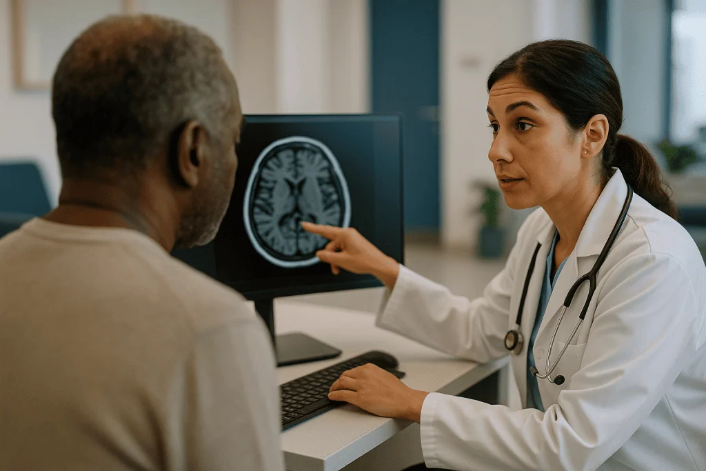
Several appearances are characteristic. The meninges, especially at the base of the brain, may show thick or nodular enhancement, a finding neuroradiologists describe as leptomeningeal or pachymeningeal involvement. The cranial nerves can appear swollen and bright. The hypothalamus and pituitary stalk, which sit at the base of the brain and regulate hormones, are a favoured site. Within the brain itself, granulomas can look like small enhancing masses or diffuse patches in the white matter. Spinal cord involvement often produces a long segment of swelling and enhancement, which is one of the features that help distinguish it from the shorter lesions typical of multiple sclerosis. Hydrocephalus, a build-up of spinal fluid inside the brain, sometimes develops when inflammation blocks its normal flow.
The limits are just as important. None of these neurosarcoidosis MRI findings is specific. Tuberculous meningitis, fungal infection, lymphoma spreading along the meninges, and other inflammatory diseases can produce near-identical pictures. A normal MRI does not rule the condition out either, particularly when the problem is in peripheral nerves or muscles, which MRI of the brain does not assess.
MRI earns its place by doing two things well: it shows that inflammation is present and where, which guides the lumbar puncture and any later biopsy, and it provides a baseline against which the team can judge, weeks or months later, whether treatment has changed anything. Repeat scans are a normal part of care, not a sign that the first was inconclusive.
Lumbar puncture for neurosarcoidosis: what the spinal fluid reveals
A lumbar puncture, often called a spinal tap, involves placing a thin needle between two bones in the lower back to draw off a small amount of cerebrospinal fluid, the clear liquid that cushions the brain and spinal cord. MedlinePlus describes the procedure as taking roughly 30 minutes, performed with local anaesthetic, with the person lying curled on their side or sitting bent forward.
The fluid is then examined in several ways. In neurosarcoidosis it commonly shows a raised white cell count dominated by lymphocytes, a type of immune cell, together with elevated protein. Glucose may be mildly low. Oligoclonal bands, which are clusters of antibodies produced inside the nervous system, are present in a proportion of people, which complicates the distinction from multiple sclerosis. The laboratory may also measure angiotensin-converting enzyme (ACE), a protein released by granulomas; a raised level in spinal fluid supports the diagnosis but is neither sensitive nor specific enough to settle it.
The most valuable role of lumbar puncture for neurosarcoidosis may be what it excludes. The same sample is cultured and tested for bacteria, tuberculosis, fungi and viruses, and sent for cytology, a microscopic search for cancer cells that would point towards lymphoma or metastatic disease. Each negative result strengthens the case.
Spinal fluid can also be entirely normal in neurosarcoidosis, which is why a clean result does not close the door. Conversely, mild abnormalities overlap with many other inflammatory conditions. Clinicians treat the fluid as one chapter of the evidence, read alongside the MRI.
Afterward, a headache that worsens on standing is the commonest side effect and usually settles with rest and fluids. The team will advise when to report anything persistent.
Blood tests, ACE and other markers: useful, but never the whole answer
People often arrive hoping for a blood test that gives a clear answer. There is not one, and understanding why saves considerable frustration.
Serum ACE is the test most associated with sarcoidosis. The enzyme is produced by the cells inside granulomas, so its level in blood tends to rise when the body carries a large burden of them. The Mayo Clinic and NHS both list it among standard investigations, and the Mayo Clinic is explicit that no single blood test confirms the condition. ACE can be normal in people with active sarcoidosis and raised in several unrelated conditions, including liver disease, diabetes and thyroid disorders. Some people also take a class of blood pressure medicine, ACE inhibitors, that lowers the measured level and makes the test uninterpretable; the team will note this when reading the result.
Calcium is checked because granulomas can activate vitamin D in an unregulated way, pushing blood and urine calcium above normal; a raised level supports the diagnosis and matters for care in its own right. Kidney and liver function tests look for involvement of those organs. A full blood count and inflammatory markers such as C-reactive protein give a sense of the overall inflammatory load, although they are frequently unremarkable.
Newer markers, including soluble interleukin-2 receptor, are measured in some centres and may be more sensitive than ACE, but availability and interpretation vary, and none has replaced tissue diagnosis in guideline practice.
The blood work also serves the exclusion side of the ledger. Tests for HIV, syphilis and tuberculosis exposure are routine, because each can cause inflammation of the meninges that mimics neurosarcoidosis and would be treated entirely differently. Reading the blood panel as a whole, rather than searching it for one decisive number, is the mindset the team brings to it.
Why a chest scan and PET-CT matter when the problem is in the brain
It can feel odd to be sent for a chest CT when your symptoms are facial weakness or blurred vision. The logic is practical. Sarcoidosis most often affects the lungs and the lymph nodes in the centre of the chest, and MedlinePlus notes it is frequently found there incidentally. Demonstrating granulomas in a safely reachable organ allows the team to make a probable diagnosis without touching the nervous system.
A chest X-ray is often the first look. It may show enlarged lymph nodes on both sides of the central chest, a pattern called bilateral hilar lymphadenopathy, or fine shadowing in the lung tissue. CT of the chest is more sensitive and better at picking out small nodes that could be sampled.
FDG PET-CT goes further. A small amount of a radioactive sugar is injected and taken up by metabolically active cells, including the immune cells inside granulomas. The combined scan then maps those hot spots across the whole body. For neurosarcoidosis, PET-CT does two jobs: it reveals occult disease in the chest, abdomen, bones or skin when standard imaging is normal, and it pinpoints the most active and most accessible target for biopsy. A lymph node that lights up brightly is more likely to yield a diagnostic sample than one that does not.
PET-CT is less useful for looking at the brain itself, because normal brain tissue consumes so much sugar that the background glow drowns out small lesions. It is also not specific; infections and cancers light up too. The scan is a map for the next step rather than a diagnosis in itself.
Eye examination by an ophthalmologist and, where indicated, a cardiac evaluation are added for the same reason: evidence of sarcoidosis in another organ changes the certainty of the neurological diagnosis.
Neurosarcoidosis biopsy: why tissue remains the anchor
Under the microscope, sarcoidosis has a recognisable signature: compact collections of activated immune cells, forming what pathologists call non-caseating granulomas. “Non-caseating” means the centre of the granuloma has not broken down into the cheese-like dead tissue typical of tuberculosis, which is one of the key distinctions the pathologist makes. Special stains and cultures on the same sample look for the organisms, fungi and foreign material that can produce similar granulomas.
Where that tissue comes from determines how certain the diagnosis is. In order of preference, teams look for the safest site that is likely to be positive. A skin lesion can be sampled in clinic with local anaesthetic. An enlarged lymph node in the neck is reachable with a needle. Lymph nodes in the chest are commonly sampled by endobronchial ultrasound, a procedure in which a flexible camera is passed into the airways and a fine needle guided into the node; the Mayo Clinic describes bronchoscopic biopsy as one of the standard routes. Lung, liver or a salivary gland are other options depending on the PET-CT map.
A neurosarcoidosis biopsy of the nervous system itself is reserved for situations where nothing else is accessible and the diagnosis materially changes care. Meningeal or brain biopsy is a neurosurgical operation with risks of bleeding, infection and neurological injury, and even then the sample may miss the affected area. When it is offered, the team will explain why they believe the information is worth the risk and what the alternatives would be.
Biopsy also has limits. A negative sample does not exclude sarcoidosis if the needle simply missed; granulomas are patchy. The pathologist’s report is read against the imaging and fluid results rather than in isolation. Tissue is the anchor of the diagnosis, not a substitute for the rest of the case.
How neurosarcoidosis is diagnosed in practice: comparing the main tests
Seeing the tests side by side clarifies why no single one is enough and why the order matters. Each answers a different question, and the gaps in one are covered by another.
| Test | What it shows | Main strength | Main limitation |
|---|---|---|---|
| MRI with contrast | Inflammation of meninges, cranial nerves, brain, pituitary region or spinal cord | Most sensitive way to locate nervous system disease; baseline for follow-up | Appearances overlap with infection, lymphoma and other inflammatory diseases |
| Lumbar puncture | Cell counts, protein, glucose, oligoclonal bands, ACE, cultures, cytology | Excludes infection and cancer cells; supports inflammatory picture | Can be normal; abnormalities are non-specific |
| Blood tests | Serum ACE, calcium, organ function, infection screens | Flags systemic involvement; rules out mimics | No blood test confirms or excludes sarcoidosis |
| Chest X-ray / CT | Enlarged chest lymph nodes, lung changes | Finds the commonest site of sarcoidosis | Can be normal in isolated neurological disease |
| FDG PET-CT | Active inflammation anywhere in the body | Finds hidden disease and the best biopsy target | Poor for brain itself; lights up in cancer and infection too |
| Biopsy (extraneural) | Non-caseating granulomas with negative stains for organisms | Gives a probable diagnosis without nervous system surgery | Patchy disease can be missed |
| Biopsy (nervous system) | Granulomas in brain, meninges or nerve | Only route to a definite diagnosis | Surgical risk; reserved for selected cases |
Read together, the pattern is clear. MRI and lumbar puncture establish that the nervous system is inflamed and narrow the list of causes. Chest imaging and PET-CT look for the disease where it is safest to prove. Biopsy converts a strong suspicion into a diagnosis the team can act on with confidence. The sequence can be compressed when symptoms are severe and stretched when they are mild and stable.
Who is usually offered the full work-up, and who is asked to wait
Not everyone with sarcoidosis and a neurological symptom is pushed through every test. Clinicians triage on two questions: how likely is neurosarcoidosis, and how urgent is it to know?
People most likely to proceed promptly through MRI, lumbar puncture and body imaging include those with a new cranial nerve palsy that does not behave like typical Bell’s palsy, those with seizures, new headaches with neurological signs, visual loss, symptoms of pituitary dysfunction such as marked thirst and urination, and anyone with signs of spinal cord disease such as leg weakness, numbness rising up the body, or bladder change. Someone already known to have sarcoidosis who develops any of these is usually investigated quickly, because the probability of a connection is high and some presentations, particularly spinal cord and hydrocephalus, can worsen rapidly.
Others are reasonably asked to wait or to proceed in stages. Mild, non-progressive numbness in the hands or feet with a normal examination may be assessed first with nerve conduction studies rather than brain imaging. Fatigue and low mood, which are common in sarcoidosis of any organ, are not by themselves reasons for a lumbar puncture. A person with a single, non-specific MRI finding and no symptoms may be followed with a repeat scan rather than an immediate biopsy.
Some tests are deferred for safety. Lumbar puncture is postponed if the MRI shows pressure effects that could make it dangerous, or if blood thinning or a clotting disorder has not been managed. PET-CT may be delayed in pregnancy. Nervous system biopsy is withheld whenever a safer site can give the answer.
Waiting, in this context, is a clinical judgement, not neglect. The team balances the small but real risks of invasive tests against the chance that they change what happens next, and that balance is reviewed whenever symptoms shift.
What the following weeks usually look like
The diagnostic phase of neurosarcoidosis rarely resolves in a single appointment. People commonly describe a period of several weeks in which results arrive piecemeal and the picture sharpens gradually. Knowing the shape of that period makes it easier to tolerate.
MRI is often performed within days when symptoms are significant, and the report may be available at the first follow-up. Lumbar puncture results come in layers: cell counts and protein within hours, cytology over a few days, and cultures for slow-growing organisms such as tuberculosis over several weeks. Chest imaging and PET-CT are scheduled around scanner availability. If a biopsy is arranged, the pathology report typically follows within one to two weeks, with special stains sometimes adding more time.
Many teams discuss the assembled results at a multidisciplinary meeting, after which the neurologist explains where the diagnosis sits on the definite, probable or possible scale and what they recommend.
Treatment decisions may begin before every result is back. When vision, spinal cord function or pressure inside the head is threatened, clinicians often start corticosteroids, medicines that broadly dampen immune activity, once infection has been reasonably excluded, because waiting for complete certainty risks permanent damage. Longer-term options include other immunosuppressant classes chosen by the treating team according to the pattern and severity of disease. The decision to start, what to use, and for how long rests entirely with the prescribing clinician, who weighs individual risks that a general article cannot.
Early follow-up usually involves repeat neurological examination and a further MRI after some weeks to months to assess whether inflammation is settling. Improvement on steroids supports the diagnosis but does not confirm it, since lymphoma and some infections also respond temporarily, which is why the exclusion work continues alongside care.
What people often get wrong about neurosarcoidosis diagnosis
A handful of misunderstandings surface again and again in clinic, and correcting them changes how people experience the process.
“If the ACE test is normal, it isn’t sarcoidosis.” It may well be. ACE can be normal in active disease and is affected by medicines and unrelated conditions. The Mayo Clinic is clear that no blood test confirms sarcoidosis; it is one supporting clue among many.
“A clear chest X-ray rules it out.” Isolated neurological disease without visible chest involvement is well recognised, which is precisely why PET-CT is used to search further afield.
“They should just biopsy the brain and be done with it.” Nervous system biopsy carries meaningful surgical risk and can miss patchy disease. Guideline practice is to prove sarcoidosis in the safest available organ and infer the connection, which is why most diagnoses sit at the probable level.
“Getting better on steroids proves the diagnosis.” It is reassuring, but lymphoma and several infections also improve, sometimes dramatically, in the short term. Teams keep the alternative explanations open for exactly this reason.
“It must be multiple sclerosis because there are oligoclonal bands.” Those antibody bands appear in a proportion of people with neurosarcoidosis and in several other conditions. MRI patterns, body imaging and tissue, not the bands alone, separate the two.
“Possible means they don’t believe me.” The label describes the evidence, not the symptoms. A person with a possible diagnosis can be treated seriously and promptly; the word signals that no tissue has been obtained, nothing more.
“A normal MRI means my symptoms aren’t neurological.” Peripheral nerve and muscle involvement do not show on brain MRI and need different tests, such as nerve conduction studies or muscle imaging.
Questions to ask your care team
Appointments move quickly and results arrive in fragments, so it helps to have a short list ready. These are the questions that tend to produce the most useful answers in a neurosarcoidosis work-up.
- Where does my diagnosis currently sit: definite, probable or possible, and what would move it up a level?
- Which alternative conditions have you excluded so far, and which are still open?
- What specifically did the MRI show, and which areas of my nervous system are involved?
- What did the spinal fluid show, and are any cultures or cytology results still pending?
- Has the search for sarcoidosis outside the nervous system been completed, and what did the chest imaging or PET-CT find?
- Is a biopsy planned, from which site, and what are the risks and the chance it gives a clear answer?
- If treatment is starting before every result is back, what are you treating for, and how will you know whether it is working?
- Which symptoms should prompt me to contact you straight away rather than wait for the next appointment?
- Will my case be discussed with respiratory, eye or heart specialists, and do I need to see any of them?
- When is my next scan or follow-up, and what will you be looking for on it?
Writing the answers down, or asking whether a family member can attend, is worthwhile. People regularly report that the sheer volume of information makes it difficult to recall, afterwards, which uncertainties the team considered settled and which remain active. Asking for a brief written summary of the diagnostic reasoning is a reasonable request that many clinicians are glad to meet.
When to call your doctor
Neurosarcoidosis can change course during the weeks of investigation, and some developments need same-day attention rather than a note for the next visit. Contact your care team promptly, or seek emergency care, if you notice any of the following.
- A new or rapidly worsening headache, especially with vomiting, drowsiness or confusion, which can signal rising pressure inside the head or hydrocephalus.
- A first seizure, or seizures becoming more frequent.
- Sudden loss or blurring of vision, double vision, or a painful red eye.
- New weakness or numbness in the legs, difficulty walking, or a loss of bladder or bowel control, which may indicate spinal cord involvement.
- Marked thirst with very frequent urination, which can reflect pituitary involvement affecting water balance.
- Fever, neck stiffness or a new rash, particularly if immune-suppressing treatment has started, since infection must be excluded quickly.
- After a lumbar puncture: a headache that persists beyond a couple of days despite lying flat and drinking fluids, fever, or new numbness or weakness in the legs.
- After any biopsy: bleeding that does not stop, increasing pain, redness or swelling at the site, breathlessness, or coughing up blood.
- Palpitations, fainting or chest discomfort, because sarcoidosis can involve the heart and may not have been fully assessed yet.
None of these signs confirms anything on its own, and many have ordinary explanations. The point of calling is that your team can decide, with the full picture in front of them, whether a scan, a blood test or an urgent review is needed. During an active work-up, erring on the side of making the call is the safer habit, and clinicians would rather hear from you than learn later that a change went unreported.
Frequently asked questions
What are the neurosarcoidosis diagnostic criteria?
The widely used consensus criteria define three levels. Definite requires granulomas in nervous system tissue plus a compatible picture and exclusion of other causes. Probable requires a compatible neurological presentation and tests together with biopsy-proven sarcoidosis elsewhere in the body. Possible applies when the clinical and imaging findings suggest the disease and alternatives have been excluded, but no tissue has been obtained.
What are the typical neurosarcoidosis MRI findings?
Contrast MRI commonly shows thickened or nodular enhancement of the meninges, especially at the base of the brain, swollen enhancing cranial nerves, involvement of the pituitary stalk and hypothalamus, enhancing lesions within the brain, or a long segment of spinal cord swelling. Hydrocephalus may be present. None of these appearances is specific, and the MRI can be normal when only peripheral nerves are affected.
Is lumbar puncture for neurosarcoidosis always necessary?
Not always, but it is performed in most cases where the MRI shows inflammation and the procedure is safe. The fluid often shows raised lymphocytes and protein, and it allows cultures, tuberculosis testing and cytology that exclude infection and lymphoma. It may be deferred if imaging shows pressure effects or if clotting has not been managed, and the treating team makes that call.
Can neurosarcoidosis be diagnosed without a biopsy?
Yes, at the possible level. When MRI and spinal fluid point to sarcoidosis-type inflammation, body imaging and blood tests are supportive, and infections, lymphoma and other causes have been reasonably excluded, clinicians may diagnose and treat without tissue. A biopsy from a safe site, such as a lymph node, upgrades the diagnosis to probable and is pursued whenever one is found.
Why do I need a chest CT or PET-CT for a brain problem?
Because sarcoidosis most often affects the lungs and chest lymph nodes, finding granulomas there allows a probable diagnosis without a risky nervous system biopsy. PET-CT maps active inflammation across the whole body, revealing hidden disease when the chest looks normal and identifying the most accessible site to sample. It is less useful for the brain itself because normal brain tissue takes up the tracer strongly.
Does a normal ACE blood test rule out neurosarcoidosis?
No. Serum ACE is raised in many but not all people with active sarcoidosis, and it can be lowered by a common class of blood pressure medicine or raised by unrelated conditions. The Mayo Clinic states that no single blood test confirms sarcoidosis. ACE is one supporting clue read alongside imaging, spinal fluid and, where possible, tissue.
How long does it take to confirm neurosarcoidosis?
There is no fixed timeline. MRI is often done within days, basic spinal fluid results arrive within hours, cytology within days, and cultures for slow-growing organisms can take several weeks. Biopsy pathology typically follows within one to two weeks. Many people experience a stepwise process over several weeks, and treatment may start before every result is back when function is threatened.
What is a neurosarcoidosis biopsy looking for?
The pathologist looks for non-caseating granulomas, compact clusters of immune cells without the central dead tissue seen in tuberculosis, along with negative special stains and cultures for bacteria, fungi and foreign material. Tissue is usually taken from a safer site such as a chest lymph node, skin or neck gland; brain or meningeal biopsy is reserved for cases where nothing else can give the answer.
Could it be multiple sclerosis instead of neurosarcoidosis?
The two are genuinely confused at times, because both cause enhancing lesions and both can produce oligoclonal bands in spinal fluid. Clues favouring sarcoidosis include meningeal enhancement, cranial nerve or pituitary involvement, long spinal cord lesions, and evidence of granulomas elsewhere on chest imaging or PET-CT. The distinction is made from the whole picture, and the treating team will explain which features weighed most.
If steroids help, does that confirm the diagnosis?
Improvement on corticosteroids supports but does not prove neurosarcoidosis. Lymphoma and some infections can also shrink or settle temporarily when immune activity is dampened. For that reason, clinicians continue to exclude alternative causes even after treatment begins, and they use follow-up MRI alongside examination to judge whether the response fits the expected pattern for sarcoidosis.
References
- Neurosarcoidosis, National Institute of Neurological Disorders and Stroke (NIH)
- Sarcoidosis, NHS
- Cerebrospinal Fluid (CSF) Analysis, MedlinePlus
- Sarcoidosis, MedlinePlus
This article is for general information only and is not a substitute for professional medical advice. Please consult a qualified doctor about your individual situation.

More from the Blog

Insomnia Treatment: Why CBT-I Comes First and How a Sleep Specialist Builds Your Plan
Insomnia is treated first with cognitive behavioral therapy for insomnia (CBT-I), a structured, short-term program that retrains sleep timing, bedroom habits and anxious thinking…
What Do Follow-Up MRI Scans Look For After Brain Tumor Treatment and How Often Are They Done?
A brain tumor follow up MRI checks whether anything has changed since the last scan: new or growing contrast enhancement, expanding signal on fluid-sensitive…

Why Do Brain Shunts Sometimes Block or Fail? What Shunt Revision Surgery Involves
Brain shunts usually fail because the tubing becomes blocked by tissue, protein or debris, because parts disconnect or break, because infection develops, or because…

SDR Recovery Timeline: Lying Flat, First Steps and the Months of Physical Therapy That Follow
A typical SDR recovery timeline runs in stages: two to three days lying flat after surgery, roughly a week in hospital, supported standing and…

Leg Stiffness That Keeps Progressing: When a Neurologist Considers an HSP Evaluation
Hereditary spastic paraplegia (HSP) is a group of inherited conditions in which leg stiffness and weakness slowly worsen over years. A neurologist referral is…

Which Tests Shape a Brain Tumor Treatment Plan? From the First MRI to the Pathology Report
A brain tumor treatment plan is built from several tests that work in sequence: a CT scan if symptoms arrive suddenly, an MRI with…