

How Is ROP Treatment Performed? Sedation, the Retinal Procedure and the Minutes It Takes
Key Takeaways
- The ETROP trial found that treating at the type 1 ROP threshold reduced unfavorable structural outcomes at nine months from 15.6 percent to 9.1 percent, which is why treatment is arranged within days of diagnosis.
- ROP laser is always performed under general anesthesia or deep sedation with continuous monitoring, because the burns and the pressure used to view the retinal edge would be painful to an awake infant.
- The laser itself takes minutes to about an hour per eye depending on how much retina lacks vessels, but dilation, anesthesia and recovery mean the total window is usually several hours.
- In BEAT-ROP, disease recurred in 6 percent of zone I eyes treated with bevacizumab versus 42 percent treated with laser, while zone II showed no significant difference, which shapes how the two options are chosen.
- Laser permanently sacrifices the oxygen-starved peripheral retina to protect central vision, and treated children have higher rates of nearsightedness, strabismus and amblyopia that require follow-up through childhood.
- Stage 5 ROP, a total retinal detachment, can sometimes be reattached surgically, but the literature reports limited useful vision afterward, making screening attendance the most preventable factor in outcomes.
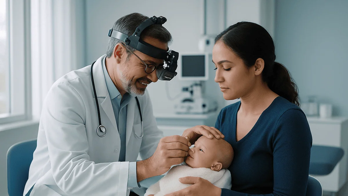
ROP laser treatment is performed while a premature baby is sedated or under general anesthesia and continuously monitored by the neonatal team, either at the bedside or in an operating room. After dilating drops and a small eyelid holder are placed, the ophthalmologist uses a headset-mounted laser to make many tiny burns on the immature outer retina, reducing the growth signals that drive abnormal blood vessels. The laser portion is typically measured in minutes to around an hour per eye.
The consent form arrives on a clipboard, between a feed and a diaper change, in a room where the loudest sound is a monitor. A baby who weighed less than a bag of sugar at birth now needs an eye procedure, and the word on the form is laser. Most parents in that moment have one question underneath all the others: what, exactly, is going to happen to my child?
The ROP laser treatment procedure has been the standard way to treat severe retinopathy of prematurity for decades, yet it remains one of the least explained events in a neonatal stay. It happens fast, it happens early, and it happens while parents are often still learning what a retina is.
This explainer walks through the sedation, the steps in the room, the minutes involved, the alternative injection treatment, and what the following weeks tend to look like. Every decision still belongs to the team caring for your baby; the aim here is that you understand the conversation when it comes.
What is retinopathy of prematurity, and why does timing matter so much?
Retinopathy of prematurity, or ROP, is a disease of the retina, the light-sensing layer at the back of the eye, in babies born too early for that layer to finish growing its blood supply. In a full-term pregnancy the retinal vessels grow outward from the optic nerve and reach the edge of the retina around the due date. A baby born at 26 weeks arrives with a large rim of retina that has no vessels at all.
Two things then go wrong. First, the higher oxygen levels of life outside the womb, including supplemental oxygen, can pause normal vessel growth. Second, as the retina keeps developing and demanding more oxygen than the stalled vessels can deliver, it releases a chemical signal called vascular endothelial growth factor, or VEGF, a protein that tells blood vessels to grow. Too much VEGF produces fragile, disorganized vessels that can bleed and pull on the retina, which is why advanced ROP can lead to retinal detachment.
Not every case needs treatment. Many babies have mild changes that regress on their own as the retina matures. According to the National Eye Institute, ROP affects roughly 14,000 to 16,000 infants born prematurely in the United States each year, and most improve without intervention; a much smaller group progresses to disease that threatens sight.
Timing is the whole story in that smaller group. Screening exams are scheduled to catch the narrow window when abnormal vessels are active but the retina is still attached. The Early Treatment for Retinopathy of Prematurity trial, known as ETROP, showed that treating at a defined threshold called type 1 ROP, rather than waiting for the older, later threshold, reduced unfavorable structural outcomes at nine months from 15.6 percent to 9.1 percent. That finding is why an ophthalmologist who finds type 1 disease will usually arrange treatment quickly, often within about 72 hours.
What actually happens during the ROP laser treatment procedure, step by step
Strip away the vocabulary and the procedure is a sequence of small, deliberate actions. Here is what a parent standing outside the room would be watching if the doors were glass.
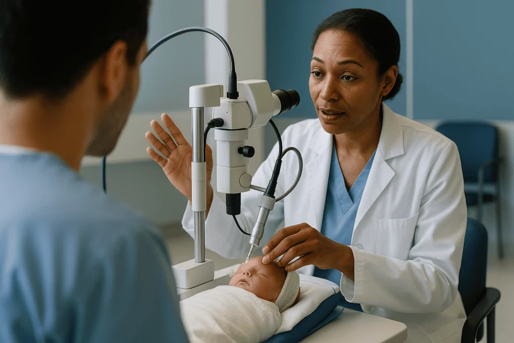
Preparation begins an hour or more before the laser. Dilating drops widen the pupil so the surgeon can see the far edge of the retina. A nurse confirms when the last feed was, checks the baby’s temperature, and ensures monitoring leads for heart rate, breathing and oxygen saturation are secure. The anesthesia or neonatal team then gives sedation or general anesthesia, discussed in the next section, and settles the baby in a stable position, sometimes with a small towel roll to support the head.
The surgeon places a lid speculum, a tiny wire device that gently holds the eyelids open, since a sedated baby cannot cooperate. Lubricating drops keep the surface of the eye moist.
The laser itself is delivered through a laser indirect ophthalmoscope, a headset that lets the surgeon view the retina through a handheld lens while aiming a fine beam through the pupil. With a blunt instrument called a scleral depressor, the surgeon gently presses the outside of the eye to bring the peripheral retina into view. Each press of a foot pedal places one spot, a pale gray-white burn roughly the width of a pinhead, on the retina that has no blood vessels. Spot follows spot until the entire avascular zone in front of the abnormal vessels is covered in a near-continuous pattern, stopping short of the vessels themselves.
The second eye is usually treated in the same session. Once both are done, the speculum comes out, an antibiotic or lubricating ointment may be applied, and the baby is moved back to the incubator to wake under observation. Parents are typically called back within the hour, although the exact rhythm depends on the unit.
Is laser treatment for ROP painful? Sedation and comfort explained
This is the question parents ask most, and it deserves a straight answer. The laser burns themselves would be uncomfortable in an awake person, and the scleral depression that brings the retinal edge into view is the part most often described as pressure or aching. For that reason, ROP laser is never performed on an unsedated infant.
Two approaches are common. General anesthesia, given by a pediatric anesthesiologist, puts the baby fully to sleep and usually involves a breathing tube for the duration, which also keeps oxygen and breathing steady. Deep sedation with pain relief, managed by the neonatal team at the bedside, keeps the baby still and comfortable without a full anesthetic; some units add a breathing tube even so, because a very small baby’s breathing can slow under sedation. Which route is chosen depends on the baby’s lung condition, weight, other medical problems and where the procedure takes place, and the choice belongs to the treating team.
Topical anesthetic drops numb the surface of the eye, and comfort measures familiar from the rest of neonatal care, such as swaddling and a quiet, dimly lit recovery space, help the waking period.
Afterward, babies commonly show signs of eye irritation for a day or two: puffy or reddened eyelids, some sensitivity to light, more fussiness than usual. Nurses assess comfort with validated infant pain scales and treat accordingly; details of any medicine given are the team’s decision and are not something this article can advise on.
What the evidence does not support is the idea that babies feel nothing simply because they are small. Pain pathways are functional well before term, which is exactly why sedation and monitoring are built into the procedure rather than added as an afterthought.
How long does ROP laser take? The minutes in the room
Parents are often told a time and then watch the clock run past it. Understanding what fills those minutes helps.
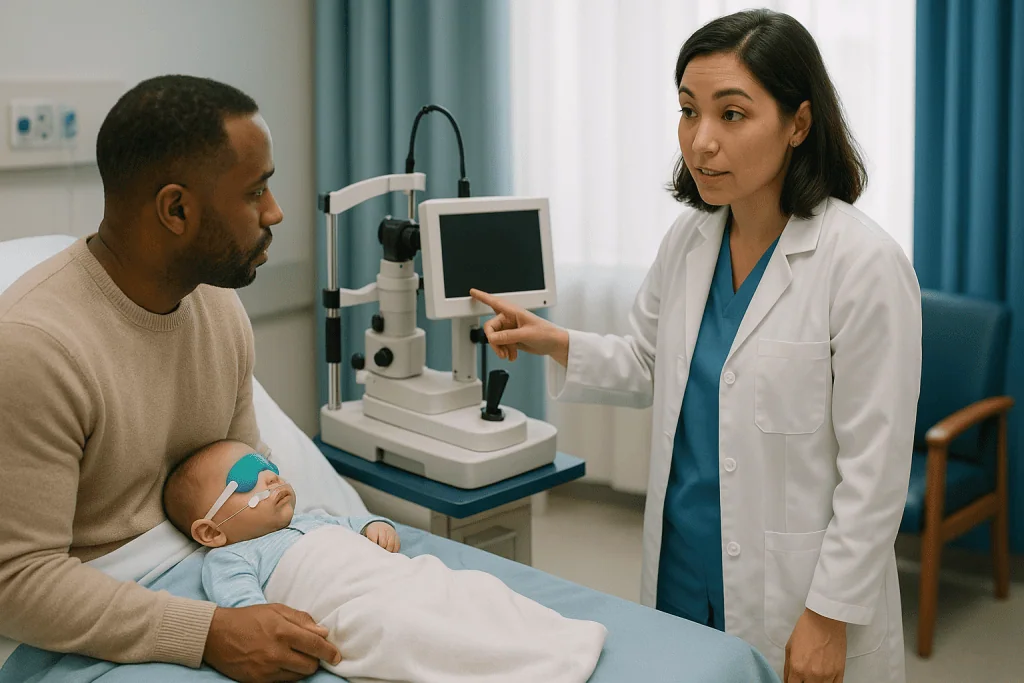
The laser portion is the shortest segment. The number of spots depends on how much of the retina lacks vessels: a baby with disease close to the optic nerve, called zone I, has far more bare retina to cover than a baby whose disease sits nearer the edge in zone II, so the treatment for the first baby takes longer. Surgeons describe the active laser time per eye as a matter of minutes for a small area up to around an hour for extensive disease, and your ophthalmologist can give a realistic estimate for your child once the exam has mapped the retina.
Everything around the laser takes longer than the laser. A typical sequence looks like this:
- Pupil dilation with drops, which may need repeating, often takes 45 minutes to an hour before the surgeon can begin.
- Induction of anesthesia or sedation, positioning and securing the airway can add a further stretch before the first spot is placed.
- Treatment of the second eye, usually done in the same session, roughly doubles the laser time.
- Recovery, including waking and stabilizing breathing, continues for an hour or more after the surgeon leaves.
Parents should therefore expect the block of time when the baby is unavailable to be considerably longer than the surgeon’s laser time. Pressing the team for a total window, rather than a laser-only figure, prevents needless worry in the waiting area.
One more point on timing: ETROP recommended treating type 1 ROP promptly, and clinicians commonly aim for within about 72 hours of diagnosis. That urgency reflects how quickly active vessels can progress, not a sign that something has gone wrong.
Who is usually treated, and who is usually asked to wait
Not every baby who is screened is treated, and not every baby with ROP is treated. The decision follows a staging system that ophthalmologists use worldwide.
Screening itself is offered to the babies most at risk. In the United States that generally means infants born weighing 1500 grams or less, or at 30 weeks of gestation or earlier, plus selected larger babies whose course has been unstable. Exams start a few weeks after birth and repeat at intervals set by what the last exam showed.
The findings are described by zone, meaning how close to the center of the retina the disease sits; stage, meaning how severe the vessel changes are, from 1 to 5; and plus disease, meaning the vessels near the optic nerve have become dilated and tortuous, a sign of high activity.
ETROP defined two groups. Type 1 ROP, which is treated promptly, includes any zone I disease with plus disease, zone I stage 3 disease with or without plus disease, and zone II stage 2 or 3 with plus disease. Type 2 ROP, which is watched closely with exams every few days to a week, includes zone I stage 1 or 2 without plus disease and zone II stage 3 without plus disease. Babies with type 2 disease are the ones asked to wait, not because their eyes are ignored but because the trial found that many such eyes regress without treatment and that the laser carries its own costs to peripheral vision.
Treatment may also be deferred, briefly, when a baby is too unstable to tolerate sedation, for example during a serious infection. In that situation the neonatologist and ophthalmologist weigh the risk of waiting against the risk of the procedure, and parents should expect that conversation to be explicit rather than assumed.
ROP laser vs anti-VEGF injection: how the two treatments compare
Since the BEAT-ROP trial, a second option has existed alongside laser: an injection of an anti-VEGF medicine, such as bevacizumab or ranibizumab, into the jelly of the eye. Anti-VEGF drugs are antibodies or antibody fragments that bind VEGF and block it from stimulating vessel growth. The injection takes moments and is done under topical anesthesia with sedation, without laser burns.
The table below summarizes the main differences as described in the trial literature and major patient-information sources. It is a summary of evidence, not a recommendation; the choice depends on the zone and stage of disease, the baby’s stability and the team’s judgment.
| Feature | Laser | Anti-VEGF injection |
|---|---|---|
| How it works | Destroys the oxygen-starved retina that produces VEGF | Blocks VEGF directly, allowing vessels to keep growing normally |
| Procedure time | Minutes to about an hour per eye | A few minutes per eye |
| Anesthesia | General anesthesia or deep sedation, often with breathing support | Sedation and topical anesthesia; general anesthesia sometimes |
| Effect on peripheral retina | Treated retina is permanently ablated | Peripheral retina preserved if vessels complete growth |
| Recurrence pattern | Less common; usually early | Can recur later; longer follow-up required |
| Reported refractive effect | Higher rates of significant nearsightedness in comparative studies | Lower rates in the same studies |
| Unknowns | Long track record | Long-term systemic and developmental effects still being studied |
BEAT-ROP reported that ROP recurred and needed retreatment in 4 percent of eyes given bevacizumab versus 22 percent given conventional laser overall; the difference was largest in zone I disease, at 6 percent versus 42 percent, and not significant in zone II disease. That is why anti-VEGF is often considered for the most posterior disease and laser remains widely used elsewhere. Some babies receive both: an injection first, then laser later if disease recurs.
How laser treatment works inside the eye
It seems counterintuitive that burning part of a baby’s retina could protect sight. The logic becomes clear once you follow the oxygen.
The peripheral retina that never received blood vessels is alive but starving. Starving tissue pumps out VEGF, and VEGF drives the abnormal vessels growing at the border between vascular and avascular retina. Those vessels are the threat: they leak, they bleed, and as they scar they contract like a drying tarp, lifting the retina off the wall of the eye.
Laser spots convert the starving tissue into small scars that no longer demand oxygen or produce VEGF. The signal fades, the abnormal vessels lose their stimulus and, over the following weeks, they regress. Meanwhile the healthy retina behind them stays where it is. The treated retina is sacrificed, which is why laser slightly narrows the visual field, but the central retina responsible for reading and recognizing faces is preserved. In the trial data that underpins the treatment, that trade was clearly worth making for eyes at the type 1 threshold.
The laser most commonly used is a diode laser at a near-infrared wavelength, absorbed by the pigmented layer under the retina. The surgeon aims for spots that are pale and confluent but not so intense that they crack or bleed, and places them all the way to the edge, including the far retina reached by scleral depression. Skipped areas are a known cause of treatment failure, so surgeons deliberately fill gaps, and part of the follow-up exam is checking for untreated islands.
Compare that with an anti-VEGF injection, which removes the signal without removing the tissue. The vessels may then finish growing to the edge, which preserves more peripheral retina but also means the VEGF source persists and can reactivate once the medicine wears off. Both approaches target the same molecule, one indirectly and one directly.
The days and weeks after ROP laser treatment
The waking period is the first milestone. Babies who had a breathing tube are usually extubated once they are breathing well, though some who were already on respiratory support before the procedure return to it. Feeds restart when the team is satisfied the baby is alert and stable; a short pause is normal and does not mean anything went wrong.
Over the first 24 to 48 hours, eyelid swelling and redness are common, and some babies keep their eyes closed more than usual. Ointment or drops prescribed by the surgeon are given on a schedule set by the team. Nurses watch for pauses in breathing or drops in heart rate, which can be more frequent for a day or so after sedation in very small babies.
The first post-laser eye exam usually falls within about a week. The surgeon is looking for the signs that treatment is working: plus disease settling, abnormal vessels flattening and fading, no new bleeding. Exams then continue, typically every one to two weeks, until the disease has clearly regressed and the retina looks stable, a process that plays out over several weeks rather than days.
Some eyes do not settle after one session. If active vessels persist or skip areas are found, additional laser may be added, or an anti-VEGF injection may be discussed. A second treatment is not a failure of the first; it is part of a plan built around repeated looking.
Once the acute phase is over, follow-up shifts from the neonatal unit to an outpatient pediatric ophthalmology clinic. The intervals stretch out, but they do not stop; children treated for ROP are usually seen through early childhood for reasons covered later in this article.
ROP laser side effects and risks in plain language
A procedure that saves central vision still carries risks, and parents deserve to hear them without either alarm or minimization.
Around the procedure itself, the main concerns come from sedation and anesthesia in a fragile infant: pauses in breathing, slow heart rate, drops in oxygen and instability of temperature or blood sugar. These are the reasons the baby is monitored continuously and why some teams prefer general anesthesia with a secured airway. Serious anesthetic complications are uncommon, but they are the risks the anesthesia consent will describe.
In the eye, the laser can occasionally burn structures other than the target retina. Burns to the iris or cornea, a temporary rise in eye pressure, small hemorrhages and, rarely, a cataract, a clouding of the lens, have all been reported. Cataract is rare with modern diode lasers but is the complication surgeons watch for most carefully at early follow-up.
Over the longer term, the most consistent finding is nearsightedness, or myopia, meaning the eye focuses close objects clearly but distant ones appear blurred. Children treated with laser have higher rates of significant myopia than untreated premature children and, in comparative studies of laser and anti-VEGF, than children who received injections. Glasses in toddlerhood are therefore common after laser. Strabismus, meaning misaligned eyes, and amblyopia, meaning a weaker eye the brain learns to ignore, occur more often in this group as well, which is another reason for continued eye checks.
Finally, the treatment can fail. Despite adequate laser, some eyes progress to retinal detachment, particularly those with aggressive zone I disease. That is not a comment on the surgeon; it reflects how severe the underlying disease was. Parents should ask what the plan would be if the first treatment does not control the vessels, so that a second step is not a surprise.
What about stage 4 and stage 5 ROP? Surgery beyond the laser
Laser and injections are treatments for active ROP before the retina detaches. When scar tissue has already begun to pull the retina away, the disease is classified as stage 4, a partial detachment, or stage 5, a total detachment. At that point the laser cannot reattach anything; the options become surgical.
Two operations are used. A scleral buckle wraps a soft silicone band around the outside of the eye to push the wall inward toward the lifted retina and relieve traction; it is later loosened or removed as the eye grows. A vitrectomy removes the vitreous gel and the scar membranes from inside the eye so the retina can settle back. Both are performed under general anesthesia by surgeons who specialize in pediatric retina, and both take longer than laser, often several hours.
Parents searching online frequently ask whether stage 5 ROP can be reversed. The honest answer from the published literature is that surgery can sometimes achieve partial or complete anatomical reattachment, but useful vision after a total detachment is limited in most cases, because the retina has been separated from its blood supply and the developing visual pathways have been deprived of input during a critical period. Stage 4 outcomes are considerably better than stage 5, especially when the central retina is still attached, which is one more reason early detection and treatment carry so much weight.
None of this means nothing can be done for a child with advanced ROP. Even limited light perception influences how a child moves through the world, and early intervention services, low-vision support and developmental therapy begin regardless of the surgical result. What the evidence does not support is any promise of restored sight after total detachment, and a team that speaks cautiously here is being truthful, not pessimistic.
Preparing your baby and yourself for the day of treatment
The practical parts of the day are mostly handled by the unit, but knowing them in advance reduces the sense of things happening around you.
Feeding is the first detail. Because sedation and anesthesia carry a small risk of milk entering the lungs, feeds are usually paused for a set period beforehand, with intravenous fluids given to keep the baby hydrated and blood sugar steady. If you are providing expressed breast milk, keep pumping to your usual schedule; the pause is temporary and the milk will be used afterward.
Consent is a conversation, not a signature. Expect two discussions: one with the ophthalmologist about the eye procedure and its alternatives, and one with the anesthesia or neonatal team about sedation. Bring written questions, since the setting makes them easy to forget. Ask whether you can be present for the induction of sedation or for the wake-up; policies vary, and some units welcome parents at one or both points.
Skin-to-skin contact before and after, when the baby’s stability allows, is comfort for both of you and is supported by neonatal units as a routine part of care. Ask the nurse when it might fit around the procedure.
Plan for your own waiting. A cup of something warm, a phone charger and a person to sit with you make a difference during a window that can stretch to two or three hours once dilation, treatment of both eyes and recovery are added together.
Finally, ask how and when you will hear the result. Some surgeons speak with parents directly afterward; others leave a note and see the family at the next exam. Knowing which to expect prevents the particular anxiety of watching a door.
What people often get wrong about ROP laser treatment
The myths around this procedure are understandable, and correcting them makes the real conversation easier.
“Laser fixes the eye, so vision will be normal.” Laser stops the disease process; it does not undo prematurity. Many treated children see well enough for ordinary school and life, but nearsightedness, misaligned eyes and reduced peripheral vision are common, and regular eye care continues for years.
“The baby is too small to feel anything.” The opposite is true, which is why sedation and monitoring are mandatory rather than optional.
“Oxygen caused this, so the unit made a mistake.” Oxygen is one factor among several, and the babies who need the most respiratory support are also the most premature. Modern units target oxygen saturation ranges precisely for this reason, but ROP still occurs in babies whose care was entirely appropriate.
“Injections are newer, so they must be better.” The evidence shows an advantage for anti-VEGF in zone I disease and no significant difference in zone II in the BEAT-ROP trial, with the trade-off of later recurrences and unanswered questions about long-term effects. Neither treatment is universally superior.
“One treatment means it’s over.” Follow-up exams continue for weeks after laser and for months after injections, and retreatment is planned for, not feared.
“Cost determines who gets treated.” This article does not discuss prices, and the decision about which treatment to use is a clinical one based on the zone and stage of disease. Financial counselors in most units can answer coverage questions separately from the medical plan.
“Screening exams are just checks; the treatment is the important part.” Screening is the treatment’s foundation. Missing a scheduled exam after discharge is one of the most preventable causes of late-detected, untreatable ROP.
Long-term follow-up: glasses, eye alignment and school-age checks
The last eye exam in the neonatal unit is the beginning of a longer relationship with a pediatric ophthalmologist. Understanding why avoids the sense that appointments are being scheduled for no reason.
Refraction, the measurement of whether the eye focuses light correctly, is checked repeatedly through the first years. Significant nearsightedness after laser can appear in infancy and progress through early childhood, and uncorrected blur during this period risks amblyopia, so glasses may be prescribed earlier than parents expect. Toddler glasses with soft frames and elastic straps are designed for exactly this situation.
Eye alignment is assessed at each visit. Strabismus is more common after ROP, and if one eye drifts, patching or glasses may be used to protect the vision in the weaker eye. The window for treating amblyopia is widest in the preschool years, which is why these checks cluster early.
The retina itself is examined periodically for the rest of childhood. Treated eyes have a slightly higher long-term risk of retinal tears or detachment, and of a condition where the retina is dragged toward the treated area, so a child who later reports new floaters, flashes or a curtain over part of the vision needs prompt review. Eyes that received anti-VEGF injections are followed on a longer schedule during infancy because recurrence can occur later than after laser, sometimes several months after treatment, and the team will specify when that risk has passed.
Developmental follow-up runs in parallel. Premature babies who needed ROP treatment are, by definition, the most premature, and many are enrolled in neurodevelopmental clinics regardless of their eyes. Vision and development inform each other: a child who cannot see the edge of a table learns to walk differently. Sharing what the eye team finds with the developmental team, and vice versa, is a reasonable request for parents to make.
Questions to ask your care team before ROP laser treatment
A good consent conversation is a two-way exchange. These questions, in roughly the order they arise, tend to draw out the information parents later wish they had asked for.
- What zone and stage is the disease in each eye, and does it meet the type 1 threshold?
- Why are you recommending laser rather than an anti-VEGF injection for my baby, or the reverse?
- Will the procedure happen at the bedside or in the operating room, and who will provide the sedation or anesthesia?
- Will my baby have a breathing tube, and is there a chance of staying on breathing support afterward?
- Roughly how long will my baby be unavailable to me, from the first drops to being settled in recovery?
- Can I be present at any point, and when and how will I hear how it went?
- Both eyes today, or one at a time, and why?
- What will you look for at the first follow-up exam, and when is it?
- If the vessels do not settle, what is the next step: more laser, an injection, or both?
- What are the specific risks for my baby given his or her lungs, weight and other conditions?
- What eyelid or eye changes should prompt me to call before the next scheduled exam?
- How long will outpatient eye follow-up continue, and who coordinates it after discharge?
- Are there early intervention or vision support services we should know about now, regardless of outcome?
Write the answers down, or ask permission to record the conversation. Nothing on this list is unusual, and a team that treats ROP regularly will have answered every one of them before.
When to call your doctor: red-flag signs after ROP treatment
While your baby is in the neonatal unit, the nurses are watching for problems and you do not need to carry that responsibility alone. Once home, or between clinic visits, the picture changes, and a short list of warning signs is worth keeping on the refrigerator.
Contact the eye team or your pediatrician promptly if you notice any of the following:
- Increasing redness, swelling or discharge from either eye more than a couple of days after treatment, or a lid that is hot, tense or spreading in redness.
- A pupil that looks white or gray instead of black in photographs or under a light, which can signal a cataract, bleeding or a detached retina.
- A cornea, the clear front window of the eye, that appears cloudy or hazy.
- An eye that seems larger than the other or unusually watery, which can accompany raised pressure.
- Eyes that shake rhythmically, drift apart or fail to follow a face by the age the team told you to expect it.
- Any missed or unscheduled follow-up exam. If you are unsure when the next appointment is, call and ask; this is the single most important item on the list.
Seek urgent care the same day, or use emergency services, if your baby has pauses in breathing, turns blue or gray around the lips, is unusually limp or difficult to rouse, has a fever, or is feeding far less than usual. These are general signs of a sick infant rather than eye-specific problems, but a recently treated premature baby should be assessed quickly.
For older children with a history of ROP, new floaters, flashing lights, or a shadow or curtain across part of the vision need a same-day eye assessment because they can indicate a retinal tear or detachment.
Every decision about what these signs mean, and what to do next, sits with the treating team. When in doubt, call; no one who cares for premature babies minds a question that turns out to be nothing.
Frequently asked questions
Is laser treatment for ROP painful for the baby?
The procedure would be painful without medication, so it is never done on an awake infant. Babies receive either general anesthesia or deep sedation with pain relief, plus numbing drops on the eye surface, and are monitored throughout. Afterward, eyelid swelling and irritability for a day or two are common and are managed by the neonatal team using standard infant comfort measures and pain assessment scales.
How long does ROP laser take from start to finish?
The active laser time ranges from minutes for a small area to around an hour per eye for extensive zone I disease, and both eyes are usually treated in one session. Add pupil dilation, anesthesia induction and recovery, and the period when your baby is unavailable commonly stretches to two or three hours. Ask the team for the total window rather than the laser time alone.
How painful is laser surgery for retinal tears in older children or adults compared with ROP laser?
Laser for a retinal tear in an older child or adult is usually done awake with numbing drops and is described as brief flashes of discomfort or a dull ache, so sedation is rarely needed. ROP laser covers a far larger area of retina and involves pressing on the eye to view its edge, which is why infants are sedated or anesthetized rather than treated awake.
Can ROP stage 5 be treated successfully?
Stage 5 means the retina has completely detached, and laser no longer helps at that point. Surgeons can perform a vitrectomy or scleral buckle to attempt reattachment, and anatomical success is sometimes achieved, but published series report that useful vision afterward is limited in most eyes. Outcomes at stage 4, a partial detachment, are considerably better, which is why early detection matters so much.
How much does laser treatment for ROP cost?
This article does not discuss prices, because costs vary widely by country, insurance arrangement and hospital and are unrelated to the clinical decision about whether treatment is needed. The choice between laser and injection is made on the zone and stage of disease. Financial counselors or social workers attached to neonatal units can explain coverage and support options separately from the medical plan.
ROP laser vs anti-VEGF injection: which is better?
Neither is universally superior. In the BEAT-ROP trial, anti-VEGF injection had a clear advantage for zone I disease, with recurrence in 6 percent of eyes versus 42 percent after laser, but no significant difference in zone II. Injections preserve more peripheral retina and cause less nearsightedness, while laser has a longer safety record and fewer late recurrences. The treating team weighs these factors for each baby.
What are the main ROP laser side effects parents should expect?
Short-term effects include eyelid swelling, redness and irritability for a day or two, plus sedation-related pauses in breathing or slow heart rate that the unit monitors closely. Longer term, treated children have higher rates of nearsightedness, misaligned eyes and amblyopia, and a modest increase in the risk of later retinal problems. Rare procedural complications include cataract and burns to other eye structures.
Will my baby need glasses after ROP laser treatment?
Many children treated with laser develop significant nearsightedness in infancy or early childhood, and glasses are often prescribed earlier than parents expect, sometimes in the first year. Correcting blur early protects the developing visual system from amblyopia. Not every treated child needs glasses, but repeated refraction checks through the preschool years are standard so that any need is caught promptly.
Why does my baby need more eye exams after treatment if the laser worked?
Regression takes weeks, and the surgeon must confirm the abnormal vessels are fading, check for skipped areas that may need more laser, and watch for rare complications. After anti-VEGF injections, recurrences can appear months later, so follow-up runs longer. Later in childhood, exams monitor refraction, eye alignment and retinal health, because all three are affected more often in children who had ROP.
Can ROP come back after laser treatment?
Yes, although less often than after injection. In BEAT-ROP, 22 percent of laser-treated eyes overall needed retreatment, mostly because active vessels persisted or areas of retina were missed, and recurrence after laser tends to occur early rather than months later. Additional laser or an injection can then be used. This is a planned contingency, which is why the first follow-up exam falls within about a week.
References
This article is for general information only and is not a substitute for professional medical advice. Please consult a qualified doctor about your individual situation.

More from the Blog

Packing for a Kawasaki Disease Admission: Fever Records, History and Comfort Items
For a Kawasaki disease hospital admission, pack a written fever record (dates, times, readings, and what was given), a one-page medical history with allergies…

Bronchiolitis in Babies: Why Small Airways Swell and How the Illness Usually Runs Its Course
Bronchiolitis in babies is caused by common respiratory viruses, most often respiratory syncytial virus (RSV), that infect the smallest airways in the lungs. The…

Neonatal Sepsis Treatment in the NICU: Blood Cultures, IV Lines and Round-the-Clock Monitoring
Neonatal sepsis treatment in the NICU usually begins the moment infection is suspected: a blood culture is drawn, intravenous antibiotics that cover the most…

Who Needs Medical Treatment for Chickenpox Beyond Comfort Care? Higher-Risk Groups Explained
Most healthy children with chickenpox need only comfort care at home, because the illness usually clears on its own within one to two weeks.…

Preparing for a First Pediatric Physical Therapy Visit: Reports, Braces and Loose Clothing
For a first pediatric physical therapy appointment, bring the referral, any recent medical or developmental reports, imaging results, and every brace, splint, orthotic, or…

Preparing Your Child for Sensory Integration Therapy: Reports to Bring and What to Wear
To prepare a child for sensory integration therapy, bring the referral letter, any developmental or diagnostic assessments, hearing and vision results, school reports, and…