

How Lymphedema Microsurgery Is Performed: Tiny Vessels, the Microscope and Operating Time

Key Takeaways
- Lymphedema microsurgery reroutes fluid rather than removing tissue: bypass joins lymphatic vessels to veins, and node transfer relocates living lymph nodes with their blood supply.
- The vessels involved are often under a millimeter wide, which is why surgeons work at ten to forty times magnification with sutures finer than a human hair.
- Indocyanine green lymphography decides candidacy: limbs showing bright linear channels can be bypassed, while diffuse glow means few vessels are left to connect.
- Operating time is driven by anatomy, with bypass taking several hours depending on the number of connections and node transfer typically longer because two surgical sites and blood vessels are involved.
- Wounds heal in weeks, but any change in limb volume unfolds over many months, and compression garments and skin care continue for life according to mainstream guidance.
- Most outcome data come from single-center case series with varied measurement methods, so no single success percentage fairly describes what surgery achieves.
Lymphedema microsurgery reroutes or rebuilds lymphatic drainage rather than cutting swelling away. Under a high-power microscope, a surgeon either joins lymphatic vessels under a millimeter wide to nearby small veins (lymphaticovenous anastomosis) or transplants a small block of lymph nodes with its blood supply into the swollen region. Operations typically take several hours, are done under general anesthesia, and are combined with lifelong compression and skin care rather than replacing them.
The sleeve came off in the consultation room and she rested her arm on the desk the way you might set down a heavy bag. Eight years since the mastectomy. Eight years of measuring, wrapping, pumping, and explaining to strangers why one forearm looked like it belonged to someone else. Then she asked the question that had taken her three appointments to voice: “Can you actually fix the plumbing?”
That is, more or less, the question behind how lymphedema microsurgery works. It is not a question about removing swelling. It is a question about drainage, about whether a system of vessels thinner than a strand of hair can be rejoined, rerouted, or supplemented with a transplanted filter, and whether hours of stitching under a microscope translate into a lighter limb months later.
The honest answer is longer than a yes or no. This article walks through what happens on the operating table, how long it tends to take, who is usually offered it, and what the evidence does and does not yet show.
How lymphedema microsurgery works in plain language
Start with the problem the surgeon is trying to solve. Lymphedema is swelling caused by lymph fluid, the clear, protein-rich liquid that leaks out of blood vessels into tissue, failing to drain back toward the heart. Normally that fluid travels through lymphatic vessels, tiny thin-walled channels, and passes through lymph nodes, the bean-shaped filters clustered in the armpit, groin, and neck. When nodes are removed or irradiated during cancer treatment, or when the vessels are malformed from birth, fluid backs up. Mayo Clinic describes this as a blockage in the lymphatic system rather than a disease of the skin or fat itself.
Microsurgery attacks the blockage in one of two ways. The first is a bypass. Lymphatic vessels that are still open but congested are stitched directly into small neighboring veins, so fluid can hop onto the venous highway and be carried away. This is lymphaticovenous anastomosis; an anastomosis is simply a surgically created connection between two tubes. The second approach is a transplant. A small cluster of lymph nodes, together with the artery and vein that keep it alive, is moved from a donor site to the swollen region, where it is thought to act as a sponge and to encourage new lymphatic channels to grow.
Neither operation removes the swollen tissue. That distinction matters and comes up again below. Both aim to restore movement of fluid. What makes the work “micro” is scale: the vessels involved are often less than a millimeter across, which is why the surgeon works through an operating microscope with sutures finer than a human hair. Cleveland Clinic lists these procedures among the surgical options a specialist may discuss when conservative therapy is no longer enough, always alongside continued compression and skin care.
Why can't you surgically remove lymphedema?
People often picture swelling as something that could be scooped out, the way a surgeon drains an abscess. Lymphedema does not behave that way. The excess fluid is not pooled in one pocket; it is distributed through the tissue under the skin, and it keeps arriving as long as the drainage problem persists. Cut away the swollen tissue today, and the same faulty plumbing refills the limb tomorrow, often with more scar tissue in the way.

Over time the fluid also changes what it sits in. Protein left in the tissue triggers inflammation, and the body responds by laying down fibrous tissue and depositing fat. The NHS notes that in later stages the swelling becomes harder and no longer pits when pressed, a sign the limb is now enlarged by solid tissue as much as by liquid. That is the point at which a purely reductive operation, such as specialized liposuction, may be considered, but it treats the consequence rather than the cause, and the patient must wear compression around the clock afterward to stop fluid from re-accumulating.
There is a third reason removal fails as a strategy. The skin of a lymphedematous limb is a poor barrier. Large open wounds in that environment heal slowly and invite cellulitis, a bacterial infection of the skin and the tissue beneath it, which Mayo Clinic identifies as one of the most common complications of lymphedema. Older debulking operations that stripped large amounts of skin and tissue left disfiguring scars and did not fix drainage.
So the modern goal is redirection. Whether by bypass or transplant, the surgeon is trying to give the fluid somewhere to go. Understanding that reframes every later question about timing, expectations, and why compression garments do not disappear the day after surgery.
Lymphaticovenous anastomosis surgery: joining vessels thinner than a hair
Lymphaticovenous anastomosis, usually shortened to LVA, is the operation most people mean when they say lymphedema microsurgery. The principle is a shunt. Lymphatic vessels in the forearm or calf normally run alongside small veins. Under the microscope, the surgeon isolates a working lymphatic vessel, opens a nearby vein of similar size, and stitches the two together so that lymph drains into the venous system.
The scale is difficult to appreciate until you see the instruments. The vessels are often 0.3 to 0.8 millimeters in diameter, roughly the thickness of a pencil lead or less, which is why some surgeons describe this as supermicrosurgery, meaning anastomosis of vessels under about 0.8 millimeters. Sutures are so fine they are nearly invisible to the naked eye and must be handled with needle holders under high magnification.
Several features make LVA appealing. Incisions are small, typically a few centimeters, and placed where a suitable pairing of lymphatic and vein has been mapped beforehand. Because no tissue is transplanted, there is no second surgical site to heal. Many programs perform it under general anesthesia, though the operation itself does not involve the chest or abdomen.
The limitation is equally important. LVA depends on lymphatic vessels that still work. In advanced lymphedema, years of pressure and inflammation can turn those vessels into scarred cords that cannot carry fluid, and there is nothing useful to connect. That is why LVA is generally discussed for earlier-stage disease, as Mayo Clinic’s treatment overview implies when it describes creating new drainage paths for people who still have functioning channels. Whether a given limb qualifies is decided by imaging, not by how it looks in clinic.
Lymph node transfer for lymphedema: moving a living filter
The second major operation is vascularized lymph node transfer, often abbreviated VLNT. Vascularized means the transplanted tissue comes with its own artery and vein, which are reconnected at the new site so the nodes stay alive rather than dying as a graft would. Think of it less as installing a part and more as relocating a small working organ.
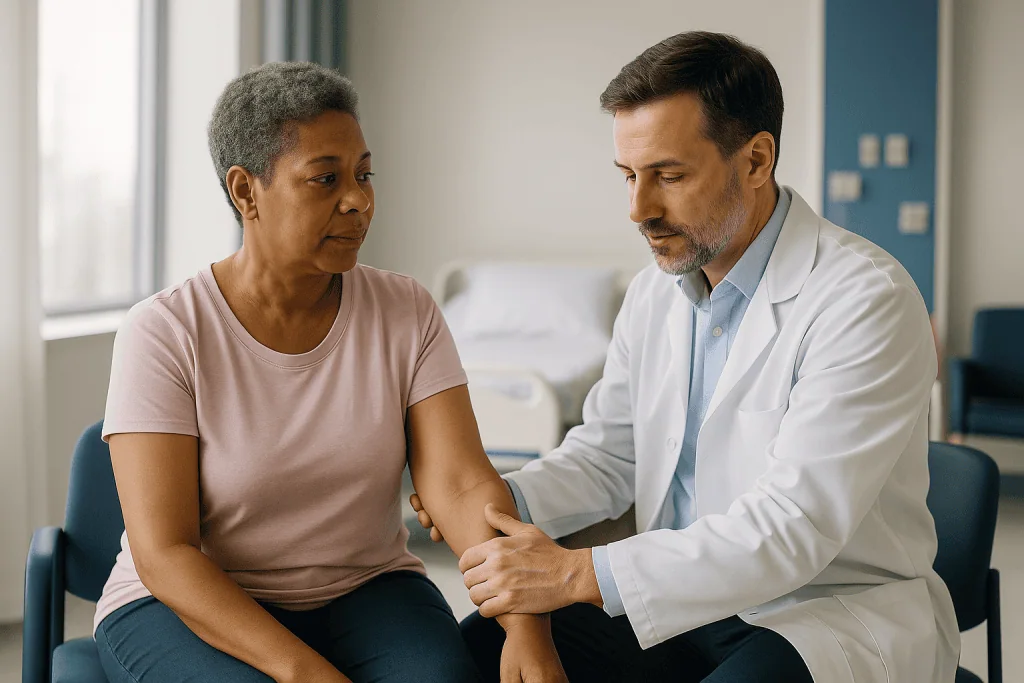
Donor sites vary. Surgeons have harvested node clusters from the groin, the side of the chest, the neck, and even from inside the abdomen near the stomach or bowel. Each option carries a trade-off between how much lymphatic tissue it provides and the risk of causing lymphedema at the donor site itself, a complication surgeons take seriously enough to map the donor drainage before choosing.
The transferred nodes are placed either at the site of the original damage, such as a scarred armpit, or lower in the limb where fluid pools most. Two mechanisms are proposed. The nodes may act as a pump and sponge, absorbing fluid and returning it through their reconnected vein. They may also release growth signals that encourage new lymphatic vessels to sprout and bridge the blocked area. Both ideas are supported by laboratory and observational work, but the relative contribution of each in a human limb remains uncertain, and honest teams say so.
Because VLNT involves reconnecting an artery and a vein, it is technically a free tissue transfer, the same family of operation used in breast reconstruction. Mayo Clinic groups it under lymph node transplant in its overview of surgical options. It is often combined with breast reconstruction in the same operation for people whose lymphedema followed breast cancer treatment, which is one reason the procedure sits within plastic and reconstructive surgery rather than general surgery.
Mapping the drainage before the first incision
No microsurgeon opens a limb hoping to stumble on a usable vessel. The operation is planned on a map drawn hours or days earlier, and that map determines whether surgery is offered at all.
The main tool is indocyanine green lymphography. Indocyanine green is a fluorescent dye that glows under near-infrared light; a tiny amount is injected into the skin of the hand or foot, and a special camera follows it as it travels up the lymphatic vessels. Healthy channels show up as bright lines. Damaged ones appear as diffuse patterns with names such as splash, stardust, and diffuse, describing how the dye spreads sideways through skin rather than moving along a vessel. A limb showing crisp linear channels is a candidate for bypass; one showing only diffuse glow has little left to connect.
Other imaging fills in the picture. Lymphoscintigraphy, a nuclear medicine scan in which a mildly radioactive tracer is tracked as it drains, remains the standard test to confirm the diagnosis and assess how sluggish the whole system is. MedlinePlus and Mayo Clinic both list it among the tests used to evaluate suspected lymphedema. Ultrasound or MRI may be used to find veins of the right size, and to check that a swollen limb is not actually swollen for another reason, such as a blood clot or heart failure.
Before any of this, the team confirms the basics. Compression, skin care, and decongestive therapy should already be in place, both because they are the foundation of lifelong management and because a limb that is as decongested as possible is easier to map and operate on. Skipping this step is one of the most common reasons a referral stalls.
How lymphedema microsurgery works under the microscope
The room looks like any other operating theater until you notice the microscope, an arm-mounted instrument the size of a small refrigerator with two eyepieces for the surgeon and often a second set for an assistant. Many teams also use a video screen so the rest of the room can follow. Magnification runs from roughly ten to forty times, enough to make a half-millimeter vessel look like a garden hose.
Once anesthesia is established, the mapped skin sites are marked and the dye camera may be used again to confirm the vessel positions. A small incision, often a few centimeters long, is made over each site. The surgeon then separates the tissue layers with fine forceps until a lymphatic vessel, recognizable by its translucent wall and the faint glow of dye inside, is found alongside a small vein.
Here the work slows. Each vessel is gently freed from surrounding tissue without crushing it. Tiny clamps hold the vein still. Under the highest magnification, the surgeon places sutures, sometimes six or eight around a vessel narrower than a grain of rice, to join the two openings. Blood must not leak back into the lymphatic; lymph must flow freely into the vein. A surgeon may check patency by watching dye pass across the join or by observing lymph wash into the vein, sometimes described as a blue or clear stream against dark blood.
For a node transfer, the sequence is longer: harvest the node cluster with its blood vessels, prepare the recipient vessels in the limb, join artery to artery and vein to vein, then confirm blood flow before closing. Every anastomosis, lymphatic or vascular, is checked before the skin is closed with fine sutures and covered with a light dressing designed to avoid pressure on the fresh connections.
How long does lymphedema microsurgery take, and what stretches the clock?
Ask three surgeons and you will hear three honest answers, because operating time is set by the anatomy found on the day rather than by a schedule. Still, some patterns hold.
Lymphaticovenous anastomosis time scales with the number of connections. Each join can take a considerable stretch of careful suturing, and a surgeon may perform anywhere from two to six or more along a limb depending on how many usable pairings the imaging revealed. A limb with several bright channels takes longer than one with two, not because the surgery is going badly but because there is more to do. Teams generally describe LVA as taking several hours, with the operation ending when the mapped vessels have been addressed rather than when a clock runs out.
Vascularized lymph node transfer is typically longer. It involves two surgical fields, the donor site and the recipient limb, and the reconnection of blood vessels that must be checked for flow before the surgeon can close. When it is combined with breast reconstruction, the combined procedure can occupy most of an operating day. Your surgical team should give you their own expected range at the pre-operative visit, and that range, not a figure read online, is the one to plan around.
Several things lengthen any lymphatic operation: heavy scarring from earlier surgery or radiation, a large limb where vessels sit deep in fat, vessels that prove too fragile once exposed, or a vein that keeps refilling with blood and must be handled differently. None of these mean failure. They are the reason microsurgery is described as slow by design. The vessels do not forgive haste, and a team that budgets generous time is protecting the connections they are building.
Who is usually offered lymphedema microsurgery, and who is asked to wait?
Surgery is not the first rung of the ladder. Mayo Clinic, the NHS, and Cleveland Clinic all describe surgery as an option to consider when swelling persists despite committed conservative treatment, not as a replacement for it. That framing shapes who is typically referred.
Candidates for bypass surgery usually share three features. First, a confirmed diagnosis, with imaging that excludes other causes of a swollen limb. Second, earlier-stage disease in which lymphatic vessels still carry dye on lymphography; the International Society of Lymphology staging that clinicians use ranges from stage 0, where the system is impaired but the limb looks normal, through stage 3, where tissue is hard and skin changes are established. Bypass is most often discussed at stages 1 and 2. Third, months of consistent compression, exercise, and skin care, which both optimizes the limb and shows the person can sustain the aftercare surgery still requires.
Node transfer may be discussed for people with more extensive damage at the original site, particularly after axillary surgery and radiation, or where few vessels remain for bypass. Debulking procedures are reserved for later stages where excess fat and fibrous tissue dominate.
People are commonly asked to wait, or are steered toward other management, when active cancer is being treated, when there is an ongoing or recent skin infection, when the limb is not yet optimized with compression, or when other conditions such as heart failure, uncontrolled diabetes, or heavy smoking would compromise wound healing and the survival of a transplanted flap. Body weight also matters: Mayo Clinic notes that obesity itself can cause or worsen lymphedema, and teams may recommend addressing it first, not as judgment but because it changes both surgical risk and the likely benefit. The final call rests with the multidisciplinary team, ideally including a lymphedema therapist.
Bypass, transfer, debulking, or therapy alone: a side-by-side view
The options are easier to weigh when laid out together. The table below summarizes what each approach does, when it is usually considered, and its main trade-off. It is a guide for conversation with your team, not a decision tool.
| Approach | What it does | Typically considered when | Main trade-off |
|---|---|---|---|
| Complete decongestive therapy | Manual lymphatic drainage, compression, exercise, skin care | All stages; foundation of care before and after any surgery | Demands daily effort for life; controls rather than corrects drainage |
| Lymphaticovenous anastomosis | Joins working lymphatic vessels to small veins | Earlier stages with visible channels on lymphography | Needs functioning vessels; may not help fibrotic limbs |
| Vascularized lymph node transfer | Transplants nodes with blood supply to the affected region | Extensive damage at the original site; few vessels for bypass | Longer operation, donor-site wound, small risk of donor-site lymphedema |
| Liposuction-based debulking | Removes excess fat and fibrous tissue | Later stages dominated by solid tissue | Does not restore drainage; requires continuous compression afterward |
Two threads run through every row. Compression never leaves the table; Mayo Clinic’s treatment overview is explicit that garments and skin care remain central regardless of surgery. And none of the operations is described in mainstream guidance as reversing lymphedema. They are tools to reduce volume, cut the frequency of infections, and make daily management lighter.
The comparison also highlights sequencing. Many teams consider bypass or transfer first and reserve debulking for a later stage, sometimes years on, if solid tissue remains. Others combine approaches. Which order makes sense depends on your imaging, your goals, and the surgeon’s assessment, which is why the same diagnosis can lead to different plans for different people.
Lymphedema surgery recovery time: what the first days and weeks usually look like
Recovery has two clocks running at once, and confusing them causes most of the disappointment people describe. One clock is wound healing. The other is lymphatic change, and it runs far slower.
After lymphaticovenous anastomosis, the wound clock is short. Incisions are small, and many programs discharge people the same day or after one night. Light dressings protect the sites, and the team will usually ask you to avoid tight compression directly over the fresh connections for a period so the delicate joins are not squeezed shut, then reintroduce garments on their schedule. Gentle movement is encouraged early; heavy lifting and strenuous use of the limb are held back for a few weeks. Stitches, if not dissolvable, come out at a follow-up visit.
After node transfer, expect a longer stay in hospital because the team monitors blood flow into the transplanted tissue in the first days, when a clot in the reconnected vessels is most likely. There are two wounds to heal, and the donor site can be more uncomfortable than the limb. Return to desk work often takes a few weeks; physically demanding work takes longer. Your team’s written instructions override any general figure.
Now the slower clock. Limb volume, if it improves, does so gradually as fluid finds the new routes and inflammation settles. Teams commonly measure the limb at set intervals over the first year or more, and some improvement in heaviness or infection frequency may be noticed before a tape measure shows a difference. Others notice little change. Compression therapy, exercise, and skin care continue throughout, and your therapist may adjust garments as the limb changes shape. Recovery, in other words, is measured in seasons, not in the days it takes a scar to fade.
Is lymphedema surgery successful? What the evidence actually shows
This is the question everyone asks, and the honest answer requires a short lesson in how evidence is graded. Most published data on lymphedema microsurgery come from case series and cohort studies, in which one center reports how its own patients fared, often without a comparison group receiving therapy alone. Randomized controlled trials, the standard for proving that a treatment causes an outcome, are few and small. Outcome measures vary from study to study: some report limb circumference, some volume by water displacement or perometry, some episodes of cellulitis, some quality-of-life questionnaires. Follow-up periods differ widely.
Within those limits, the pattern reported across series is that many people experience reduced limb volume, fewer skin infections, and a lighter, more comfortable limb, while a meaningful minority see little measurable change. Earlier-stage disease with intact vessels tends to be associated with better bypass results; more advanced, fibrotic limbs less so. Because the numbers depend so heavily on patient selection, measurement method, and how long people were followed, this article deliberately does not quote a single success percentage. Any figure you encounter should be read alongside the stage of disease studied and whether a control group existed.
Mainstream patient resources reflect this caution. Mayo Clinic describes surgical options without promising results and frames them as ways to make the condition more manageable. The NHS similarly presents surgery as an option in selected cases while emphasizing that lymphedema is a long-term condition managed over a lifetime.
What this means for you is concrete. Ask your team what outcomes they track, how they define improvement, and what they have observed in people whose imaging looks like yours. A surgeon who answers with ranges, caveats, and their own audited figures is giving you better information than one who offers a single reassuring number.
Risks, complications, and the alternatives your team should raise
Microsurgery is low-drama compared with major cancer operations, but it is still surgery on a limb whose skin and immune defenses are already compromised. The risks worth understanding fall into three groups.
General surgical risks apply: bleeding, wound infection, reactions to anesthesia, and blood clots in the deep veins, the last of which is minimized with early walking and any measures your team prescribes. Because lymphedematous skin is prone to cellulitis, a wound infection can escalate faster than in a healthy limb, which is why teams stress meticulous skin care before and after.
Procedure-specific risks differ. For bypass, the main concern is that the tiny connections clot or scar closed over time, quietly undoing the work; a temporary increase in swelling can also occur while tissues settle. For node transfer, the transplanted flap depends on its reconnected blood vessels, and a clot there in the first days can lead to loss of the tissue and a return to the operating room. The donor site carries its own wound-healing risks and a small but recognized risk of causing new lymphedema in that region, which surgeons try to avoid through careful mapping.
Then there is the risk of disappointment, which deserves to be named. Some people undergo a technically successful operation and see no change they can feel or measure.
Alternatives are part of a fair conversation. Optimized complete decongestive therapy, newer compression technologies and pneumatic compression pumps, supervised exercise, and weight management all have supporting evidence and remain the backbone of care, as MedlinePlus and Cleveland Clinic outline. Debulking procedures exist for solid-tissue swelling. Choosing to continue with therapy alone is a legitimate decision, not a failure, and a good team will say so before you sign a consent form.
What people often get wrong about lymphedema microsurgery
The myths cluster around the same hopes, so it is worth naming them plainly.
“Surgery means I can throw away the sleeve.” Almost never. Mainstream guidance from Mayo Clinic and the NHS treats compression as lifelong. Some people find they can step down to lighter garments or wear them less often after surgery, and that is a genuine gain, but the decision is made by measurement over months with your therapist, not assumed on the day you go home.
“They remove the swelling.” Bypass and transfer move fluid; they do not take tissue out. Only debulking removes tissue, and it does so without repairing drainage. Knowing which category your operation belongs to prevents a mismatch between what you expect and what the surgeon is doing.
“If it worked, I’d see it in a week.” The incisions heal in weeks; the limb changes, if it changes, over many months. Judging the operation at the first dressing change is like judging a garden the day after planting.
“It’s experimental, so no one should do it.” The techniques are established in reconstructive surgery and are offered in mainstream centers; what remains limited is high-grade comparative evidence about who benefits most. Those are different statements.
“Later is fine; I’ll wait until it’s really bad.” For bypass, waiting can close the window, because vessels that could have been connected become scarred. That is not an argument to rush, but it is an argument to have the imaging conversation earlier rather than later.
“Lymphedema is just cosmetic.” Recurrent cellulitis, heaviness that limits work and sleep, and the daily hours of self-care are medical problems. Framing the condition as vanity undersells what people carry, and it delays referrals that should happen.
Questions to ask your care team before deciding
A consultation goes better when you arrive with questions that force specific answers. The list below is a starting point; write down the replies and compare them across visits.
- Based on my lymphography, do I still have functioning lymphatic vessels, and which operation does that make me a candidate for?
- Which stage of lymphedema do you consider my limb to be, and how does that affect what you expect surgery to achieve?
- How many connections or which donor site are you planning, and what would change that plan once you are operating?
- How long do you expect my operation to take, and how long will I be in hospital afterward?
- What outcomes do you measure, over what period, and what have you seen in people whose imaging and stage resemble mine?
- What does my compression and skin-care routine look like in the first weeks, and who coordinates it with my lymphedema therapist?
- What are the specific risks in my case, including at the donor site, and what would prompt a return to the operating room?
- What happens if the operation does not reduce my swelling? What would the next step be?
- Is there anything I should optimize first, such as fitness, weight, smoking, blood sugar, or an unsettled skin infection?
- If I decide to continue with therapy alone for now, what signs would suggest revisiting surgery, and is there a point after which bypass would no longer be possible?
Notice what the questions share: they ask for ranges, mechanisms, and plans for the case where things go less well. A team that welcomes them is signaling the kind of long-term partnership this condition requires. Bring a companion if you can; the volume of detail in a microsurgery consultation is hard to hold alone, and a second set of ears often catches the answer you missed.
When to call your doctor
Most recoveries are quiet. A few situations are not, and in a limb already prone to infection, waiting to see if things settle is the wrong instinct. Call your surgical team, or seek urgent care if you cannot reach them, for any of the following.
- Spreading redness, warmth, or a hot, painful area on the limb, especially with fever, chills, or feeling generally unwell. Mayo Clinic identifies cellulitis as a serious complication of lymphedema, and it can progress rapidly.
- A sudden increase in swelling, tightness, or pain in the operated limb that is clearly different from the gradual fluctuation you know.
- Bleeding from an incision that does not stop with gentle pressure, or a wound that opens, oozes cloudy fluid, or smells unpleasant.
- After node transfer, any change your team asked you to watch for at the flap site, such as color change, coolness, or a dressing suddenly soaked, particularly in the first days.
- Pain, swelling, or tenderness in the calf or thigh, or chest pain and sudden breathlessness, which can signal a blood clot and need emergency assessment.
- Numbness, weakness, or color change in the hand or foot beyond the operated area.
- A fever without an obvious cause in the weeks after surgery.
Between visits, a low threshold for contact is reasonable. Your team would rather hear about a false alarm than miss an infection in a limb that cannot fight one well. Keep the ward or clinic number where you can find it, and know which after-hours route they want you to use.
Longer term, the same rule applies to the condition itself. New skin thickening, recurrent infections, or swelling that stops responding to your usual routine are reasons to book a review with the lymphedema service, whether or not surgery is on the table.
Frequently asked questions
What is microsurgery and how does it treat lymphedema?
Microsurgery is surgery performed under an operating microscope on structures too small to handle by eye, often vessels under a millimeter across. For lymphedema, it either connects congested lymphatic vessels to nearby small veins so fluid can drain into the bloodstream, or transplants a small cluster of lymph nodes with its own artery and vein into the swollen region. Both approaches aim to improve drainage rather than remove swollen tissue, and both are used alongside continued compression and skin care.
Why can't you surgically remove lymphedema?
Because the swelling is caused by fluid that keeps arriving through a blocked drainage system, cutting tissue away does not stop it from refilling. Over time protein-rich fluid also triggers fibrous tissue and fat deposition, so the limb becomes enlarged by solid tissue as well as liquid. Large excisions in lymphedematous skin heal poorly and invite infection. Modern surgery therefore redirects fluid or, in later stages, removes excess fat with liposuction while compression is worn continuously afterward.
Is lymphedema surgery successful?
It can reduce limb volume, cut infection episodes, and make daily management lighter for many people, but results vary and some see little change. Most published evidence comes from case series using different measurement methods and follow-up periods, with few randomized trials, so a single success rate would be misleading. Earlier-stage disease with working vessels tends to be associated with better bypass results. Ask your team what outcomes they track and what they have observed in cases resembling yours.
How long does it take to heal from lymphedema surgery?
Incisions from a bypass are small and usually heal within a few weeks, with many people home the same day or after one night. Node transfer involves a longer hospital stay so the team can monitor blood flow to the transplanted tissue, and a donor-site wound that also needs to heal. Changes in limb volume, if they occur, develop gradually over months as fluid finds new routes. Your team’s written recovery plan takes precedence over any general timeline.
What is lymphaticovenous anastomosis surgery like on the day?
It is typically done under general anesthesia through a few small incisions placed over vessels mapped beforehand with fluorescent dye. The surgeon isolates a lymphatic vessel and a neighboring small vein under high magnification and stitches them together, repeating this at several sites along the limb. The operation takes several hours depending on how many connections are made. Dressings are light to avoid pressure on the joins, and gentle movement is encouraged early while heavy use is delayed.
How does lymph node transfer for lymphedema differ from bypass?
Bypass works with vessels already in the limb, joining them to veins. Lymph node transfer moves a block of nodes from elsewhere in the body, commonly the groin, chest wall, neck, or abdomen, along with its artery and vein, which are reconnected at the new site so the tissue stays alive. Transfer is a longer operation with two surgical sites and is often considered when the original drainage area is heavily scarred or few vessels remain to bypass.
Will I still need compression garments after lymphedema microsurgery?
In most cases, yes. Mainstream guidance treats compression and skin care as lifelong parts of managing lymphedema, and surgery is offered as an addition rather than a replacement. Some people are able to step down to lighter garments or shorter wearing times after their limb has been measured over months, but that adjustment is made with a lymphedema therapist based on evidence from your own limb, not assumed at discharge.
What does lymphedema surgery recovery time look like for going back to work?
After bypass surgery, people with desk-based jobs often return within a couple of weeks once incisions are settled, while physically demanding work waits longer to protect the delicate connections. After node transfer, the hospital stay is longer and two wounds must heal, so a return to full duties commonly takes several weeks. Individual timing depends on the operation, the donor site if any, and your overall health, and your surgical team will set the schedule.
Who is usually not a candidate for lymphedema microsurgery?
People with active cancer treatment underway, a current or recent skin infection, a limb not yet optimized with compression therapy, or conditions that impair healing such as uncontrolled diabetes, heart failure, or heavy smoking are commonly asked to wait or to pursue other management. Advanced, fibrotic limbs with no working vessels on lymphography are usually unsuitable for bypass, though transfer or debulking may still be discussed. The multidisciplinary team makes the final decision.
What imaging is done before lymphedema microsurgery?
Indocyanine green lymphography is central: a fluorescent dye injected into the skin is tracked with a near-infrared camera to show which lymphatic vessels still carry fluid and where they run. Lymphoscintigraphy, a nuclear medicine scan, confirms the diagnosis and gauges overall drainage. Ultrasound or MRI may locate suitable veins and rule out other causes of swelling such as a blood clot. Together these tests determine whether surgery is offered and which procedure fits your anatomy.
References
This article is for general information only and is not a substitute for professional medical advice. Please consult a qualified doctor about your individual situation.

More from the Blog
Is Body Aesthetics Surgery Right for You? Weight Stability, Skin Quality and Overall Health
A body aesthetics surgery candidate is usually an adult whose weight has held steady for months, who does not smoke, whose long-term conditions are…

Does Hi Def Liposuction Build Muscle? Separating Fact From Marketing on Sculpted Abs
No. Hi def liposuction does not build muscle. It removes fat from the thin layer under the skin and sculpts what remains so that…

CoolSculpting vs Liposuction: What Each Can Remove, Downtime and Who Suits Which Approach
Cryolipolysis (the technique widely known by the trade name CoolSculpting) and liposuction both target pockets of fat under the skin, but they differ in…
Which Patients Suit Fat Grafting? Donor Areas, Skin Quality and Volume Goals Explained
A good fat grafting candidate is generally a healthy, non-smoking adult at a stable weight who has enough surplus fat in a donor area…

Does Laser Liposuction Hurt? Anesthesia Options and Managing Soreness in the First Days
Laser liposuction is usually done under local anesthesia, so most people feel pressure, warmth and tugging rather than sharp pain during the procedure. Soreness…
How Vaginoplasty Is Performed: Anesthesia, Tissue Repair and Time in the Operating Room
Vaginoplasty is performed under general anesthesia and takes several hours. In the most common technique, penile inversion, surgeons remove the testes, preserve the nerves…