

How Prior Radiation or Tumor Surgery Shapes a Scalp Reconstruction Plan
Key Takeaways
- Radiation injures the small blood vessels and collagen-producing cells of scalp skin, and those changes are largely permanent, which is why a field treated years ago is still planned around as damaged tissue.
- On irradiated scalp or exposed bone, skin grafts often fail for lack of a blood supply beneath them, so surgeons frequently move up the reconstructive ladder to flaps that bring their own circulation.
- Old surgical scars divide the arteries that feed the scalp, so a local flap that would be safe on untouched skin may be unreliable if it has to cross a previous incision.
- Whether hair returns after head radiation depends chiefly on the dose the follicles received; lower doses usually allow regrowth within months, while higher doses can destroy follicles permanently, according to Mayo Clinic.
- Grafts and flaps taken from other parts of the body carry no scalp hair, so any hair-bearing coverage has to come from preserved scalp, and hair transplantation into irradiated or grafted skin is supported mostly by case series rather than large trials.
- Reconstruction is often staged around cancer care, with temporary coverage until margins are confirmed or radiation decisions are made, because a large flap placed too early can hide residual disease.
Prior radiation or tumor surgery changes a scalp reconstruction plan because irradiated skin heals more slowly, has fewer working blood vessels, and may sit over damaged bone. Surgeons often prefer bringing in healthy tissue from elsewhere, such as a flap, rather than relying on grafts or tissue expansion in treated skin, and they time the repair around cancer follow-up. The treating team decides case by case.
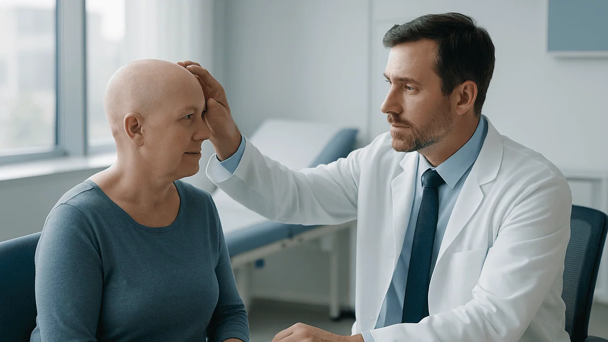
The mirror is usually where it starts. A man in his sixties, a year past treatment for a skin cancer on the crown, tilts his head under the bathroom light and notices that the patch never quite closed. The skin is shiny, thin, a little pink at the edges, and it aches when he sleeps on that side. His dermatologist has already said the word “reconstruction.” He has heard “radiation” and “surgery” often enough to know that they are now part of his story, but nobody has explained how they change the next chapter.
That is the question this article takes seriously. Scalp reconstruction after radiation, or after earlier tumor surgery, is not the same operation as fixing a fresh wound on untouched skin. The tissue remembers. Blood supply, elasticity, bone health, even the way hair follicles behave, have all been altered.
Understanding those changes makes the surgeon’s reasoning far less mysterious, and it gives you sharper questions to ask.
Why a scalp that has been treated before is a different starting point
Think of untreated scalp as a well-stocked workshop. It has a rich network of arteries running in from five directions, a tough fibrous layer called the galea that gives it strength, and a loose gliding layer underneath that lets skin move a little over the skull. Surgeons closing a small wound on this kind of scalp have plenty of tools: stretch the edges, borrow from a neighbor, slide a piece across.
Radiation and previous surgery quietly remove tools from that workshop. Radiation therapy uses high-energy beams to damage the DNA of cancer cells, but it also injures the small blood vessels and the collagen-producing cells in nearby normal tissue. Mayo Clinic describes the skin effects as ranging from early redness and peeling to longer-term changes that can persist after treatment ends. The result, months or years later, is skin that is thinner, stiffer, drier, and slower to close a wound because fewer capillaries reach it.
Previous surgery leaves its own footprint. Every incision divides some of those incoming vessels. A scar that runs across the scalp can cut off blood flow to the tissue on one side of it, so a flap that would have been safe on a fresh scalp may be unreliable when it has to cross old surgical lines.
Put those two histories together and you have a scalp where the usual first-choice repairs carry more risk of breaking down. That is why a reconstructive surgeon spends so much time on your history before drawing a single line on your head. Which fields were irradiated, how long ago, where the earlier incisions ran, whether bone was exposed or removed: each answer closes or opens a door. None of this means reconstruction is off the table. It means the plan has to respect what the tissue has already been through.
How scalp reconstruction after radiation actually works
Strip away the jargon and the goal is simple: cover exposed tissue or bone with living, well-supplied skin that will stay closed, protect the skull and brain, and look as natural as the situation allows. Scalp reconstruction after radiation pursues that goal by importing blood supply rather than hoping the treated area can supply its own.
In practice the surgeon first assesses the defect, meaning the hole or non-healing area that needs covering. Its size, depth, and location matter, but on an irradiated scalp the surrounding tissue matters just as much. The team will often examine the skin for signs of radiation damage: telangiectasia (tiny visible surface vessels), fibrosis (hardening from excess scar tissue), and loss of hair follicles. Imaging such as CT or MRI may be used to check whether the underlying bone is intact or shows osteoradionecrosis, a condition in which irradiated bone loses its blood supply and begins to die.
The reconstruction itself is chosen to match those findings. If healthy, non-irradiated scalp lies close by, the surgeon may design a local flap, a piece of skin and tissue rotated on its own blood vessels from a neighboring area. When the entire top of the head has been treated, or bone is exposed, the team is more likely to recommend a free flap, tissue transferred from a distant site such as the forearm, back, or thigh, with its artery and vein reconnected under a microscope to vessels in the neck or temple.
Skin grafts, thin sheets of skin lifted from another part of the body without their own blood supply, need a healthy wound bed to survive. On irradiated tissue or bare bone that bed is often missing, so grafts are used more selectively. Each of these choices is a judgment call that belongs to the treating team, based on your particular history.
What happens when skin cancer is removed from the scalp?
Many people who end up needing reconstruction started with a skin cancer, so it helps to understand that first step. Scalp skin cancers, most commonly basal cell carcinoma, squamous cell carcinoma, and melanoma, are removed with a margin of normal-looking tissue around them. The size of that margin depends on the type and depth of the cancer, and it is why a small spot can leave a surprisingly large wound.
One technique often used on the scalp is Mohs surgery, in which the surgeon removes thin layers of tissue and examines each one under a microscope during the same visit, continuing only until no cancer cells remain. Mayo Clinic notes this approach is designed to remove the cancer while sparing as much healthy tissue as possible, which is valuable on a surface where every millimeter of hair-bearing skin counts.
What happens next depends on what is left behind. Small, shallow wounds may be closed directly or allowed to heal on their own from the base, a process called secondary intention healing that can take weeks. Larger or deeper wounds, especially those exposing the skull, usually need a planned closure with a flap or graft.
Here the cancer history begins to shape the reconstruction. If the pathology report shows aggressive features, or if the margins are close, the oncology team may recommend radiation afterward. That possibility changes the surgeon’s calculations, because a repair that will later be irradiated needs to be robust enough to tolerate it. Some teams choose to stage the reconstruction, providing temporary coverage while waiting for final pathology, so that the definitive repair is not undermined by an unexpected need for further treatment. This is one reason “why can’t you just close it now” often has a thoughtful answer.
How radiation changes skin, blood supply, and bone over time
Radiation damage unfolds in two acts, and reconstructive surgeons plan around both. The first act is acute. During and shortly after a course of radiotherapy, the skin in the treated field can become red, sore, dry, and itchy, and may peel or blister; the NHS explains that these reactions usually settle in the weeks after treatment finishes. Hair in the field typically falls out during this phase because follicles are rapidly dividing and highly sensitive to radiation.
The second act is chronic and far quieter. Over months and years, the small vessels that were injured do not fully recover. Fibroblasts, the cells that produce collagen, become dysfunctional and lay down dense, disorganized scar. The skin thins, loses its oil glands and sweat glands, and can develop the fine visible vessels mentioned earlier. It bruises and tears easily. Most importantly for surgery, it delivers less oxygen to a healing wound, so incisions may gape, and flaps designed within the field may not survive.
Bone is also affected. The outer table of the skull, when included in a high-dose field, can lose its blood supply and become brittle or exposed. Once exposed to air and bacteria, irradiated bone rarely regrows skin over itself. Surgeons may need to remove the devitalized portion, a procedure called debridement, before any soft tissue can be brought in to cover it.
The time since radiation matters as well. Tissue continues to change for years, so a scalp irradiated a decade ago may behave differently from one treated last year. Surgeons ask for the radiation records, including the field diagram and total dose, not to judge the earlier treatment but to map exactly which tissue they can trust and which they cannot. Decisions about how much bone to remove and what to cover it with rest entirely with the team that has examined you.
Who is usually offered scalp reconstruction after radiation, and who is asked to wait
Reconstruction is generally offered when a scalp wound cannot be expected to close on its own, when bone or the protective membrane around the brain is exposed, or when a chronic wound in irradiated skin has stopped improving despite good wound care. It may also be considered when a healed area is so thin and fragile that it repeatedly breaks down, or when a large hairless scar affects daily life and the person wishes to explore options.
Timing, though, is often the more delicate question. Several groups are commonly asked to wait or to proceed in stages:
- People whose cancer treatment is not yet complete, because a reconstruction placed before further surgery or radiation may be damaged or may obscure recurrence.
- Those whose recent radiation is still in the acute phase, when skin is inflamed and healing capacity is at its lowest.
- Anyone with an active wound infection, which must be controlled before healthy tissue is brought in.
- People with uncontrolled diabetes, significant smoking exposure, or poor nutrition, since each further reduces wound healing; Cleveland Clinic lists these among factors that raise the risk of skin graft failure.
- Those taking medicines that impair healing, such as certain anti-inflammatory or anticancer drugs, where the prescribing clinician may adjust timing rather than the medicine.
None of these is a permanent barrier. Waiting is usually a way of stacking the odds in favor of the repair surviving. In the meantime, teams often use dressings, negative pressure wound therapy (a sealed dressing attached to gentle suction that helps some wounds contract), or a temporary skin substitute to protect the area.
Who decides? Ideally a multidisciplinary team, meaning the reconstructive surgeon, the oncologist, and often a dermatologist or radiation oncologist, reviewing your case together. If you have not heard from more than one specialty, it is reasonable to ask whether such a discussion has happened.
Which reconstruction options exist, and how do they compare?
Reconstructive surgeons talk about a “ladder,” starting with the simplest closure and stepping up as the wound demands. On a previously treated scalp, the lower rungs are often skipped. The table below summarizes the main options in plain terms; the right choice for any individual depends on findings only the treating team can assess.
| Option | What it involves | Where it tends to fit after radiation or prior surgery | Main trade-offs |
|---|---|---|---|
| Secondary intention healing | Wound is dressed and allowed to fill in and contract on its own | Small, shallow wounds outside the treated field | Slow; unreliable on irradiated tissue or bare bone |
| Primary closure | Edges are brought together with sutures | Small defects with mobile surrounding scalp | Tension on stiff, scarred skin can cause breakdown |
| Skin graft | Thin sheet of skin placed on a prepared wound bed | Wounds with healthy underlying tissue, sometimes after drilling or removing outer bone to create a bleeding surface | No hair; thin cover; needs good blood supply beneath it |
| Local or regional flap | Nearby scalp rotated or advanced on its own vessels | Medium defects where untreated scalp is close by | Must avoid crossing old scars or heavily irradiated skin; leaves a secondary wound |
| Tissue expansion | A balloon placed under healthy scalp is gradually filled to grow extra skin | Staged repair when time allows and adjacent scalp is healthy | Multiple visits; expanders are generally avoided under irradiated skin |
| Free flap | Tissue from a distant site with its artery and vein reconnected microsurgically | Large defects, exposed bone, or scalps irradiated over a wide area | Longer operation; donor-site wound; usually hairless cover |
The pattern is clear: as radiation exposure and previous surgery increase, the plan shifts toward options that import a fresh blood supply. That shift is not about the surgeon being cautious for its own sake; it reflects the biology of tissue that can no longer heal itself reliably.
Scalp reconstruction after tumor surgery: timing the repair around cancer care
People whose defect came from a tumor removal often face two clocks running at once. The wound wants closing. The cancer care wants certainty. Reconciling them is one of the more thoughtful parts of the plan.
When margins are confirmed clear at the time of removal, as often happens with Mohs surgery, reconstruction can frequently follow immediately or within days. When the specimen has gone to a laboratory and results will take longer, many teams place a temporary dressing or skin substitute and wait. Closing a wound with a large flap before the pathology is known risks burying residual cancer under healthy tissue, where it is harder to detect.
If postoperative radiation is recommended, the sequence matters even more. Radiation oncologists generally prefer to start within a defined window after surgery, and the reconstructive surgeon needs the repair healed enough to tolerate the beams. A flap with robust blood supply generally withstands radiation better than a thin graft, so knowing that radiation is coming can push the team toward a sturdier option from the outset.
Recurrence surveillance is the third consideration. Some flaps bring in bulky tissue that can hide a returning tumor beneath it. Others keep the reconstructed surface thin and easy to examine. The oncology team’s view on how closely the site needs watching may influence the design.
Finally, previous tumor surgery leaves scars that constrain future flap design. A rotation flap has to be based on intact vessels, and if an earlier operation divided them, the surgeon may look elsewhere. Bringing every old operative note and pathology report to your consultation is one of the most useful things you can do; it lets the team see the map of what has already been done rather than guessing.
Does hair grow back after head radiation?
It depends chiefly on the dose the follicles received, and honest clinicians will say so. Hair follicles are among the most radiation-sensitive structures in the skin because their cells divide rapidly. In lower-dose fields, follicles are stunned rather than destroyed. Mayo Clinic explains that hair loss from radiation therapy usually begins within a few weeks of starting treatment and that hair often regrows after treatment ends, although it may return with a different texture or thickness.
MedlinePlus describes a similar picture: hair in the treated area may fall out, and regrowth, when it happens, typically begins a few months after therapy finishes. The new hair can be finer, grayer, or curlier than before, and it may not fully match the surrounding hair.
At higher doses, or when radiation was combined with surgery that removed the follicle-bearing layer, the loss can be permanent. The follicle’s stem cells sit near the base of the hair shaft; if enough of them are destroyed, no new hair can form. Mayo Clinic notes that whether radiation hair loss is permanent depends on the dose delivered. Your radiation oncologist can tell you which category your treatment fell into, because the dose to the scalp surface will be recorded in your plan.
Why does this matter for reconstruction? Because it sets realistic expectations. A flap brought in from the thigh will not grow scalp hair, and an irradiated area that lost its follicles permanently will not regrow them regardless of how well the wound heals. Surgeons may position hair-bearing scalp where it is most visible and place hairless tissue where it can be covered, but they cannot create follicles where none survive. Options to address the appearance afterward are covered in the next section.
Will hair grow back after surgery on the scalp, and can a hair transplant help?
Surgery affects hair in a few distinct ways, and separating them helps. Along an incision, follicles are cut and a thin hairless scar forms; on a well-designed closure this line can be narrow enough to hide. Around a flap, some hair may shed temporarily because the follicles were stressed during the operation, a process similar to the shedding some people notice after any major illness. This kind of shedding usually recovers as the flap settles.
Hair does not return where the follicle-bearing layer has been removed or replaced. A skin graft has no follicles of its own. A free flap from the back or arm carries its donor site’s hair pattern, which on a scalp means essentially none. These areas will remain hairless unless further steps are taken.
That is where questions about hair transplantation come in, and where caution is warranted. Hair transplantation moves individual follicular units from a dense donor area, usually the back of the head, into thinner areas. On healthy scalp it is a well-established procedure. On irradiated or reconstructed scalp, the picture is more complicated. Grafted follicles need a good blood supply to survive, and both radiation-damaged skin and thin skin grafts may offer less of it. Some surgeons stage the process, first improving the recipient bed or waiting until a flap has matured, and then testing a small area before committing to a larger session.
The evidence here is drawn mostly from case series rather than large trials, so anyone considering it should expect an honest discussion of uncertainty rather than firm promises. Non-surgical options, including scalp micropigmentation (tattooing tiny dots that mimic shaved hair) and hair systems, are also legitimate routes to consider. Whichever direction appeals, the timing and suitability should be discussed with the team that performed the reconstruction, since they know the blood supply of your particular repair.
How long does it take to recover from scalp surgery?
Recovery has a rhythm, and knowing it reduces anxiety in the early days. The exact course depends on the procedure, so treat the following as typical ranges rather than a schedule for you.
In the first few days after a flap or graft, the priority is protecting the repair. Dressings stay in place; you will likely be asked to keep your head elevated, avoid pressure on the site, and skip strenuous activity. For a free flap, hospital staff monitor the color and warmth of the tissue closely, because problems with the reconnected vessels usually announce themselves early.
Skin grafts follow their own timetable. MedlinePlus notes that a graft typically begins to develop its own blood supply within a few days and that the donor site, where the skin was taken, usually heals within about one to two weeks for thinner grafts. The grafted area itself remains fragile for longer and is often kept covered while it toughens.
Suture or staple removal, when used on the scalp, is commonly scheduled around one to two weeks after surgery according to MedlinePlus guidance on flaps and grafts, though your team will set the date. Swelling and numbness around a flap are expected and can persist for months; sensation returns gradually as small nerves regrow.
Irradiated tissue slows every part of this. Wound edges may take longer to seal, and the team may keep you on protective dressings beyond the usual window. Tissue expansion, when used, adds a series of outpatient fill appointments over weeks before the second operation.
By the second and third months, most people are back to ordinary activities, with the flap softening and scars beginning to fade. Scar maturation continues for a year or more. Follow-up visits during this time serve two purposes at once: checking the reconstruction and continuing cancer surveillance.
What can go wrong in an irradiated scalp, and what are the alternatives?
Every reconstruction carries risk, and a previously treated scalp raises several of them. Naming them plainly is part of informed consent, not a reason for alarm.
Wound dehiscence, meaning the edges of the closure pull apart, is more common when stiff irradiated skin is placed under tension. Partial flap loss can occur when the tip of a local flap farthest from its blood supply does not receive enough oxygen. Free flaps carry a small risk of failure of the microsurgical vessel connection, which is why they are watched so intently in the first days. Skin grafts can fail to take if fluid collects beneath them, if infection develops, or if the underlying bed is poorly supplied, all of which are more likely on irradiated tissue.
Infection is a particular concern where bone has been exposed, because irradiated bone has limited ability to fight bacteria. Persistent exposed bone may require further debridement. Chronic non-healing ulcers can develop years later in the irradiated field even after a successful repair, which is one reason lifelong sun protection and skin checks are advised.
Alternatives always exist, even if none is perfect. Continued conservative wound care with specialist dressings may be appropriate for some smaller wounds. Negative pressure wound therapy can help prepare a bed for later grafting. Hyperbaric oxygen therapy, in which a person breathes pure oxygen in a pressurized chamber, is sometimes used to support healing in irradiated tissue; the evidence is mixed and it is not suitable for everyone, so it should be regarded as an adjunct that some teams consider rather than a proven standalone treatment. Prosthetic coverings can protect a stable but hairless area without surgery.
The right balance between these risks and alternatives is individual. A frank conversation about what would happen if the first plan did not work is a sign of a team that has thought the case through.
What people often get wrong about scalp reconstruction after radiation
Misunderstandings cluster around a few themes, and clearing them up early spares frustration later.
“The radiation was years ago, so the skin is back to normal.” Radiation changes in skin and bone are largely permanent and can even progress over time. Surgeons treat a field irradiated fifteen years ago with the same respect as one irradiated last year, and sometimes more, because the chronic vessel damage has had longer to develop.
“A skin graft is the simple, safe choice.” On healthy tissue it often is. On irradiated scalp or bare bone, grafts frequently fail for lack of a blood supply beneath them. What sounds like the smaller operation can end up being the less reliable one.
“Hair will grow through the new skin eventually.” Follicles cannot migrate into a graft or a flap from elsewhere on the body. Hair grows only where follicle-bearing scalp has been preserved or moved.
“If the wound has not healed in a few months, something must have been done wrong.” Slow healing is the expected behavior of irradiated tissue. It is a reason to reassess the plan, not evidence of error.
“Reconstruction means the cancer is finished with.” Reconstruction addresses the wound; surveillance for recurrence continues on its own schedule. The two run in parallel, which is why follow-up appointments matter even when the scalp looks well.
“Bigger flaps are always better.” Bulkier tissue can hide recurrence and may need thinning later. The best flap is the one that matches the defect and the surveillance plan, which is a judgment for the treating team.
Each of these corrections points the same way: the biology of treated tissue sets the rules, and good plans work within them rather than against them.
Questions to ask your care team before scalp reconstruction
Consultations move quickly, and it is easy to leave with more uncertainty than you arrived with. Writing questions down beforehand changes the dynamic. These are the ones that tend to unlock the most useful conversations.
- Which parts of my scalp were in the radiation field, and how does that affect where you can safely take tissue from?
- Have you reviewed my previous operative notes and pathology reports, and do my old incisions limit any options?
- Is the cancer treatment complete, or is there a chance I will need more surgery or radiation after this repair? How would that change the plan?
- Are you considering a graft, a local flap, tissue expansion, or a free flap for me, and what makes one more suitable than the others in my case?
- Is any bone exposed or damaged, and will part of it need to be removed?
- Will the reconstructed area have hair? If not, what options exist later, and when could they be considered?
- What does recovery typically look like for this procedure, and how might my radiation history slow it?
- What would you do if the flap or graft did not survive?
- Are there factors I can improve beforehand, such as smoking, blood sugar control, or nutrition, and how much time would that take?
- How will you and the oncology team check the site for recurrence after it is covered?
- Who do I call, day or night, if I am worried about the wound after I go home?
You are entitled to ask for a second opinion within the same health system or elsewhere, and a confident team will not be troubled by the request. Bring a family member or friend if you can; a second set of ears catches details that anxiety filters out. The answers you receive should sound like reasoning, not reassurance, because the honest version of this conversation includes uncertainty.
When to call your doctor after scalp surgery or during radiation-related healing
Most recoveries proceed without drama, but an irradiated scalp gives you less margin for waiting things out. Your team will give you specific instructions; the signs below are widely recognized reasons to make contact promptly rather than at the next scheduled visit.
Call the same day if you notice increasing redness spreading beyond the wound edges, new or worsening pain after an initial improvement, warmth around the site, a foul smell, or cloudy or thick drainage. These can indicate infection, which irradiated tissue handles poorly.
Seek urgent advice if a flap changes color, becoming pale, dusky purple, or dark, or if it feels cold compared with surrounding skin. In free flaps especially, these changes can signal a problem with the blood vessels and are time-sensitive. Bleeding that soaks through a dressing and does not slow with gentle pressure also warrants a call.
Contact your team without delay if the wound edges separate, if you can see bone or a grayish membrane at the base of the wound, or if a previously healed area breaks open. Fever, chills, or feeling generally unwell alongside any wound change should be treated as a red flag.
Emergency care is appropriate for severe headache with confusion, drowsiness, or vomiting after surgery that involved exposed skull, for clear fluid leaking from the wound that does not look like ordinary drainage, or for sudden weakness or difficulty speaking. These are uncommon, but they need immediate assessment.
Between visits, slower concerns still deserve a conversation: a graft that stays fragile and cracks repeatedly, a new lump or crusted spot within or near the treated field, or numbness that seems to worsen rather than improve. Cancer surveillance depends partly on you noticing change, so no question about the site is too small to raise with the team following you.
Frequently asked questions
Does hair grow back after radiation to the head?
Often, but not always. Mayo Clinic explains that hair loss from radiation usually starts within a few weeks of treatment and that regrowth, when it occurs, typically begins a few months after therapy ends, sometimes with a different texture or color. At higher doses the follicles can be permanently destroyed. Your radiation oncologist can tell you which category your treatment fell into from the recorded dose to the scalp.
How long does skin graft on scalp healing usually take?
A skin graft generally starts to develop its own blood supply within a few days and the donor site for a thin graft heals in roughly one to two weeks, according to MedlinePlus. The grafted surface itself stays fragile for longer and is often kept protected for several weeks. On irradiated scalp, healing tends to be slower and graft survival less predictable, so your team may extend dressings and follow-up.
What does scalp flap surgery recovery involve day to day?
The first days focus on protecting the flap: head elevation, no pressure on the site, dressings in place, and close checks of the tissue’s color and warmth. Sutures or staples are commonly removed around one to two weeks after surgery per MedlinePlus guidance. Swelling and numbness can persist for months as small nerves regrow. Most people return to ordinary activities within a couple of months, while scars keep maturing for a year or more.
Is a hair transplant after radiation possible on a reconstructed scalp?
Sometimes, with important caveats. Transplanted follicles need a good blood supply to survive, and irradiated skin, skin grafts, and some flaps may not provide enough. Surgeons who consider it usually wait until the reconstruction has matured and may test a small area first. The evidence comes mainly from case series, so expect an honest discussion of uncertainty. The team that performed your reconstruction is best placed to judge suitability and timing.
What happens when skin cancer is removed from the scalp?
The cancer is cut out with a margin of normal-looking tissue, often using Mohs surgery, in which layers are checked under a microscope until no cancer remains. Small wounds may be closed directly or allowed to heal from the base. Larger wounds, or those exposing bone, need a planned repair with a graft or flap. If pathology suggests radiation will be needed afterward, the reconstruction is often staged or designed to withstand it.
Why can't the surgeon just use a skin graft on my irradiated scalp?
Skin grafts have no blood supply of their own and depend entirely on the wound bed beneath them. Radiation reduces the number of working capillaries in that bed, and bare bone offers almost none, so grafts placed there frequently fail. Flaps bring their own artery and vein, which is why surgeons often prefer them on treated scalp. Your surgeon can explain which findings on your scalp point one way or the other.
How long after radiation should scalp reconstruction wait?
There is no single interval. Surgeons generally avoid operating during the acute phase, when skin is inflamed and healing capacity is lowest; the NHS notes skin reactions usually settle in the weeks after treatment finishes. Beyond that, the decision weighs whether cancer treatment is complete, whether bone is exposed, and whether infection is controlled. Waiting is usually about improving the odds of the repair surviving rather than a fixed rule.
Will the reconstructed area of my scalp ever look normal?
It can improve substantially, but honesty matters: tissue moved from the arm, back, or thigh will not grow scalp hair or match its color exactly, and scars keep fading for a year or more. Surgeons place hair-bearing scalp where it is most visible when they can. Later options such as scalp micropigmentation, hair systems, or in selected cases hair transplantation can address appearance. Your team can describe what is realistic for your particular repair.
Can radiation damage the skull bone under my scalp?
Yes. High-dose fields can reduce the blood supply to the outer layer of the skull, leaving it brittle and, if exposed, unable to regrow skin cover. This condition is called osteoradionecrosis. Surgeons may need to remove the damaged portion before bringing in soft tissue, and imaging such as CT is often used to assess it. Whether and how much bone is removed is a decision for the treating team based on your scans.
Does reconstruction mean my cancer follow-up is finished?
No. Reconstruction addresses the wound; surveillance for recurrence continues on its own schedule set by the oncology team. Some flaps can make the area harder to examine, so surgeons and oncologists often coordinate on flap design and follow-up. You remain an important part of that surveillance: report any new lump, crusted spot, or non-healing area within or near the treated field between visits.
References
- NHS: Radiotherapy side effects
- MedlinePlus: Skin graft
- MedlinePlus: Skin flaps and grafts – self-care
- Cleveland Clinic: Skin graft
This article is for general information only and is not a substitute for professional medical advice. Please consult a qualified doctor about your individual situation.

More from the Blog
Temple Hair Transplants and Hairline Design: The Details That Make It Look Real
A temple hair transplant moves individual follicles, almost always single-hair grafts, into the temple recessions and temporal points to rebuild a receded hairline. A…
The First Two Weeks After a Hair Operation: Swelling, Scabs and Milestones Day by Day
In the first two weeks after a hair operation, most people move through three overlapping stages: forehead swelling that peaks around days two to…
Which Female Hair Transplant Side Effects Are Normal? Redness, Scabbing and Swelling Explained
After a female hair transplant, mild redness, pinpoint scabs over each graft, tightness and swelling that drifts toward the forehead are expected in the…
Getting Ready for a Beard Transplant: Skin Checks, Mapping the Beard Line and What to Pause
Beard transplant preparation usually spans several weeks and has three parts: a skin check of the face and donor scalp for active acne, folliculitis,…
What Female Hair Transplant Results Look Like Month by Month: From Shedding to Settled Density
A female hair transplant results timeline typically unfolds in stages: swelling and crusting in the first week, shedding of most grafted hairs between roughly…
Types of Hair Transplant: FUE, DHI, Sapphire and FUT: Which Fits Whom
The main types of hair transplant are FUT (a strip of scalp is removed and divided into grafts), FUE (follicles are extracted one by…