

IMRT Planning Day Explained: Simulation CT, Immobilization Mask and Skin Marks

Key Takeaways
- An IMRT simulation delivers no treatment radiation; the CT is purely for mapping and the scan portion itself takes only minutes.
- Planning scans are taken on a flat couch in treatment position because density values and geometry from that exact setup drive the dose calculation.
- Immobilization masks are warm thermoplastic mesh that hardens in minutes, and for head and neck treatment the alignment marks usually go on the mask rather than the skin.
- Radiotherapy tattoos are freckle-sized permanent dots used as laser alignment coordinates, not indicators of where the beam enters or how large the tumor is.
- Between simulation and the first session, an oncologist contours targets, a dosimetrist optimizes beam intensities and a physicist verifies the plan on the actual machine.
- Weight change, new swelling or a lifted mark should be reported promptly, because the plan is only valid for the anatomy captured on planning day.
An IMRT simulation planning CT is the mapping appointment before radiation treatment begins. You lie in the exact position you will use for every session while a CT scanner images the treatment area; the team may make a custom immobilization mask or mold and place tiny skin marks or pin-point tattoos as alignment references. No radiation treatment is given that day. The scan becomes the three-dimensional blueprint from which your individual IMRT plan is built.
The letter says “CT simulation, please arrive fifteen minutes early, bring a list of medicines.” What it does not say is what a simulation actually is, whether it hurts, or why a scan you have already had several times needs repeating. So the night before, many people lie awake wondering if the mask will feel like a trap and whether “skin marks” means something permanent.
Here is the reassurance up front: an IMRT simulation planning CT is a measuring day, not a treatment day. Nothing is aimed at a tumor. Instead, a radiation therapist positions you, the scanner records where everything sits, and the team gathers the reference points that let every future session line up to within a few millimeters of the plan.
This explainer walks through the appointment minute by minute, including the mask, the marks, the wait that follows, and the questions worth asking before you leave.
What actually happens at an IMRT simulation planning CT appointment?
Intensity-modulated radiation therapy, or IMRT, is a form of external-beam radiation that shapes the beam into many small segments of varying strength so the dose wraps around a target while sparing nearby tissue. Because the plan is so tightly tailored, it needs an equally precise map. That map is built on simulation day.
You will usually be greeted by a radiation therapist, the professional who runs the treatment machines, and sometimes by a dosimetrist, the specialist who later calculates how the beams will be arranged. After identity checks and a review of your consent, you change if needed and lie on a flat, hard couch that mimics the treatment table. The therapists position you using laser lights projected onto your skin and, depending on the area, a headrest, arm supports, a knee cushion or a molded cradle.
Once you are settled, the couch slides through a CT scanner, which takes cross-sectional X-ray images of the region. The scan itself is quick, often a matter of minutes, though the whole appointment runs longer because positioning and device-making take time. The National Cancer Institute describes simulation as the step where your team determines the treatment area and marks your body so that the same position can be reproduced every day.
Before you get up, the team places alignment marks and photographs your setup. You leave with nothing more than a few small dots, possibly a fitted mask stored at the department, and a date for your first session. The real work of planning, described later, happens after you have gone home.
Why does IMRT depend so heavily on a planning CT?
Think of IMRT as a sculptor working blind. The machine, a linear accelerator that generates high-energy X-rays, cannot see the tumor during treatment. Everything it does is executed from a plan built in advance. That plan is only as good as the picture it was drawn on, and the picture is your planning CT.
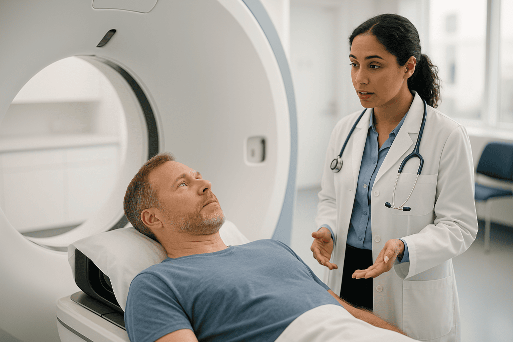
Three features make this scan different from a diagnostic one. First, it is taken in treatment position, on a flat couch rather than a curved one, with the same supports you will use each day. A scan taken with your arms at your sides is useless if you will be treated with them raised, because organs and skin shift. Second, the images carry density information. CT measures how much each tiny volume of tissue attenuates X-rays, and planning software uses those values to calculate how the beam will deposit energy as it passes through lung, bone, fat or muscle. Third, the scan is geometrically linked to the room. Laser lines in the simulator correspond to laser lines at the treatment machine, so a mark on your skin translates to a coordinate in the software.
Mayo Clinic describes IMRT as delivering radiation from many angles with beams whose intensity is modulated to conform to the shape of the target. Conforming to a shape presupposes knowing the shape in three dimensions. That is what the planning CT provides, and why your team will insist on it even if you have a recent diagnostic scan on file. Those earlier images help doctors decide whether radiation is appropriate; the simulation scan tells the machine exactly where to point.
Who is usually asked to have a CT simulation for radiation, and who is asked to wait?
Almost everyone scheduled for IMRT will have a simulation, whether the target is in the head and neck, prostate, breast, brain, lung or elsewhere. The MedlinePlus overview of radiation therapy notes that external-beam treatment is planned individually, and simulation is that planning step in practice. Some departments also use it for simpler three-dimensional conformal radiation, so a simulation appointment does not by itself tell you which technique has been chosen.
Timing matters, and several situations lead a team to postpone rather than proceed. If you have had recent surgery, they may wait for swelling to settle so the anatomy on the scan matches the anatomy weeks later. If chemotherapy or hormone therapy is planned first, simulation is usually scheduled toward the end of that phase, because the tumor may shrink and the plan should reflect its final size. A significant change in body weight, a new drain, or a wound that is still healing can also prompt a delay or a repeat scan later.
People who are pregnant, or who might be, must tell the team before the scan; the CT carries an X-ray dose and treatment planning for pregnant patients follows separate pathways. Those with pacemakers or implanted defibrillators are not excluded, but the device is documented so that beams can be arranged away from it. Metal implants, such as hip replacements or dental work, create artifacts on CT and may lead the team to adjust technique.
The decision to simulate now, later or not at all always rests with the radiation oncologist, the physician who leads your radiation care, weighing the overall treatment sequence rather than any single scan.
How should you prepare for a radiotherapy planning scan?
Preparation depends entirely on where the treatment will be aimed, and the instructions your department sends take precedence over anything general. A few patterns recur, though, and knowing the reasoning makes them easier to follow.
For pelvic targets such as the prostate, cervix or rectum, you may be asked to arrive with a comfortably full bladder and, sometimes, an empty bowel. A full bladder pushes small-bowel loops up and away from the beam; consistent filling means the organ sits in the same place on planning day and treatment days. The NHS radiotherapy guidance notes that you may be asked to follow specific bladder or bowel instructions for this reason. If you are given a drinking routine, it is a positioning tool, not a hydration test, and the aim is repeatability.
For head and neck or brain treatment, shave or trim according to instructions, remove earrings and hair clips, and expect the mask to be made over bare skin. Dentures may need to come out or stay in depending on how you will be treated, so ask rather than guess.
For the chest and breast, wear a top that is easy to remove. You may be asked not to use deodorant, lotion or powder on the area, because some products leave residue that interferes with marking.
Bring a list of your medicines and any allergies, particularly to contrast dye or iodine. Eat normally unless told otherwise; fasting is usually required only if intravenous contrast is planned. If you take regular pain relief and lying flat is uncomfortable, ask your team in advance whether to take it as usual, and follow their answer rather than adjusting on your own.
How is a radiation therapy mask made, and what does it feel like?
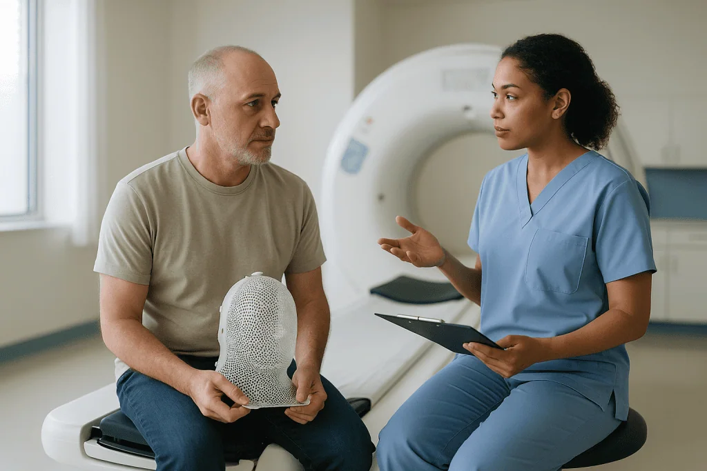
The immobilization mask, sometimes called a shell, is a sheet of thermoplastic mesh that becomes soft in warm water and hardens as it cools. Its job is simple: to hold your head, and often your shoulders, in exactly the same position at every session, so that the millimeter-scale precision of IMRT is not undone by a small turn of the neck.
Making it takes a few steps. You lie on your back with your head in a molded rest. A therapist lifts a warm, pliable sheet from a water bath, drapes it over your face and neck, and gently presses it around your nose, chin and forehead while it sets. It feels like a warm, damp towel that gradually tightens. Most masks have holes throughout the mesh so you can breathe normally, and many departments can cut openings for the eyes or mouth if that helps you cope. The setting time is a matter of minutes, during which you are asked to stay still and breathe steadily. Once it has hardened, the mask is clipped to the couch, the CT is taken through it, and it comes off.
Skin marks for head and neck treatment usually go on the mask, not on you, which is one reason people treated in this area often have no tattoos.
The same principle extends to other body regions. Vacuum-formed bags mold to your back or legs, breast boards angle the chest, and thermoplastic body shells sometimes cover the abdomen. Each device is labeled with your name and kept in the department between sessions. If the mask ever feels noticeably tighter or looser, mention it: weight change can alter the fit, and the team may remake it.
Radiotherapy tattoo marks and skin marks: what they are and why they matter
At the end of the scan, the therapists mark the points where the room lasers cross your skin. These are alignment references, not treatment boundaries. On treatment days the lasers are matched to the marks first, then imaging on the machine fine-tunes the position.
There are two main approaches. Temporary marks are drawn with a skin pen and covered with clear waterproof dressings, which you are asked to leave in place and keep from peeling. Permanent marks are tiny tattoos: a drop of ink placed with a fine needle, producing a dot about the size of a freckle. The NHS describes these as small permanent marks, often only two or three, made with a pin-prick that people generally find uncomfortable for a second rather than painful.
Why permanent? A pen mark that fades or washes off midway through a multi-week course would force a repeat setup, and re-drawing a mark from memory reintroduces the error that the whole system is designed to remove. A tattoo cannot smudge, so it is trusted as a fixed coordinate.
Some departments now offer alternatives, including ink that fluoresces only under ultraviolet light or surface-guidance cameras that track the body’s contour without marks. Availability varies and the choice is a departmental one, so if the idea of tattoos troubles you, ask what your center uses rather than assuming.
Caring for marks is uncomplicated: do not scrub them, do not apply lotions over pen marks, and never try to redraw a fading line yourself. If a dressing lifts or a mark disappears, tell the therapists before your next session so they can relocate it properly from the plan.
Contrast dye, breath-hold and 4D CT: extras you might be offered
Not every simulation is a plain scan. Depending on the site, your team may add one or more techniques that sharpen the picture or account for movement.
Intravenous contrast is an iodine-based dye injected through a small cannula in the arm. It brightens blood vessels and some tumors on CT, helping the oncologist tell tumor from normal tissue when drawing the target. You may feel a warm flush and a metallic taste for a minute; that is expected. Tell the team about kidney problems, prior contrast reactions or metformin use, because these affect whether contrast is appropriate. Oral contrast, a drink taken before the scan, is sometimes used to outline the bowel for abdominal targets.
Movement is the other challenge. Lungs, liver, breast and some pelvic structures shift with every breath. A four-dimensional CT, or 4D CT, records images across the breathing cycle so planners can see the full envelope of motion and design margins that cover it. You breathe normally while a small sensor on your chest tracks the rhythm. Alternatively, you may be coached in a breath-hold technique, holding a deep breath for a short interval while images are acquired. For left-sided breast treatment, a deep inspiration breath-hold pulls the heart down and away from the chest wall, which is one reason the technique is widely used.
Fiducial markers, tiny inert seeds placed in or near a tumor before simulation, are used for some prostate and liver treatments. They show clearly on CT and on daily machine imaging, providing an internal reference alongside the external skin marks.
None of these extras changes the fundamental nature of the day. They add minutes, not risk of treatment, and your team will explain any that apply to you.
Planning day versus treatment day: a side-by-side view
Confusion between the two appointments is common, and it drives a lot of unnecessary anxiety. The table below lays out what is the same, what differs and what you should expect from each.
| Aspect | Simulation planning CT | IMRT treatment session |
|---|---|---|
| Purpose | Map anatomy and record the reproducible position | Deliver the planned dose to the target |
| Machine | CT scanner with a flat couch and lasers | Linear accelerator with on-board imaging |
| Radiation given | Diagnostic-level X-rays for imaging only | Therapeutic dose according to the plan |
| Mask or mold | Made and fitted | Reused at every visit |
| Skin marks | Placed at the end | Aligned to lasers, then verified by imaging |
| Typical duration | Longer, because of device-making and setup | Shorter; the NHS notes most of the time is positioning, with the beam on for only minutes |
| Frequency | Usually once, occasionally repeated if anatomy changes | Typically daily on weekdays, per NCI and NHS descriptions |
| What you feel | Nothing from the scan; mild pressure from devices | Nothing from the beam; skin and site effects build over weeks |
Two points deserve emphasis. First, the simulation involves no treatment, so you will not feel tired or sore from radiation afterward. Second, the machine you see on planning day is not the one you will be treated on, so do not be alarmed on your first session when the room looks different. The couch, lasers and your own devices are the constants that carry across.
What happens between the planning scan and your first IMRT session?
The quiet stretch after simulation feels, to many people, like the team has gone silent. In fact this is the busiest phase of planning, and it happens entirely on computers.
Your radiation oncologist first outlines, slice by slice on the CT, the target volume and every nearby organ at risk: spinal cord, salivary glands, heart, bowel, bladder, kidneys, whichever apply. This process, called contouring, may take hours for complex head and neck cases and is often informed by fusing your planning scan with a diagnostic MRI or PET scan.
A dosimetrist then works with planning software to arrange the beam angles and intensity patterns that meet the doctor’s prescription while keeping organs below agreed limits. IMRT planning is an iterative optimization: the computer proposes a solution, the planner adjusts priorities, and the cycle repeats until the trade-offs are acceptable. The oncologist reviews and approves the plan.
A medical physicist, the scientist responsible for machine accuracy, performs quality assurance. For IMRT this often includes delivering your plan to a measuring device on the actual linear accelerator to confirm the machine produces what the software calculated.
The NHS radiotherapy guidance explains that treatment does not usually begin on the same day as planning and that there is often a gap while the plan is prepared. The length of that gap varies by complexity and department, and your team will give you a realistic estimate rather than a fixed promise.
On the first treatment day, expect extra time. Therapists verify your position with imaging, compare it with the planning CT and make small couch shifts before the first beam is delivered.
Comfort, anxiety and claustrophobia: getting through the mask and the stillness
The physical demands of simulation are modest, but the psychological ones are real. Lying still on a hard surface while a mask sets over your face asks something of anyone, and admitting that is not weakness.
Practical measures help more than people expect. Ask whether you can see and handle a sample mask beforehand; the mesh is more open than most imagine. Some departments allow a short practice lie-down in the position before the warm sheet is applied. Tell the therapists if you are prone to panic in enclosed spaces so they can talk you through each step, keep a hand on your shoulder, and pause if you raise a hand. Many find that keeping eyes closed and counting slow breaths in fours is enough; others prefer eyes open through cut-out windows.
If lying flat causes breathlessness, pain or coughing, say so at the start. A slight change in headrest or a knee support can often solve it, and if it cannot, the team needs to know before the plan is built around a position you cannot hold.
For people with significant anxiety, the radiation oncologist may discuss a short-acting anxiolytic medicine before the session. Whether this is appropriate, and how it is used, is a decision for the prescribing clinician, weighed against the need to lie still and travel safely home. Do not take anything you have at home on your own initiative before the scan.
Children and some adults with cognitive impairment may be simulated under sedation or general anesthesia, arranged through the anesthesia team, with the same devices and marks applied while they sleep. The Cleveland Clinic overview of radiation therapy notes that the team’s goal throughout is a position you can reliably hold, not a test of endurance.
What are the risks of the simulation, and are there alternatives?
Set against a course of radiation, the planning day carries very little risk, but a precise explainer should still name what exists.
The CT scan uses ionizing radiation at diagnostic levels, far below the treatment doses that follow. The MedlinePlus radiation therapy page places this in context: imaging exposures are small, and the benefit of an accurate plan clearly outweighs them for someone already committed to treatment. Pregnant patients are the exception and must disclose the possibility before scanning.
Intravenous contrast can cause allergic-type reactions ranging from itching to, rarely, severe responses, and can strain kidneys that are already impaired. This is why the team screens kidney function and allergy history first. A cannula site can bruise.
Tattoo marks carry a needle-prick and a permanent dot, with a very small risk of local infection if the site is scratched. Mask-making occasionally causes brief warmth or a feeling of pressure; skin irritation is uncommon. The most consequential “risk” is a plan built on a position you cannot reproduce, which is why honesty about discomfort matters more than stoicism.
As for alternatives: there is no substitute for a planning scan if IMRT is to proceed, because the technique is defined by its computer plan. Alternatives exist at the level of technique rather than the scan itself. MRI-based planning is used in some centers alongside or, rarely, instead of CT. Surface-guided systems and ultraviolet ink reduce or remove the need for visible tattoos. Simpler radiation techniques may need fewer immobilization devices but still require a scan.
Which approach suits you is a judgment for the treating team, based on tumor site, available equipment and your own circumstances.
What people often get wrong about IMRT simulation and planning
Misunderstandings about planning day travel fast between waiting-room chairs. A handful come up repeatedly.
“The simulation is my first dose.” It is not. The scan takes pictures; no treatment beam is involved, and you will feel no effects from radiation afterward. The word “simulation” simply means rehearsing the position.
“The dots show where the beam goes.” The marks are alignment coordinates, usually at the center of the setup or on the sides of the body. Beams enter from many angles in IMRT and the treated volume is defined in the software, not on the skin. Do not read tumor size into the spacing of your tattoos.
“My recent CT could have been used instead.” Diagnostic scans are taken on curved couches, in different positions, sometimes with contrast phases that distort density values. Planning requires treatment position, a flat couch and known geometry, so the scan must be repeated.
“The mask is only for people with brain tumors.” Masks are standard for head and neck cancers, brain targets and some upper-spine or upper-chest treatments. Having one made says nothing about prognosis.
“If I lose weight the plan still works.” Substantial weight change alters where the skin and organs sit relative to the marks and can loosen a mask. Teams routinely re-simulate or adapt the plan when this happens, so report noticeable changes rather than hoping they do not matter.
“Tattoos fade like pen marks.” They do not. They are permanent, which is precisely why they are used. If permanence troubles you, raise it before the scan, because alternatives may be available.
Getting these right will not change your plan, but it will change how the day feels.
Questions to ask your care team before and on planning day
A good simulation appointment is a two-way conversation, and the therapists expect questions. Bringing a short written list keeps nerves from crowding them out. The following are worth considering, grouped by when they help most.
Before the appointment, ask what preparation is required for your specific site, particularly around bladder and bowel, eating, skin products and what to wear. Ask whether contrast is planned and what you should disclose about kidney function or allergies. If you have a pacemaker, defibrillator, insulin pump or other implanted device, ask how it will be documented and protected.
On the day, ask whether a mask or mold will be made and whether you can see one first. Ask how your position was chosen and what to do if you cannot hold it comfortably. Ask whether your marks will be permanent tattoos, pen marks under dressings, or a mark-free surface-guided system, and how to care for whatever is used. Ask who to contact if a dressing lifts or a mark fades.
Before leaving, ask roughly how long the planning phase will take and how you will be told your start date. Ask whether other scans, such as MRI or PET, will be fused with your planning CT. Ask what the first treatment day will look like and whether it will run longer than subsequent sessions. If you are unsure, ask whether the plan is IMRT specifically or another technique, and what that means for the number of sessions.
Finally, ask how weight change, new symptoms or a change in other treatments should be reported during the planning gap. Writing down the answers, or bringing someone who can, is the simplest safeguard against forgetting them on the drive home.
When to call your doctor after your simulation appointment
Because no treatment is delivered on planning day, serious problems afterward are unusual. A few situations nonetheless warrant a prompt call to your radiation team or, where noted, emergency services.
Seek emergency help immediately if, within hours of receiving intravenous contrast, you develop difficulty breathing, swelling of the face, lips or tongue, widespread hives, or feel faint. These can indicate a serious allergic reaction. Call your team the same day if you notice a spreading red or itchy rash, or if urination drops markedly over the following day or two, particularly if you have known kidney disease.
Contact the department promptly, before your next visit, if a pen mark has washed off or a dressing has fully lifted; if a tattoo site becomes hot, red, swollen or leaks fluid; or if the cannula site develops increasing pain, redness tracking up the arm, or a hard cord under the skin.
Tell your team as soon as possible about any change that could affect the plan: significant weight loss or gain, new swelling at the treatment site, a new or worsening wound, a new drain or feeding tube, or a change in other cancer treatments. The plan is built on the anatomy captured that day, and your team needs to know if the anatomy is changing.
Call urgently, regardless of the simulation, if you experience new severe headache, sudden weakness or numbness, uncontrolled bleeding, chest pain, or a fever with chills, since these relate to the underlying illness rather than the scan and should not wait for a scheduled appointment.
When in doubt, ring. Radiation departments expect these calls and would rather adjust a plan early than discover a problem on the first day of treatment.
Frequently asked questions
What is a radiotherapy planning scan and how is it different from my diagnostic CT?
A radiotherapy planning scan is a CT taken in your exact treatment position on a flat couch with room lasers, and it is used to build your radiation plan rather than to diagnose. Diagnostic scans are acquired on curved tables, often in different postures and contrast phases, so their geometry and density values cannot be used for dose calculation. That is why the scan is repeated even if you had imaging recently.
Does the IMRT simulation planning CT hurt?
The scan itself is painless and you feel nothing from the imaging X-rays. Discomfort, when it occurs, comes from lying still on a hard surface, from a mask setting over the face, from a contrast cannula, or from the brief needle-prick of a tattoo mark. Tell the therapists about any pain or breathlessness in position, because a comfortable, repeatable posture is essential to the plan.
How long does a CT simulation for radiation take?
The appointment is longer than a routine scan because positioning, device-making and marking take most of the time; the CT acquisition itself lasts minutes. The NHS radiotherapy guidance describes planning as a separate session before treatment starts, with duration depending on whether a mask or mold is made and whether contrast or breath-hold techniques are used. Your department will give you a realistic estimate.
Is a radiation therapy mask claustrophobic?
Many people expect it to be worse than it is, because the mesh is open and you can breathe freely, but some do find it difficult. Ask to see a sample mask, request eye or mouth cut-outs if offered, and tell the therapists so they can talk you through and pause if you signal. If anxiety is severe, your radiation oncologist can discuss options, which remain the prescribing clinician’s decision.
Are radiotherapy tattoo marks permanent, and can I refuse them?
Yes, they are permanent freckle-sized ink dots, chosen precisely because they cannot smudge or wash off during a multi-week course. Some departments offer alternatives such as ultraviolet-visible ink or surface-guidance camera systems that track your body contour without marks. Availability varies, so raise the question before the scan; your team will explain what is possible at your center.
Why do I need a full bladder for my planning CT?
For pelvic targets, a comfortably full bladder lifts small bowel away from the treatment area and keeps the bladder in a consistent position from planning day through each session. The NHS notes that specific bladder or bowel instructions are sometimes given for this reason. Follow your department’s exact routine, because the aim is reproducibility rather than any particular volume.
Will I feel side effects after the simulation appointment?
No, because no treatment dose is delivered on planning day. You may notice a warm flush during contrast injection, mild bruising at a cannula site, or slight tenderness where a tattoo was placed. Fatigue, skin changes and site-specific effects belong to the treatment course, which begins later after the plan is built and verified.
What happens if I lose weight after my planning CT?
Substantial weight change can shift skin marks relative to internal organs and can loosen a mask, both of which reduce setup accuracy. Teams routinely watch for this using daily imaging on the treatment machine and may remake devices or repeat the simulation and adapt the plan. Report noticeable changes promptly rather than waiting to be asked.
Can I have my IMRT treatment on the same day as the planning scan?
Usually not. After the scan, the oncologist contours the target and organs, a dosimetrist optimizes the beam arrangement, and a physicist checks the plan on the machine. The NHS explains that treatment typically starts after a gap while planning is completed. The interval varies with complexity and department workload, and your team will tell you when to expect your start date.
Do I need a mask for every type of IMRT?
No. Masks are standard for head and neck cancers, brain targets and some upper-spine or upper-chest treatments where small movements matter most. For breast, lung, abdominal and pelvic treatment, other devices such as vacuum-formed bags, breast boards, arm supports or knee cushions are used instead. Having a mask made reflects the anatomy involved, not the seriousness of your diagnosis.
References
This article is for general information only and is not a substitute for professional medical advice. Please consult a qualified doctor about your individual situation.

More from the Blog

Why Chronic Lymphocytic Leukemia Is Often Watched First, and What Triggers Treatment
Chronic lymphocytic leukemia is often watched rather than treated at first because trials have not shown that starting treatment early helps people live longer,…

The Days Before Radiation Therapy Starts: Simulation, Skin Marks and a Checklist to Follow
Preparing for radiation therapy usually means attending a planning appointment called a simulation, where you lie in your treatment position for a CT scan,…
Bleeding, Fever or Pain After Endometrial Cancer Surgery: Which Signs Need a Call
After a hysterectomy for endometrial cancer, call your surgical team the same day for bleeding heavier than a light period or that suddenly restarts,…

When Does Leukemia Treatment Include a Stem Cell Transplant, and Who Is Assessed for It?
A stem cell transplant becomes part of leukemia treatment when the disease is judged likely to return after chemotherapy alone, or when it has…
Staging Before Cancer Treatment Starts: How Imaging and Pathology Set the Plan
Cancer staging before treatment describes how large a tumor is and whether it has reached nearby lymph nodes or distant organs, most often using…

Surgery or Radiation for Prostate Cancer? How Specialists Compare the Two Paths
For localized prostate cancer, surgery and radiation therapy are both standard treatments, and the largest long-term randomized trial found deaths from prostate cancer were…