

Is Glaucoma Only About High Eye Pressure? Misconceptions That Delay Treatment
Key Takeaways
- Glaucoma is defined by damage to the optic nerve, not by an eye pressure reading; a typical pressure range is about 10 to 21 mmHg, yet people develop glaucoma inside that range and others with higher readings never do.
- Normal tension glaucoma is a recognized form of open-angle glaucoma in which the nerve is damaged at normal pressure, and lowering pressure further still slows its progression.
- Public health estimates suggest about half of people with glaucoma in the United States do not know they have it, because the common chronic form causes no symptoms until vision loss is advanced.
- Every proven glaucoma treatment, whether eye drops, laser trabeculoplasty or surgery, works by lowering eye pressure to protect remaining vision; none can restore nerve fibers already lost.
- There is no recognized clinical rule of 5 for glaucoma in any major guideline; pressure targets and progression are judged individually from repeated nerve imaging and visual field tests.
- No food has been shown to worsen glaucoma; drinking a liter of fluid in a few minutes and very high caffeine intake cause only brief pressure rises, while leafy greens are associated with lower risk in observational studies.
No. Glaucoma is damage to the optic nerve, the cable that carries images from the eye to the brain, and high eye pressure is its most important risk factor rather than its definition. Some people develop glaucoma with pressures in the normal range, and some people with raised pressure never develop it. Because early glaucoma usually causes no symptoms, regular eye examinations, not pressure alone, are what catch it early.
The letter from the optometrist is polite and a little puzzling. Your eye pressure, it says, was perfectly normal. Yet the same letter asks you to see an ophthalmologist about possible glaucoma. If pressure is normal, you think, how can there be a problem? Many people have the opposite experience: a reading above the usual range, a worried week, and then a specialist who says the nerve looks healthy and no treatment is needed yet.
Both scenes come from the same misunderstanding, and it is one of the most persistent glaucoma myths: that glaucoma simply means high eye pressure. The truth is more interesting and more useful. Glaucoma is a disease of the optic nerve. Pressure is the lever doctors can pull, but it is not the thing itself.
That distinction matters because the people most likely to lose sight to glaucoma are rarely those with the highest readings. They are the ones who waited, or who never knew there was anything to check.
What glaucoma actually is: an optic nerve disease, not a pressure reading
Think of the optic nerve as a fiber-optic cable running from the back of each eye to the brain. The National Eye Institute describes it as a bundle of more than a million nerve fibers, each one carrying a small piece of the picture you see. Glaucoma is a group of conditions in which those fibers are gradually lost. The nerve head, the spot where the cable leaves the eye, develops a characteristic hollowed-out look that eye doctors call cupping, and the fibers that die do not grow back.
Where does pressure come in? The front of the eye is filled with a clear fluid called aqueous humor. It is made continuously behind the iris, flows through the pupil, and drains out through a spongy tissue called the trabecular meshwork, which sits in the angle where the iris meets the cornea. When fluid drains more slowly than it is produced, pressure inside the eye rises, and the delicate fibers at the nerve head are the first to suffer.
In open-angle glaucoma, by far the most common form in the United States according to the National Eye Institute, the drainage angle looks open but the meshwork works inefficiently, like a sink that empties slowly. In angle-closure glaucoma the iris itself blocks the angle, and pressure can climb quickly. Secondary glaucomas follow another cause, such as inflammation, injury or long-term corticosteroid use. Congenital glaucoma is present from birth.
What unites all of these is the damaged nerve and the pattern of vision loss it causes, not a particular number on a pressure gauge. That is why a diagnosis rests on examining the nerve and testing the field of vision, with pressure as one piece of evidence among several.
Why eye pressure still matters if glaucoma is not defined by it
Saying that glaucoma is not simply high pressure is not the same as saying pressure is unimportant. Quite the opposite. Pressure inside the eye is the single risk factor doctors can reliably change, and lowering it is the only approach that has been shown in clinical trials to slow glaucoma damage. The Mayo Clinic puts it plainly: treatment works by reducing pressure, whatever the starting level.
Eye pressure is measured in millimeters of mercury, written mmHg, the same unit used for blood pressure. The Cleveland Clinic gives a typical range of roughly 10 to 21 mmHg. Numbers above that range are described as ocular hypertension, which simply means raised eye pressure. It is a risk state, not a disease. Many people with ocular hypertension are monitored for years without ever developing nerve damage.
Two details explain why a single reading can mislead. First, pressure fluctuates through the day, so one afternoon measurement is a snapshot, not a verdict. Second, the thickness of the cornea, the clear front window of the eye, affects how the gauge reads. A thick cornea can make pressure look higher than it is; a thin cornea can make it look lower and is itself listed by the Mayo Clinic as a risk factor for glaucoma.
The useful mental model is this: pressure is the dose of stress the nerve is receiving, and each nerve has its own tolerance. A reading that one person’s nerve shrugs off may be slowly wearing down another’s. The job of a glaucoma examination is to work out which situation you are in, and the job of treatment is to bring the stress below your nerve’s tolerance.
Normal tension glaucoma: can you have glaucoma without high eye pressure?
Yes, and it happens often enough to have its own name. Normal tension glaucoma, sometimes called low tension glaucoma, is open-angle glaucoma in which the nerve shows typical damage even though measured pressure stays inside the usual range. The National Eye Institute lists it among the recognized types and notes that some people develop glaucoma without ever having pressure above normal.
Why would a nerve fail at a pressure that most eyes tolerate? Researchers have several working explanations, none of them proven to be the whole answer. The nerve head may be structurally more fragile in some people. Blood flow to the nerve may be marginal, which is why conditions such as migraine, very low blood pressure at night and circulation disorders show up repeatedly as associations in the medical literature. The pressure surrounding the nerve behind the eye may also differ from person to person, changing the net force the fibers experience.
Here is the part that surprises many patients: the treatment is still to lower pressure. Even when the starting number is normal, taking it lower has been shown to slow the loss of nerve fibers in people with this diagnosis. Your clinician is not chasing an abnormal number; they are reducing the stress on a nerve that has proved it cannot cope with the stress it already has.
Normal tension glaucoma is the cleanest rebuttal to the idea that a pressure check is the same as a glaucoma check. An air-puff test at a shopping-mall screening can come back reassuringly normal while nerve fibers are quietly disappearing. Only an examination of the optic nerve, usually with a visual field test and imaging, can tell the difference.
The glaucoma myths that keep people from getting their eyes checked
Myths about glaucoma are not harmless trivia. Each one gives a person a reason to postpone an appointment, and postponement is the mechanism by which this disease does its worst work. Three beliefs do most of the damage.

The first is the one in our title: if my pressure is normal, I do not have glaucoma. Normal tension glaucoma disproves it, and so does the reverse case of ocular hypertension without damage. Pressure is evidence, not a diagnosis.
The second is that glaucoma announces itself. People wait for pain, redness, blurring or a visible change before booking an appointment. Chronic open-angle glaucoma, the common kind, produces none of these in its early years. The National Eye Institute and the CDC both estimate that about half of Americans with glaucoma do not know they have it, a figure that only makes sense if the disease is silent for a long time.
The third is that glaucoma is strictly an older person’s disease, so there is no reason to think about it before retirement. Glaucoma is indeed most common in later life; the NHS notes it is most frequent among people in their 70s and 80s. Yet it occurs at every age, including in infants, and risk starts rising noticeably from the 40s, earlier in people with a family history or African, Hispanic or Asian heritage.
A fourth myth deserves an honorable mention: that a quick vision test at a glasses appointment counts as a glaucoma check. Reading letters on a chart tests central vision, which glaucoma spares until late. A proper check means someone looks at your optic nerve.
Every one of these beliefs feels reasonable, which is exactly why they persist. The antidote is not alarm; it is a routine examination that does not depend on how your eyes feel.
Why there are usually no early signs of glaucoma
People searching for the early signs of glaucoma are asking a sensible question with an uncomfortable answer. For the common chronic form, the honest reply is that there usually are none you would notice, and understanding why is more protective than any checklist.
Glaucoma tends to damage the nerve fibers serving the edges of vision first, leaving the sharp central area for last. The brain is extraordinarily good at filling in gaps. When a patch of your peripheral field goes dark in one eye, the other eye often still covers it, and even when both eyes have patches, the brain papers over them with a plausible guess at what should be there. Patients routinely arrive with a visual field test showing substantial loss and report, truthfully, that their sight seems fine.
By the time someone does notice, the picture often looks like clumsiness rather than an eye problem: missing a step, brushing a doorframe with a shoulder, failing to see a car in the side mirror. The Mayo Clinic describes patchy blind spots in side vision and, later, difficulty with central vision as features of advanced open-angle disease. These are not early signs. They are late ones, and the fibers behind them are already gone.
Angle-closure glaucoma is the exception that proves the rule. When the drainage angle closes suddenly, pressure spikes within hours and the eye becomes painful, red and blurred, often with nausea and colored halos around lights. That form is dramatic precisely because it is fast; the chronic form is dangerous precisely because it is slow.
So the practical message about early signs is this: do not wait for them. The NHS advises a routine eye test at least every 2 years for adults, and more often if an optometrist or ophthalmologist recommends it based on your risk.
Who is usually treated, and who is usually asked to watch and wait
Not everyone who hears the word glaucoma in a clinic leaves with a prescription, and that can feel confusing after the alarm of being referred. Clinicians sort people into broad groups, and the decision about treatment follows from the group rather than from the pressure number alone.
People with established glaucoma, meaning visible nerve damage or a matching visual field defect, are generally offered treatment. The reasoning is straightforward: the nerve has already shown that it cannot tolerate its current pressure, and the only intervention known to slow further loss is to lower it.
People labeled glaucoma suspects are in a gray zone. The nerve might look unusually cupped but be perfectly healthy for that person, or a field test may show a small irregularity that could be noise. For many of these patients the plan is monitoring: repeat imaging and field tests over time to see whether anything changes. Watching and waiting is an active decision, not neglect.
People with ocular hypertension, raised pressure and a normal nerve, are split again. Those with additional risk factors are more often treated to prevent damage from starting. The Mayo Clinic’s list of factors that raise risk includes age over 55, Black, Asian or Hispanic heritage, a family history of glaucoma, diabetes, migraine, high blood pressure, sickle cell anemia, thin corneas, very high nearsightedness or farsightedness, previous eye injury or surgery, and long-term use of corticosteroid medicines, especially eye drops. Someone with modestly raised pressure and none of these may reasonably be followed without drops.
Children with suspected glaucoma follow a separate pathway involving pediatric specialists, because the disease and its treatment differ in developing eyes.
The common thread is that your treating team weighs your nerve, your field, your pressure, your corneal thickness and your risk profile together. Two people with identical pressures can rightly receive different plans.
What actually happens at a glaucoma examination
A glaucoma check is less about one dramatic test than about building a picture from several angles. Knowing what each piece measures makes the appointment far less mysterious, and helps you understand why a normal pressure does not end the conversation.
Tonometry measures pressure. The familiar air-puff device is one version; the more precise clinic method uses numbing drops and a small probe that gently touches the cornea. Pachymetry measures corneal thickness, so the pressure reading can be interpreted correctly.
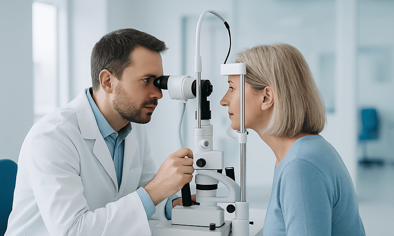
The heart of the examination is looking at the optic nerve. After the pupil is widened with drops, the clinician uses a bright light and magnifying lens to examine the nerve head for cupping, thinning of its rim and tiny hemorrhages. Increasingly this is paired with optical coherence tomography, a painless scan that uses light waves to map the thickness of the nerve fiber layer to within a few thousandths of a millimeter. Comparing scans over time is one of the most sensitive ways to detect progression.
Visual field testing, or perimetry, checks function rather than structure. You stare at a central target while faint lights flash around your peripheral vision and press a button each time you see one. The machine builds a map of any blind spots. Many people find it tiring, and results on a first attempt are often unreliable simply because the task is unfamiliar, which is one reason clinicians repeat it before drawing conclusions.
Gonioscopy uses a special mirrored contact lens to view the drainage angle directly and decide whether it is open or narrow. The Mayo Clinic lists all of these as standard components of a glaucoma assessment.
None of these tests hurts, and none of them can be replaced by a pressure reading alone. Together they answer the real question: is this nerve healthy, and is it staying that way?
How glaucoma treatment works: drops, laser and surgery
Every established glaucoma treatment does the same thing by a different route: it lowers pressure inside the eye, either by producing less aqueous fluid or by helping more of it drain. Which route is chosen, and in what order, is a decision for the prescribing clinician based on the type of glaucoma, how advanced it is, your other health conditions and your preferences.
| Approach | How it lowers pressure | What patients commonly ask about |
|---|---|---|
| Pressure-lowering eye drops | Different classes either reduce fluid production (for example beta blockers, carbonic anhydrase inhibitors) or increase outflow (for example prostaglandin analogs) | Remembering a daily routine; stinging or redness; whether drops affect the rest of the body |
| Laser trabeculoplasty | Low-energy laser applied to the trabecular meshwork improves its drainage | Whether it hurts (usually not); the Mayo Clinic notes the full effect may take a few weeks to appear; it can be repeated in some cases |
| Laser iridotomy | Creates a tiny opening in the iris so fluid can bypass a narrow or closed angle | Mainly used for angle-closure types; often done preventively in the second eye |
| Incisional or implant surgery | Creates a new drainage pathway (trabeculectomy) or places a small tube or stent to channel fluid out | Recovery time, follow-up intensity, risk of pressure going too low or scarring over |
Drops have long been the usual first step, and the NHS describes them as the main treatment. UK national guidance now also lists laser trabeculoplasty as a reasonable first option for many people with newly diagnosed open-angle glaucoma, so the order is no longer fixed. Surgery is generally reserved for eyes where drops and laser have not controlled the pressure, or where disease is advanced at diagnosis.
Two points matter more than any single technique. Treatment protects the vision you still have; it does not restore what is lost. And drops only work when they reach the eye, which is why the quiet daily act of using them as prescribed has a larger effect on outcomes than any difference between products.
Can glaucoma be reversed once the nerve is damaged?
This is the question behind a great deal of internet searching, and it deserves a straight answer. With current medicine, glaucoma cannot be reversed. Nerve fibers in the optic nerve are part of the central nervous system, and like fibers in the brain and spinal cord they do not regenerate once lost. The National Eye Institute, the NHS and the Mayo Clinic all state that vision lost to glaucoma cannot be restored.
That sounds bleak until you turn it around. Because damage is permanent, every fiber preserved by treatment is a permanent gain. Glaucoma is one of the few sight-threatening diseases where the main intervention is proven to slow progression, and most people who are diagnosed and treated in time keep useful vision for life. The goal of treatment is not to get something back; it is to make sure the picture you have today still looks much the same in twenty years.
Research into protecting or regrowing nerve fibers is active. Scientists are studying neuroprotective compounds, gene therapy and stem-cell approaches in laboratories and early trials. None has yet reached routine clinical care, and no supplement, exercise, diet or device currently sold to the public has been shown in controlled trials to repair the optic nerve. Where the evidence is uncertain, the responsible thing is to say so rather than to borrow hope from products that have not earned it.
A related misunderstanding: some people feel their vision improve after starting drops and conclude the nerve is healing. More often the explanation is that a very high pressure was causing mild corneal swelling or discomfort, and relieving it sharpened things slightly. The field defect itself does not shrink.
If you have already lost some sight to glaucoma, low-vision services can help you use what remains more effectively. Your treating team can refer you.
What percentage of glaucoma patients go blind?
People ask this question hoping for a single number, and the honest answer is that no single reliable number exists, because the risk depends enormously on when the disease is found and whether it is treated.
What the evidence does support is this. The World Health Organization and the National Eye Institute describe glaucoma as a leading cause of irreversible blindness worldwide and in the United States. That statistic reflects two things at once: the disease is common, with the National Eye Institute estimating roughly 3 million Americans affected, and a large share of cases go undetected until late. The CDC’s estimate that about half of people with glaucoma are unaware of it is the hinge on which the blindness figures turn.
Among people who are diagnosed while they still have good vision and who keep up with treatment and monitoring, blindness in both eyes is uncommon. Studies following treated patients over decades generally find that most keep useful sight, and that those who do progress to severe loss tend to have been diagnosed late, to have very high or difficult-to-control pressure, or to have had gaps in treatment. The Johns Hopkins and Cleveland Clinic patient resources make the same point in plain language: early detection and consistent treatment are what change the odds.
Beware of any website that quotes a precise percentage without saying which population it describes. A figure drawn from a hospital clinic in a region with little access to eye care tells you nothing about your own risk if you are being monitored and treated.
The more useful question is not what percentage go blind but what separates those who do from those who do not. The answer, consistently, is time to diagnosis and adherence to the plan. Both are within reach.
What is the rule of 5 for glaucoma?
Search engines suggest this phrase often, so it is worth saying clearly: there is no recognized clinical rule of 5 for glaucoma in any major guideline, including those from the National Eye Institute, the NHS or professional ophthalmology bodies. If you have encountered it, it most likely comes from a teaching mnemonic, a clinic’s informal shorthand or a misremembered statistic, and different sources use it to mean different things.
Some people use the number to recall that there are five broad categories of glaucoma: open-angle, angle-closure, normal tension, secondary and congenital. Others attach it to an eye-pressure threshold, suggesting that readings a few points above a target, or a rise of around 5 mmHg between visits, should prompt a closer look. Still others have borrowed it from a rule used in lens and dental fitting that has nothing to do with glaucoma at all.
None of these is a decision rule that your treating team will apply. Glaucoma management is individualized: the pressure target for one person may be far below the normal range because their nerve has shown it cannot cope, while for someone with ocular hypertension and a robust nerve a higher level may be acceptable for years. Progression is judged from repeated nerve imaging and visual field tests rather than from any fixed number of points.
Why mention a rule that does not exist? Because tidy rules are seductive, and they can lead people to reassure themselves inappropriately. Someone who believes a small pressure rise is harmless until it crosses an imagined threshold may skip a follow-up that mattered.
The closest thing to a real rule in glaucoma care is far less catchy: regular examinations, pressure interpreted in context, and treatment adjusted to your own nerve’s response. If a clinician mentions a rule of thumb, ask what evidence it rests on and how it applies to you.
What foods worsen glaucoma, and is there a fastest way to reduce pressure?
Two popular searches, two short answers, and then the nuance that matters.
No food has been shown to cause glaucoma or to make it progress faster. What the research does show is that a few habits nudge eye pressure up or down for a short time. Drinking a large volume of fluid very quickly, roughly a liter in a few minutes, produces a temporary pressure rise, which is why some clinicians suggest spreading fluids through the day rather than drinking a lot at once. Very high caffeine intake raises pressure slightly for an hour or two in some studies, though moderate coffee drinking has not been linked to glaucoma in large population cohorts. Neither effect is large enough to justify a special diet, and the evidence is considered low certainty.
On the positive side, diets rich in leafy green vegetables have been associated with lower glaucoma risk in observational studies, possibly through dietary nitrates and blood flow to the nerve. Association is not proof, and no diet replaces treatment. A heart-healthy pattern that also helps blood pressure and diabetes, two conditions on the Mayo Clinic’s glaucoma risk list, is a sensible, evidence-respecting choice.
As for the fastest way to reduce glaucoma: in the chronic form, speed is the wrong goal. Pressure-lowering eye drops typically begin working within hours, and laser trabeculoplasty builds its effect over weeks, according to the Mayo Clinic; what protects sight is steady control over years, not a quick drop today. The one situation where fast genuinely matters is acute angle-closure glaucoma, where pressure spikes suddenly. That is an emergency treated in hospital, not something to manage at home.
If you want to influence your own pressure, the highest-yield actions are the unglamorous ones: use prescribed drops exactly as directed, tell your clinician about every medicine you take including steroid creams and inhalers, and keep every follow-up appointment.
What the first weeks after a glaucoma diagnosis usually look like
A new diagnosis arrives with paperwork and a sense that life has changed. In practice the weeks that follow are quieter than most people expect, and knowing the shape of them removes a good deal of the dread.
If drops are prescribed, the first days are about building a habit. Many people tape the bottle to the bathroom mirror or set a phone alarm, because the most common reason drops fail is simply that they are forgotten. Your clinician or pharmacist can show you the technique: tilt the head back, pull the lower lid down to form a pocket, let one drop fall in, then close the eye gently and press lightly on the inner corner for a minute so less medicine drains into the nose and bloodstream. Mild stinging or redness in the first week is common and usually settles; anything worse is worth reporting rather than enduring.
If laser trabeculoplasty is chosen, the procedure itself is done in an outpatient setting with numbing drops and usually takes minutes. Vision may be blurry and the eye slightly achy for a day. The Mayo Clinic notes the full pressure-lowering effect may take a few weeks to show, so the first follow-up is partly to check for a short-term pressure rise and partly to begin judging the response.
Across all starting treatments, expect a return visit to measure pressure and confirm the plan is working. The Mayo Clinic describes glaucoma care as requiring regular follow-up for life, with visits spaced according to how stable the disease is; the NHS similarly describes ongoing clinic monitoring. Early on, visits tend to be closer together, then stretch out as things settle.
Between visits, carry on as normal. Reading, screens, exercise and flying do not harm the nerve. The only new rules are the drops and the calendar.
What people often get wrong: more glaucoma myths, corrected
Beyond the big three that delay diagnosis, a cluster of smaller glaucoma myths shapes how people live with the disease once they have it. Each has a tidy correction.
Reading, screens and close work damage the eyes. They do not. Eye strain is real, but it does not raise pressure in any lasting way or harm the optic nerve. Glaucoma gives you no reason to put down a book.
If my pressure is now normal on drops, I can stop. The drops are the reason it is normal. Open-angle glaucoma is a chronic condition; pressure typically returns to its untreated level within days to weeks of stopping. Any change to a prescribed medicine, including a pause because of side effects, should be discussed with the prescribing clinician first.
Glaucoma runs in families, so if no one in my family has it, I am safe. Family history raises risk substantially, but most people diagnosed have no known relative with the disease. The reverse is also true: a relative’s diagnosis should prompt earlier and more frequent checks, not panic.
Glaucoma surgery restores vision. Surgery, like drops and laser, lowers pressure to protect remaining sight. It does not bring back lost fibers.
Marijuana treats glaucoma. Cannabis does lower eye pressure for roughly a few hours, which is where the idea comes from. The effect is too brief and too variable to protect the nerve around the clock, and no professional ophthalmology body recommends it as treatment.
Only people with high pressure need worry. We have spent an article on this one. Normal tension glaucoma exists, and so does ocular hypertension without damage.
Exercise is dangerous for glaucoma. Regular moderate aerobic activity is associated with slightly lower eye pressure and is generally encouraged. The specific exception is prolonged head-down positions, such as certain inverted yoga poses, which raise pressure while held; people with glaucoma are often advised to ask their clinician about these.
Questions to ask your care team
A glaucoma appointment moves quickly, and the questions that matter most tend to surface in the car park afterward. Writing a few down in advance changes the conversation. These are the ones that experienced patients say they wish they had asked at the start.
- Do I have glaucoma, or am I a glaucoma suspect, or do I have ocular hypertension? The three mean different things for treatment and follow-up.
- What type of glaucoma is it, and is the drainage angle open or narrow? Angle-closure types carry different risks and sometimes a preventive laser for the other eye.
- What is my current pressure, what target are you aiming for, and why that number for me?
- How much damage is already present, and in which eye? Ask to see the nerve scan and the visual field printout; most people understand their disease better once they have looked at the map.
- How will we know whether treatment is working, and how often will the tests be repeated?
- What side effects should I watch for with this treatment, and which ones mean I should call rather than wait for the next visit?
- Are there other medicines I take, including steroid creams, inhalers or tablets, that could be affecting my pressure?
- Should my parents, siblings or children be examined, and from what age?
- Is laser a reasonable alternative or addition to drops in my case?
- If my vision is already affected, is a referral to low-vision services or an occupational therapist appropriate?
- Is it safe for me to drive, and does the visual field result need to be reported to the licensing authority where I live?
You are entitled to ask any of these more than once. Glaucoma is a decades-long relationship with your eye clinic, and the plan will be revisited as the picture changes. The decisions sit with your treating team, but they are made better by a patient who understands the reasoning.
When to call your doctor
Most glaucoma care happens on a calendar, not in a crisis. A few situations, however, need prompt attention, and knowing them in advance means you will not hesitate when it counts.
Seek emergency care the same day, or call emergency services, if you develop sudden severe eye pain or deep ache around one eye together with blurred vision, a red eye, colored halos around lights, headache, nausea or vomiting. The Mayo Clinic lists this combination as the picture of acute angle-closure glaucoma, in which pressure rises rapidly and hours matter. Do not wait to see whether it passes.
Contact your eye clinic or doctor promptly, within a day or two, if you notice a sudden new patch missing from your vision, a curtain or shadow over part of your sight, a sudden shower of floaters or flashes of light, or sudden loss of vision in one eye. These may signal problems other than glaucoma, such as a retinal detachment, but they all need examination quickly.
Call your clinic rather than waiting for the next appointment if prescribed eye drops cause severe stinging, swelling of the eyelids, breathing difficulty, a slowed or irregular heartbeat, dizziness or fainting; some drop classes can affect the heart and lungs, and your prescriber may wish to adjust the plan. Call too if you have missed doses for more than a few days for any reason, if you are about to start a steroid medicine for another condition, or if you have had an eye injury.
After laser or surgery, your team will give specific instructions. In general, worsening pain, increasing redness, discharge or a drop in vision in the treated eye in the days afterward should be reported straight away.
Finally, if you have not had your optic nerve examined and you are over 40, have a family history of glaucoma, or belong to a higher-risk group, the right time to book is now rather than when something feels wrong. Glaucoma rarely gives notice.
Frequently asked questions
Can you have glaucoma without high eye pressure?
Yes. Normal tension glaucoma is a recognized type of open-angle glaucoma in which the optic nerve shows typical damage even though measured pressure stays in the usual range. Researchers think a more fragile nerve or marginal blood flow may explain why. Treatment still involves lowering pressure, because that remains the only approach proven to slow progression. A normal pressure reading alone therefore cannot rule glaucoma out; the nerve has to be examined.
What percentage of glaucoma patients go blind?
There is no single reliable figure, because the risk depends heavily on when glaucoma is found and whether it is treated. Glaucoma is a leading cause of irreversible blindness worldwide, largely because around half of cases go undiagnosed until late. Among people diagnosed with good vision who keep up treatment and monitoring, blindness in both eyes is uncommon. Early detection and consistent follow-up are what change the odds.
What is the rule of 5 for glaucoma?
No major guideline recognizes a rule of 5 for glaucoma. The phrase appears to come from informal teaching mnemonics or misremembered statistics, and different sources use it to mean different things, from five types of glaucoma to a pressure change of around 5 mmHg. Clinicians do not manage glaucoma by a fixed number; pressure targets and signs of progression are judged individually from repeated nerve imaging and visual field tests.
What foods worsen glaucoma?
No food has been shown to cause glaucoma or make it progress faster. A few habits raise eye pressure briefly: drinking a large volume of fluid very quickly, and very high caffeine intake in some studies. Neither effect is large or lasting enough to require a special diet. Diets rich in leafy green vegetables are associated with lower risk in observational research, though this does not prove cause and does not replace prescribed treatment.
What is the fastest way to reduce glaucoma?
For chronic open-angle glaucoma, speed is not the goal; steady pressure control over years is what protects sight. Prescribed eye drops generally begin lowering pressure within hours, and laser trabeculoplasty builds its effect over a few weeks. The one situation where speed truly matters is acute angle-closure glaucoma, a sudden painful pressure spike that is a medical emergency treated in hospital. Any change to treatment should be made with your prescribing clinician.
Can glaucoma be reversed?
No. Optic nerve fibers lost to glaucoma do not regenerate, so vision that has gone cannot be restored with current medicine. The purpose of treatment is to lower eye pressure and preserve the vision you still have, and most people diagnosed in time keep useful sight for life. Research into protecting or regrowing nerve fibers is active, but no supplement, diet or device currently available has been shown to repair the nerve.
What are the early signs of glaucoma?
For the common chronic form, there usually are no early signs you would notice. Glaucoma removes peripheral vision first and the brain fills in the gaps, so people often have measurable field loss while feeling their sight is normal. Noticeable changes such as missing steps or not seeing cars in side mirrors are late signs. The acute angle-closure form is different and causes sudden pain, redness and blurring. Routine examination is the only reliable way to detect early disease.
If my eye pressure is normal, do I still need a glaucoma check?
Yes, if you are due one. A pressure test on its own is not a glaucoma check, because normal tension glaucoma damages the nerve at normal pressures. A proper examination includes looking at the optic nerve, often with a scan of the nerve fiber layer, and testing the visual field. The NHS recommends a routine eye test at least every 2 years for adults, with more frequent checks for people at higher risk.
Does normal tension glaucoma need treatment?
Usually, yes. Although the pressure is already in the normal range, lowering it further has been shown to slow the loss of nerve fibers in people with normal tension glaucoma. The clinician is not correcting an abnormal number; they are reducing stress on a nerve that has already shown it cannot tolerate its current pressure. Treatment choices and targets are individual decisions made with your treating team.
Is glaucoma only an older person's disease?
No. Glaucoma is most common in later life and the NHS notes it is most frequent among people in their 70s and 80s, but it occurs at every age, including in infants with congenital glaucoma. Risk begins rising noticeably from the 40s, and earlier for people with a family history, with African, Hispanic or Asian heritage, with diabetes, or who use corticosteroid medicines long term. Age is one risk factor among several.
References
This article is for general information only and is not a substitute for professional medical advice. Please consult a qualified doctor about your individual situation.

More from the Blog

Vitrectomy Recovery With a Gas Bubble: Face-Down Positioning, Drops and Weekly Milestones
After a vitrectomy with a gas bubble, the surgeon usually asks you to hold a specific head position, often face down, for several days…
How OCT and Fundus Examination Map Macular Disease Before Any Treatment Is Planned
Optical coherence tomography (OCT) and a dilated fundus examination together map macular disease by showing the retina in two complementary ways: the fundus exam…

Low Pressure, Leaks and Infection After Glaucoma Surgery: What Your Team Watches For
After glaucoma surgery, care teams watch most closely for eye pressure that drops too low (hypotony), a leak from the small drainage blister called…

Farsightedness Myths: Does Reading in Dim Light or Screen Time Make It Worse?
No. Reading in dim light and spending long hours on screens can tire the eyes and cause temporary blur or headache, but neither changes…
Keratoconus Treatment Ladder: Lenses, Cross-Linking, Rings and When Transplant Is Discussed
Keratoconus treatment options form a ladder rather than a single choice. Glasses and soft lenses help early; rigid gas-permeable, hybrid or scleral lenses restore…
Can a Contact Lens Get Lost Behind Your Eye? Contact Lens Myths, Corrected
No. A contact lens cannot travel behind your eye. The conjunctiva, the thin membrane lining the inside of the eyelids, folds back onto the…