Glaucoma Treatment
Glaucoma treatment aims to lower eye pressure and protect the optic nerve to slow or prevent vision loss. Care may include eye drops, laser therapy, or surgery depending on disease severity.
Quick answer
Glaucoma is a group of eye diseases in which the optic nerve is progressively damaged, most often by raised pressure inside the eye. Treatment lowers that pressure using prescription eye drops, laser procedures or surgery. It cannot restore vision already lost, but it can slow or halt further damage, which is why early diagnosis and lifelong monitoring matter.
Glaucoma: Protecting the Vision You Still Have
Glaucoma is a group of eye diseases in which the optic nerve, the cable that carries visual information from your eye to your brain, is progressively damaged, most often by pressure inside the eye that the nerve cannot tolerate. Glaucoma treatment lowers that pressure with prescription eye drops, laser procedures or surgery, and it is intended for anyone whose optic nerve already shows damage or sits at clear risk of it. Treatment cannot restore vision that has been lost. What it can do is slow or halt further loss, which is why glaucoma care is built around one principle: protect the vision you still have.
If you have recently been told that your eye pressure is high, that your optic nerve looks damaged, or that a visual field test shows changes you cannot yet perceive in daily life, the diagnosis can feel abstract. You may read comfortably, drive without difficulty and feel entirely well. At the same time you are probably weighing practical questions: whether drops will be enough, whether laser treatment hurts, whether surgery is unavoidable, and what happens if you do nothing. This page walks through what glaucoma is, how it is diagnosed, how each form of treatment works, what recovery looks like, and, just as importantly, what treatment honestly can and cannot achieve.
What is glaucoma?
Glaucoma is progressive damage to the optic nerve, usually linked to raised pressure inside the eye, that leads to gradual and permanent loss of vision. That is the working glaucoma definition ophthalmologists use, and each part of it matters. Progressive means the disease worsens over time if left untreated. Usually linked to pressure means that pressure is the main treatable factor, but not the whole story, some people develop nerve damage at pressure levels other eyes tolerate without difficulty. Permanent means that optic nerve fibres which have died do not grow back, so every treatment aims at preventing further damage rather than repairing what is gone.
Glaucoma is often called a silent disease because peripheral vision is usually lost first, slowly, and the brain fills in the gaps. Many people lose a substantial amount of side vision before anything feels wrong. That is also why glaucoma is described as a group of diseases rather than a single one: the mechanisms differ, the speed differs and the right treatment differs, but the endpoint, optic nerve damage, is shared.
What Is Glaucoma Treatment?
Glaucoma treatment is medical, laser or surgical care designed to reduce intraocular pressure, commonly called eye pressure, and slow damage to the optic nerve. Because some patients develop glaucoma even when their eye pressure sits within the statistically normal range, treatment planning always rests on two pillars: measuring pressure and directly assessing the health of the optic nerve. Lowering a number on a pressure test is not the goal in itself. The goal is to preserve functional vision, for reading, driving, working, recognising faces, moving safely and staying independent.
Eye pressure depends on the balance between production and drainage of a clear fluid inside the eye called aqueous humour. This fluid is not the same as tears. It is produced continuously inside the eye and normally drains away through small channels near the base of the iris. If drainage slows or becomes blocked, pressure rises. Over months and years, raised or poorly tolerated pressure damages optic nerve fibres, leading first to loss of peripheral vision and, in advanced disease, to loss of central vision as well.
Treatment may involve prescription eye drops that reduce fluid production or improve drainage, laser procedures that help fluid exit the eye more effectively, or surgery that creates a new drainage pathway or places a tiny drainage device. Many patients end up with a combination of these approaches over the years. Which option is right depends on the form of glaucoma, the severity of nerve damage, the target pressure needed to protect that particular nerve, and how the eye has responded to previous therapy.
One point deserves emphasis: glaucoma treatment is long term. Even after pressure has been successfully lowered, you will need monitoring, because glaucoma can progress silently despite treatment that once worked well. A properly structured plan includes regular pressure measurement, optic nerve imaging, visual field testing and adjustment of therapy whenever the pattern of results demands it.
What causes glaucoma?
The major cause of glaucoma is intraocular pressure that is too high for the optic nerve to tolerate, and in most cases that pressure rises because the eye’s natural drainage channels stop letting fluid out efficiently. The drainage tissue may function poorly despite looking open, as in the most common adult form, or the drainage angle itself may be narrow or physically blocked. Either way, fluid keeps being produced while its exit is restricted, pressure climbs, and the delicate nerve fibres at the back of the eye begin to fail.
Not every case fits that pattern. In normal-tension glaucoma the nerve deteriorates at pressures most eyes tolerate, which suggests that blood supply to the nerve and individual nerve vulnerability also play a role. Glaucoma can also develop secondary to something else: eye injury, inflammation inside the eye, long-term steroid use, an advanced cataract, diabetes-related eye disease, pigment dispersion or pseudoexfoliation syndrome. Identifying the cause matters, because a secondary glaucoma is treated by addressing both the pressure and, where possible, whatever is driving it.
Glaucoma Symptoms and Early Warning Signs
Glaucoma symptoms are, in the most common form of the disease, largely absent until a significant amount of damage has already occurred. This is the uncomfortable truth about open-angle glaucoma: it does not hurt, it does not blur reading vision for a long time, and it does not announce itself. Peripheral vision narrows so gradually that most people compensate without noticing. Many diagnoses are made during a routine eye examination in a person who feels completely well.
What are the early warning signs of glaucoma?
For most people with open-angle glaucoma there are no reliable early warning signs, which is exactly why regular eye examinations matter more than watching for symptoms. When changes do become noticeable, they tend to include:
- a gradual narrowing of side vision, bumping into door frames, missing objects or people approaching from the side
- difficulty adapting to dim light or judging steps and kerbs
- patchy blind spots in the visual field, usually noticed only when one eye is covered
- in later stages, tunnel-like vision with the centre still relatively clear
Angle-closure glaucoma behaves differently and can produce symptoms. Some people have intermittent episodes of eye discomfort, temporary blurring, coloured halos around lights, or headache in dim lighting, settings in which the pupil widens and the narrow drainage angle closes further. A sudden angle-closure attack causes severe eye pain, redness, nausea, vomiting and rapid loss of vision. Ophthalmologists treat an acute attack as an emergency, because very high pressure can injure the optic nerve within hours rather than years.
Who May Need Glaucoma Treatment?
You may need glaucoma treatment when testing shows elevated eye pressure, visible optic nerve damage, visual field loss, or an eye structure, such as a dangerously narrow drainage angle, that puts you at risk of pressure spikes. Some people begin treatment before any damage exists, purely to prevent it. Others start after a routine examination reveals changes they had never felt. A third group arrives with symptoms: blurred vision, halos around lights, eye pain, headache or sudden visual disturbance, particularly in the acute forms of the disease.
Screening deserves particular attention because of how silent the common form is. Regular examination is especially important for people over 40, those with a family history of glaucoma, people with diabetes or high myopia, patients who use steroid medications, and anyone with a previous eye injury or eye surgery. If you belong to more than one of these groups, the case for periodic checks is stronger still, even when your vision feels entirely normal.
Diagnosis begins with a comprehensive ophthalmological examination. The ophthalmologist measures intraocular pressure, examines the optic nerve directly, evaluates the drainage angle, checks corneal thickness and assesses the retina and visual function. Common tests include optical coherence tomography to image the optic nerve and retinal nerve fibre layer, computerised visual field testing, gonioscopy to view the drainage angle, pachymetry to measure corneal thickness, which affects how pressure readings should be interpreted, and photography to document structural change over time. None of these tests involves entering the eye; they are examinations, not procedures.
Patients who already carry a diagnosis often seek further care for specific reasons: pressure that stays above target despite medication, drops that cause intolerable side effects, genuine difficulty using drops consistently, or test results showing progression. Others want a second opinion before committing to laser therapy or surgery, particularly when both eyes are involved or previous treatment has not achieved stable control. All of these are legitimate reasons for a fresh, thorough evaluation.
Is glaucoma hereditary?
Glaucoma frequently runs in families, and a family history is one of the strongest known risk factors for developing the disease. Having a parent or sibling with glaucoma does not mean you will develop it, but it does mean your baseline risk is meaningfully higher than that of someone with no affected relatives, and that examinations should generally start earlier and happen more regularly. Several genes have been linked to different forms of glaucoma, and congenital glaucoma in particular can have a strong inherited component. If you have been diagnosed, telling your close relatives is one of the most useful things you can do for their eye health.
Types of Glaucoma Addressed by Treatment
Glaucoma treatment is not one procedure for one disease. It covers a group of conditions that damage the optic nerve through pressure-related mechanisms or impaired fluid drainage, and identifying the exact type is essential because the treatment choices differ substantially.
Primary open-angle glaucoma
Primary open-angle glaucoma is the most common form in many adult populations and is usually chronic and slowly progressive. The drainage angle appears open on examination, but fluid does not pass through the drainage tissue efficiently enough, and pressure creeps up. Treatment typically begins with eye drops or laser therapy, with surgery considered when pressure control is insufficient or the disease is already advanced at diagnosis.
Normal-tension glaucoma
Normal-tension glaucoma describes optic nerve damage that progresses even though pressure readings are not particularly high. These patients still benefit from pressure lowering, reducing pressure below the eye’s individual tolerance remains the treatment with the best evidence, but the ophthalmologist will usually also look at circulation, blood pressure patterns, sleep apnoea risk, migraine history and other factors that may affect the nerve’s blood supply.
Angle-closure glaucoma
Angle-closure glaucoma involves a drainage angle that is narrow or physically blocked. Laser treatment to create a small opening in the iris is often recommended, either preventively in an eye with a dangerously narrow angle or urgently in certain angle-closure situations. In some eyes, cataract surgery or other lens-based procedures form part of the plan, because the position and thickness of the natural lens can contribute to the narrowing.
Secondary glaucoma
Secondary glaucoma develops as a consequence of another eye or medical condition. Causes include eye trauma, inflammation inside the eye, steroid use, advanced cataract, diabetes-related eye disease, pigment dispersion, pseudoexfoliation syndrome, tumours and previous eye surgery. Treatment must address both the pressure and, wherever possible, the underlying cause, controlling inflammation, for instance, or reviewing the medical need for a pressure-raising medicine together with the doctor who prescribed it.
Congenital and childhood glaucoma
Congenital or childhood glaucoma is less common but needs specialised evaluation. Infants and children may present with enlarged eyes, cloudy corneas, light sensitivity, watering or concerns about vision. Surgical treatment is often required, and early diagnosis carries extra weight in children because the visual system is still developing, untreated pressure affects not just the eye but how the brain learns to see.
Advanced glaucoma
Advanced glaucoma requires particularly careful planning because the remaining optic nerve reserve is limited. The target pressure is usually set lower, treatment often needs to be more intensive, and the physician has to balance the case for stronger pressure reduction against the safety of a vulnerable eye and the patient’s overall condition. Advanced disease is still very much treatable; the margin for error is simply smaller.
How Glaucoma Treatment Is Performed
Glaucoma care follows a logical sequence rather than jumping straight to a procedure. A typical pathway looks like this:
- Confirming the diagnosis and identifying the exact type of glaucoma.
- Setting a target pressure for each eye individually: an eye with early damage may need moderate reduction, while an eye with advanced damage usually needs a lower target.
- Choosing first-line therapy: drops, laser or, in selected cases, earlier surgery.
- Verifying that the pressure response actually reaches the target, not just that treatment has started.
- Monitoring the optic nerve and visual field over time and adjusting the plan whenever the results show change.
Preparation and evaluation
Before any treatment, the ophthalmologist reviews your medical history, current medications, allergies, previous eye surgery and family history of glaucoma. If you already use glaucoma drops, the team will go through the exact names, the dosing schedule and any side effects, redness, burning, dry eye, breathlessness, fatigue or slow pulse. This review matters more than it might seem: some pressure-lowering drops are unsuitable for patients with asthma, heart rhythm problems, low blood pressure or certain neurological conditions, and the safest choice depends on knowing your whole medical picture.
Diagnostic imaging then establishes a baseline. Optic nerve and retinal nerve fibre imaging shows the structure of the nerve; visual field testing measures how well you actually see across your field of vision; gonioscopy lets the doctor inspect the drainage angle directly. These tests are generally non-invasive, and their real value is comparative, glaucoma management depends on detecting change over time, and change can only be detected against a properly documented starting point.
Medication-based treatment
Most patients begin with prescription eye drops. These medicines work by reducing the amount of fluid the eye produces, increasing the amount that drains away, or both. Some are used once daily, others more often, and combination drops can reduce the number of bottles you need to manage. Oral medication is sometimes used for short periods when pressure must come down quickly, but long-term tablets are less common because of their systemic side effects.
How you use the drops genuinely affects how well they work. You will usually be shown how to instil a drop without touching the bottle tip to the eye, how long to wait between different medications so one does not wash out the other, and how to close the eye gently or press near the inner corner afterwards to limit absorption into the bloodstream. If drops are hard to use, difficult to obtain where you live, or causing side effects you cannot live with, say so plainly, laser or surgical options may reasonably be considered earlier, and an honest conversation is better than quietly missed doses.
Laser treatment
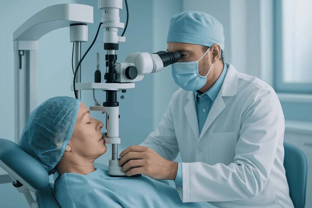
Laser therapy has a place in several glaucoma situations. In open-angle glaucoma, laser treatment applied to the natural drainage tissue can improve outflow. It is performed as an outpatient procedure with numbing drops: you sit at a laser microscope similar to the equipment used during a routine examination, the treatment itself takes a short time, and most patients return to normal light activities soon afterwards. Some people feel brief discomfort or see flashes of light during treatment.
In narrow-angle or angle-closure glaucoma, a laser is used differently, to create a tiny opening in the iris so that fluid can move more freely and the angle is less likely to close. This may be recommended preventively in an eye with a dangerously narrow angle or urgently in certain angle-closure situations.
It helps to be clear about what laser treatment is not. It is not operating-theatre surgery, it involves no large incision, and nothing is removed from the eye. Anti-inflammatory drops may be prescribed afterwards, pressure is usually checked after the procedure, and your ophthalmologist will tell you exactly what should happen with your existing glaucoma drops, decisions about continuing or adjusting them belong to the treating doctor, based on the measured response.
Surgical treatment
Surgery is considered when medication and laser therapy do not lower pressure enough, when the disease is progressing despite treatment, when pressure is very high, or when a patient cannot safely or reliably continue medication. The purpose of glaucoma surgery is to create a controlled route for fluid to leave the eye, taking pressure off the optic nerve.
Several surgical approaches exist. Traditional filtering surgery creates a new drainage pathway under the surface of the eye. Tube or shunt procedures place a small drainage implant that channels fluid out of the eye in a controlled way. Minimally invasive glaucoma procedures suit selected patients, often those with mild to moderate disease, or those undergoing cataract surgery at the same time. The choice depends on the type and severity of glaucoma, the anatomy of the eye, previous operations, cataract status and how much pressure reduction is actually needed.
Glaucoma surgery is usually performed under local anaesthesia with sedation, although the anaesthetic plan varies with the patient and the procedure. Some operations take less than an hour; more complex eyes take longer. Most patients go home the same day, but close follow-up in the early healing period is not optional, postoperative visits are when the surgeon checks eye pressure, inflammation, the healing of the drainage area and any early sign of complications, and this is where much of the final result is actually secured.
Technology used in modern glaucoma care
Modern glaucoma management relies on precise measurement and long-term comparison. Diagnostic technology includes high-resolution optic nerve and retinal nerve fibre imaging, computerised visual field analysis, corneal thickness measurement, drainage angle assessment and retinal photography. Together these tools let the ophthalmologist distinguish stable disease from progression, the single most important judgement in glaucoma care, and choose treatment intensity accordingly.
During procedures, operating microscopes, microsurgical instruments, laser platforms and fine drainage devices are used depending on the treatment. The practical value for you is accuracy: better visualisation of delicate structures, more controlled delivery of treatment and more individualised planning. Technology supports the physician’s judgement; it does not replace it, and it certainly does not replace careful follow-up.
Recovery after treatment
Recovery depends on which treatment you have. After a medication change, you may notice mild irritation, redness, or, with certain drops, gradual changes in eyelash growth or iris colour; follow-up confirms whether the pressure has reached the intended level. After laser therapy, mild irritation, light sensitivity or blurred vision can occur briefly, and most people resume routine activities quickly, sometimes with advice to avoid strenuous effort for a short while.
After surgery, recovery is more structured. You will usually use antibiotic and anti-inflammatory drops, avoid rubbing the eye, and stay away from heavy lifting, swimming and dusty environments until the surgeon clears you. Vision can fluctuate during healing, this is expected, not a sign of failure, and eye pressure may need careful adjustment through the early weeks.
Can Glaucoma Be Cured?
No, with current medicine, glaucoma is not curable, because optic nerve fibres that have died cannot be regenerated. Anyone asking whether glaucoma is curable deserves that answer stated plainly, because the honest alternative is better than it sounds: glaucoma can usually be controlled, often for decades, and controlled glaucoma means preserved vision. Drops, laser and surgery all work by lowering pressure so the remaining nerve fibres are protected; none of them rebuilds what has been lost.
You may come across personal accounts titled along the lines of “how I cured my glaucoma”. Read them carefully. What they almost always describe is well-controlled pressure and stable test results: a genuinely good outcome, but control, not cure. The distinction matters practically: a person who believes they are cured may stop monitoring, and glaucoma that progresses unwatched progresses silently. Stable disease under treatment still needs regular checks.
Can you stop glaucoma from progressing?
In many eyes, yes, lowering eye pressure to an individually set target and keeping it there can slow progression substantially, and in a good number of patients the disease remains stable for the long term. Whether that is achievable in your case depends on how much nerve damage existed at diagnosis, how low the pressure needs to go, how consistently treatment is used, and how the eye responds. Stopping progression is the entire purpose of glaucoma treatment, and it is a realistic goal for most patients who are diagnosed in time and followed properly. It is a goal pursued through measurement and adjustment, not a one-off fix.
Why Acting Early Matters
Glaucoma-related vision loss is usually permanent. Once optic nerve fibres are damaged, current treatments cannot regenerate them, which is why early diagnosis and consistent treatment sit at the centre of preserving vision. Waiting for symptoms is a poor strategy, because many forms of glaucoma progress silently for years before noticeable loss develops.
Delay allows peripheral vision loss to advance unremarked. You may not register what is disappearing, because the brain adapts and the unaffected areas of vision compensate. Over time, tasks such as driving, using stairs, navigating crowded places and noticing objects to the side become harder. In advanced glaucoma, central vision itself comes under threat.
In acute angle-closure glaucoma, delay is dangerous on a different timescale. Very high pressure can injure the optic nerve and other structures of the eye rapidly, which is why ophthalmologists handle a full-blown attack as an emergency rather than a routine appointment.
Acting early does not automatically mean having surgery early. It means completing a proper evaluation, understanding your individual risk, starting therapy appropriate to your disease and confirming through testing that it is actually stable. For many patients, timely drops or laser therapy delays or reduces the need for anything more invasive. For others, an already vulnerable nerve, dangerously high pressure, earlier surgery is the safer choice. The point is that the decision is made deliberately, with full information, rather than forced later by damage that could have been prevented.
Benefits of Glaucoma Treatment
The benefits of treatment all trace back to the same aim: protecting the optic nerve, preserving daily function and reducing the risk of future vision loss.
| Benefit | What It Means for You |
|---|---|
| Lower eye pressure | Reducing intraocular pressure decreases stress on the optic nerve and is the most proven way to slow glaucoma progression. |
| Protection of remaining vision | Treatment aims to preserve the vision you still have, including the peripheral vision that matters for mobility and safety. |
| Individualised treatment options | Care may involve drops, laser therapy, surgery or a combination, matched to your diagnosis, severity and response to previous treatment. |
| Reduced medication burden in selected patients | Laser or surgical treatment may help some patients rely less on multiple daily eye drops, although ongoing monitoring remains necessary. |
| Better long-term disease monitoring | Regular imaging and visual field testing detect progression earlier and guide timely changes in treatment. |
Recovery Timeline After Glaucoma Treatment
Recovery varies with the type of treatment, drops, laser or surgery, but the following timeline gives a realistic sense of what most patients experience.
| Time Period | What Patients Can Expect |
|---|---|
| Day 1 | After laser treatment, mild irritation or blurred vision is common. After surgery, the eye is usually protected, drops are started and activity restrictions are explained. |
| First Week | Follow-up visits check eye pressure and healing. Heavy lifting, swimming, eye rubbing and dusty environments are usually off-limits, especially after surgery. |
| First Month | Vision may fluctuate after surgery as the eye heals and inflammation settles. Medication schedules are adjusted based on pressure readings and clinical findings. |
| Longer Term | Ongoing monitoring with pressure checks, optic nerve imaging and visual field testing remains essential, because glaucoma is a chronic condition. |
Factors That Influence Outcomes
A good glaucoma result rests on more than one pressure reading. The most influential factors are the type of glaucoma, how much optic nerve damage existed at diagnosis, the baseline pressure, the target pressure required, your age, corneal thickness, drainage angle anatomy and whether other eye diseases are present. Severity at the start deserves particular weight: patients diagnosed early carry more optic nerve reserve, which makes long-term preservation of vision more achievable. Advanced glaucoma can absolutely still be treated, but the margin for further damage is smaller and follow-up has to be correspondingly tighter.
Adherence strongly affects outcomes. Drops only work when used consistently and correctly. If you forget doses, cannot tolerate side effects or have trouble obtaining a medication where you live, the plan may need to change, and it is far better that it changes deliberately than that it fails quietly. Whenever your care moves between doctors, it is worth asking for a written treatment plan and copies of your test results, so the next ophthalmologist can continue from a documented baseline rather than starting over.
The eye’s healing response matters after surgery. Some eyes scar more aggressively, which can reduce the effectiveness of a drainage procedure over time. Previous eye surgery, inflammation, trauma, diabetes-related changes and certain secondary glaucomas all make surgical planning more complex, and the surgeon adjusts technique, postoperative medication and visit frequency around these risks.
General health influences glaucoma care more than most patients expect. Low blood pressure at night, vascular disease, sleep apnoea, migraine, diabetes and steroid use can each affect optic nerve vulnerability or eye pressure, and a careful medical history helps the ophthalmologist choose safer medications and identify where coordination with other physicians is needed. Blood pressure sits on both sides of this equation: the optic nerve depends on adequate blood supply, and cardiovascular health affects the safety of any procedure. If you are treated for hypertension, make sure the ophthalmology team knows, because both the choice of pressure-lowering eye drops and any anaesthetic planning take it into account.
Finally, outcomes depend on monitoring over time. Glaucoma is judged through patterns: pressure trends, optic nerve appearance, imaging changes, visual field progression. A treatment that works well at first may need modification years later. That is not a failure of care: it is what responsible management of a chronic optic nerve disease looks like.
What should you avoid if you have glaucoma?
The single most important thing to avoid is inconsistent treatment, missed drops and skipped follow-up visits do more measurable harm than any lifestyle factor. Beyond that, a few practical points are worth knowing. Steroid medicines in any form can raise eye pressure in susceptible people, so every doctor who treats you should know you have glaucoma before prescribing; decisions about any medicine you already take belong to the prescribing doctor, never to guesswork. After surgery, avoid rubbing the eye and follow the surgeon’s activity restrictions precisely. Some activities, prolonged head-down positions in certain exercises, for example, can temporarily raise eye pressure, and whether they matter in your case is a question your ophthalmologist can answer against your actual pressure readings and disease stage. Evidence on caffeine, screen use and similar everyday habits is far weaker than internet lists suggest, so it is reasonable to ask before restricting your life unnecessarily.
How Acibadem Approaches Glaucoma Care
Glaucoma care works best when diagnosis, treatment and follow-up are planned together rather than handled as separate events. Acibadem plans glaucoma care around the individual patient: the type of glaucoma, the level of optic nerve damage, pressure trends, previous treatments, general health, lifestyle and the realistic possibilities for follow-up over the years ahead.
Ophthalmologists evaluate glaucoma using modern diagnostic tools covering eye pressure, optic nerve structure, visual field function and drainage angle anatomy. When cases are complex, care may involve discussion among the relevant specialists, glaucoma surgeons, retinal specialists, cataract surgeons, cornea specialists, anaesthesiology teams and internal medicine physicians when systemic conditions affect the treatment choice. This matters in glaucoma because prior test results, medication lists and follow-up needs must be understood precisely before any treatment decision is made.
Treatment planning is individual. Some patients are best managed with optimised drops and follow-up testing. Others can appropriately have laser therapy. Patients with advanced or uncontrolled glaucoma may need surgery and closer monitoring in the early postoperative period, and the expected timeline is explained before treatment, so the recovery period can be planned realistically rather than improvised. Physicians follow evidence-based international treatment principles while adapting them to the individual eye: defining a target pressure, choosing therapy on severity and anatomy, monitoring for progression and discussing benefits and risks in plain terms.
Continuity is treated as part of the treatment itself. Imaging and visual field results document the eye before and after treatment, and patients leave with practical instructions, how to use drops correctly, which activities to limit after a procedure and how future monitoring can continue against the documented baseline. For glaucoma, rigorous care means diagnosis, treatment, recovery and follow-up planning connected as one process rather than a series of isolated appointments.
Living With Glaucoma Over the Long Term
Glaucoma care is most effective when it is proactive, measured and individualised, and that remains true long after any single treatment ends. Living well with glaucoma comes down to a rhythm: using treatment as prescribed, attending scheduled checks even when everything feels fine, and understanding that stability is something the tests confirm rather than something you can feel. Most people with well-managed glaucoma continue reading, working, travelling and driving for as long as their visual fields permit; the disease changes the maintenance schedule of your eyes more than it changes daily life.
Your own records are a genuinely useful tool. Eye pressure measurements over time, visual field printouts, optic nerve imaging, a current medication list and the details of any previous laser treatment or surgery together tell the story of your disease far better than any single appointment can. Keeping copies, and bringing them to every new ophthalmologist you see, protects you against decisions made on incomplete information, whether you are changing doctors, moving countries or seeking a second opinion about a recommended operation.
Finally, keep the honest frame in view. Vision already lost to glaucoma does not come back, and no treatment changes that. But progression can very often be slowed or halted, functional vision can be preserved for decades, and every element of modern glaucoma care, drops, laser, surgery, imaging, monitoring, exists to serve that one measurable purpose. The earlier the disease is found and the more consistently it is managed, the more of your vision stays yours.
Preparation
- Before treatment, an ophthalmologist performs a comprehensive eye examination, including eye pressure measurement, optic nerve evaluation, and visual field testing. Patients should share all medications, allergies, and previous eye surgeries. Contact lenses may need to be removed before laser or surgical procedures.
Aftercare
- After treatment, prescribed eye drops should be used exactly as instructed to control pressure and reduce inflammation. Patients should avoid rubbing the eye and attend follow-up visits to monitor eye pressure and optic nerve health. Temporary blurred vision or mild discomfort may occur after laser or surgical care.
Frequently Asked Questions
What affects the cost of glaucoma treatment?
Cost is influenced by the type and severity of glaucoma, diagnostic tests, whether treatment involves drops, laser, or surgery, the need for implants or devices, anaesthesia, follow-up visits, and travel or language support for international patients.
How can I get a personalised quote for glaucoma care in Turkey?
You can request a free consultation and share recent eye pressure measurements, visual field tests, optic nerve imaging, medication history, and any prior surgery details. A specialist review helps determine the likely treatment pathway and a personalised quote.
Is glaucoma treatment usually a single procedure or ongoing care?
Glaucoma is usually a long-term condition that needs monitoring even after successful laser or surgery. The care plan may include regular pressure checks, optic nerve assessment, visual field testing, and medication adjustments if needed.
Does a package usually include glaucoma medicines and follow-up?
Package content varies by hospital and treatment plan. Some packages may include consultation, diagnostic testing, procedure coordination, and selected follow-up, while medicines, additional tests, or extended monitoring may be quoted separately.
How does the treatment option affect the quote?
Eye drops, laser procedures, filtering surgery, drainage devices, and minimally invasive glaucoma procedures involve different facility needs, equipment, surgeon time, and follow-up requirements. Suitability and cost can only be confirmed after specialist assessment.
Medically reviewed by the Acıbadem International Medical Board August 31, 2026
See our medical review board →
Update history
- PublishedJune 8, 2026
- Medical review approvedAugust 31, 2026
- Last content updateSeptember 8, 2026
Trusted care for international patients
From the Acibadem Blog
Visual Field Tests and Optic Nerve Scans in Glaucoma: What the Results Tell Your Doctor
A glaucoma visual field test maps how well each eye sees across its whole field, especially at the edges where glaucoma usually starts. An…