

Visual Field Tests and Optic Nerve Scans in Glaucoma: What the Results Tell Your Doctor

Key Takeaways
- Missing many lights during a visual field test is expected, because the machine deliberately shows lights fainter than you can see in order to find your threshold.
- The pattern deviation plot and pattern standard deviation reveal glaucoma's localized damage after uniform dimming from a cataract or small pupil has been stripped away.
- The most recognizable glaucoma defects, the arcuate scotoma and the nasal step, follow the arching path of nerve fiber bundles and respect the horizontal midline.
- A single abnormal field is a hypothesis; specialists look for the same defect in the same place on repeat testing and for a trend across a series.
- OCT scans tend to show nerve fiber thinning before the visual field changes, while perimetry keeps tracking loss after scan measurements reach their floor.
- Glaucoma can occur with eye pressure in the typical range, and pressure above the range does not by itself mean glaucoma, which is why both tests are needed.
A glaucoma visual field test maps how well each eye sees across its whole field, especially at the edges where glaucoma usually starts. An optic nerve scan (OCT) measures the thickness of the nerve fiber layer that glaucoma thins. Neither test diagnoses glaucoma alone; doctors compare both against eye pressure, the appearance of the nerve, and earlier results, looking for a pattern and a trend over time.
The room is dim, your chin is resting on a plastic cup, one eye is patched, and someone has just handed you a clicker with the instruction to press it every time you see a light. Some lights are bright. Some are so faint you are not sure you saw anything. By minute four, you have started to wonder whether you are failing. That small, oddly stressful half hour is the glaucoma visual field test, and for many people it is the first time the word glaucoma feels personal.
Then comes a printout that looks like a weather map, and often a second scan that traces the optic nerve in colored bands. Your ophthalmologist studies both for a long moment and says something like “we need to see this again in a few months.”
That sentence is not evasion. It is the honest state of the science. This article explains what those two tests measure, what the printouts show, and why a single result rarely settles anything.
Why would a doctor order a glaucoma visual field test?
Glaucoma is a group of conditions in which the optic nerve, the cable carrying images from the eye to the brain, is slowly damaged, usually in connection with fluid pressure inside the eye. The part that makes it hard to catch is described plainly by the NHS: early glaucoma typically causes no symptoms at all. The brain fills in missing patches of vision so smoothly that a person can lose a meaningful slice of their side vision without noticing a thing.
A doctor cannot ask you whether your peripheral vision is fading, because you would honestly answer no. So they measure it. A glaucoma visual field test, formally called perimetry, is the only routine test that captures what the nerve is actually delivering to your awareness, point by point, across the whole field of view.
Doctors order it in several situations. The most common is that an eye pressure reading, a look at the optic nerve, or a family history has raised a question. Mayo Clinic lists pressure above the usual range, a thinner cornea, a suspicious-looking nerve, and a first-degree relative with glaucoma among the reasons for closer evaluation. A second reason is that a diagnosis has already been made and the team needs to know whether treatment is holding the line. A third is that the nerve looks unusual but may simply be built that way, and the field test helps show whether the unusual shape is costing any vision.
The test answers a functional question: what can this eye see? The optic nerve scan discussed later answers a structural one: what does the nerve look like? Glaucoma care leans on both because each catches things the other misses.
How a glaucoma visual field test actually works
Most clinics use automated static perimetry. “Automated” means a computer, not a technician, decides where and how brightly to flash each light. “Static” means the lights appear in fixed positions rather than moving in from the edge. You sit facing a white bowl, one eye covered, and stare at a central target. Lights of varying brightness appear at dozens of preset points. You click when you see one. You do not move your eye to look for it, which is the single hardest habit to break.
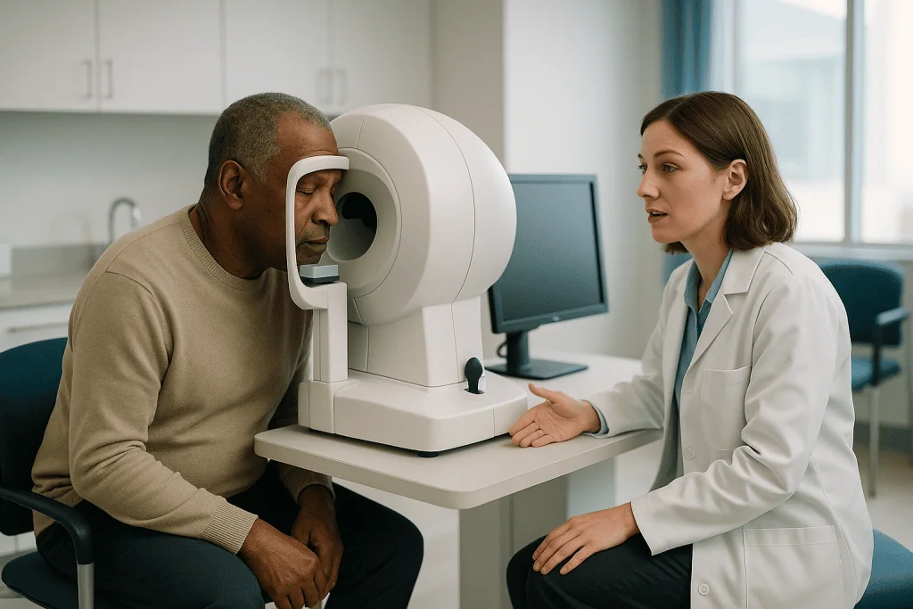
The machine is not testing whether you can see a bright light. It is hunting for the dimmest light you can detect at each location, called your threshold. It does this by making a light brighter in steps until you respond, then fainter until you stop responding, and settling on the boundary. Because the process is a search, many lights are meant to be missed. Missing lights is the test working as designed, not evidence that you are doing badly.
Along the way the software slips in checks. It flashes lights into your natural blind spot, the place where the optic nerve leaves the eye and no image can form; if you click, you were probably looking around. It occasionally makes a sound with no light; a click there counts as a false positive. It re-presents a light that you already saw clearly; missing it counts as a false negative. These checks become the reliability indices printed at the top of the report, and they matter as much as the map itself.
The NIH-hosted practical guide on visual field testing notes that a standard threshold test of one eye commonly takes several minutes, and that fatigue and unfamiliarity noticeably degrade results. That is why the first test in a person’s life is often treated as practice rather than truth.
What the machine is really measuring: the map behind the lights
The printout usually contains several panels, and each is trying to answer a different question. The grayscale map is the one patients look at first because it looks like a picture of vision: light areas are sensitive, dark areas are not. Doctors treat it with caution. It is a smoothed illustration, and it exaggerates small dips.
The panels that carry the real weight are the deviation plots. The total deviation plot compares your sensitivity at each point with what is expected for a healthy eye of your age. The pattern deviation plot goes one step further: it subtracts any general dimming across the whole field, such as that caused by a cataract or a small pupil, and shows only the localized losses. Glaucoma tends to produce localized, uneven damage, so the pattern deviation plot is where it becomes visible.
Two summary numbers sit alongside. Mean deviation (MD) is a single figure describing how far the whole field sits below the age-expected average; a more negative number means more overall loss. Pattern standard deviation (PSD) describes how irregular the field is; a higher number means some areas are much worse than others, which is the signature glaucoma leaves rather than the smooth dimming of a cloudy lens.
The last common panel is a probability map, where shaded squares mark points whose sensitivity falls below what is seen in a given small percentage of healthy people. A few isolated shaded squares appear in many normal tests. A cluster of them, side by side, in a shape that matches how nerve fibers run through the retina, is what makes an ophthalmologist sit up. Shape and clustering, not the total count of dark squares, tell the story.
Can a visual field test detect glaucoma on its own?
The short answer is no, and it is worth understanding why, because the reason shapes every decision that follows. A visual field test measures function, meaning what you perceive. Many things besides glaucoma can dent that function: a cataract, a droopy eyelid, migraine, a stroke affecting the visual pathway, certain retinal conditions, and a tired or distracted patient. The test reports a pattern of loss; it does not say what caused it.

What the field test does extremely well is confirm that a suspicious-looking nerve is actually costing vision, and show how much. That is why Mayo Clinic describes the diagnostic work-up as a set of tests taken together: measuring eye pressure, examining the optic nerve, testing the visual field, measuring corneal thickness, and inspecting the drainage angle where fluid leaves the eye. No single element carries a diagnosis. Glaucoma can exist with entirely normal pressure, and many people with pressure above the typical range never develop nerve damage. The field test is one witness among several.
The test also has a timing limitation that specialists are candid about. Standard perimetry detects damage once a fair number of nerve fibers have already stopped working, because the retina has redundancy and the brain compensates. In earliest disease, the optic nerve scan discussed later may show thinning while the field remains normal. Later in the disease the reverse can happen: the scan bottoms out because there is little tissue left to measure, and the field test becomes the more sensitive tracker of change.
So the honest framing is this: a visual field test is essential to diagnosing and monitoring glaucoma, and insufficient by itself. Your doctor is reading it alongside everything else in your chart.
What is the most common visual field defect in glaucoma?
Glaucoma does not remove vision at random. It damages bundles of nerve fibers, and those bundles follow a specific route across the retina, arching above and below the central point like the two halves of a parenthesis. Because the damage follows the wiring, the resulting blind patches, called scotomas, have recognizable shapes.
The classic and most frequently cited early defect is the arcuate scotoma: a curved blind zone that sweeps from near the natural blind spot around toward the nasal side of the field, tracking a damaged fiber bundle. In its early form it may appear as a short curved segment or as a small paracentral scotoma, a blind patch sitting just off center, within the region you use for reading faces and text. Because these sit close to the middle of vision, they can be surprisingly bothersome when they eventually reach awareness.
Another hallmark is the nasal step, a sharp difference in sensitivity above versus below the horizontal midline on the nose side of the field. It occurs because the upper and lower fiber bundles do not cross the horizontal line, so damage to one side creates a step rather than a smooth gradient. Ophthalmologists prize this sign because conditions affecting the brain’s visual pathways respect the vertical midline instead, which helps separate an eye problem from a neurological one.
A generalized, uniform dimming of the field, by contrast, points away from glaucoma and toward the lens or the pupil. This is why pattern standard deviation, the measure of unevenness, is so informative. Glaucoma is a disease of local, structured loss, and the printout is being read for structure. As the NIH-hosted practical guide stresses, a defect that fits the nerve fiber anatomy is far more persuasive than a scattering of missed points that does not.
What does it mean if you fail a field vision test?
People often leave the room convinced they failed because they missed so many lights. Take that worry off the table first: the software is designed to show you lights you cannot see. Missing a substantial share of them is normal for everyone with healthy eyes, because the machine is searching for your threshold.
What clinicians actually mean by an abnormal or unreliable field is more specific. An unreliable test is one whose internal checks show the result cannot be trusted: too many clicks during the blind-spot probes, too many clicks with no light shown, or too many misses of lights you had clearly seen earlier. This is a comment on the test session, not on your eyes. The usual response is simply to repeat it, sometimes with a brief re-explanation of the instructions. First-time results are especially prone to this learning effect, and specialists frequently disregard a first field entirely if a second, better one follows.
An abnormal but reliable test shows a cluster of reduced-sensitivity points in a pattern that fits an anatomical explanation. Even then, a single abnormal field is a hypothesis rather than a verdict. Standard practice is to look for the same defect in the same place on a repeat test before calling it established. Fields fluctuate from day to day for reasons as ordinary as fatigue, an uncorrected refractive error, a rim of the trial lens intruding, or a lowered upper eyelid.
If the defect is confirmed, the next questions are about cause and pace. The optic nerve scan, the appearance of the nerve, eye pressure, and your history all feed into whether glaucoma is the likely explanation. Your treating team, not the printout, makes that call, and they will usually tell you how confident they are and what would change their mind.
Are visual field tests reliable? Reading the fine print
Questions about visual field test reliability are fair, because the test depends on a human being paying attention to faint lights for several minutes. Its known weaknesses are worth stating plainly rather than papering over.
Variability is the largest. The same eye tested twice in a week can produce noticeably different maps, and the variability is larger in areas that are already damaged. This is why glaucoma specialists do not react to one field. They collect a series and look for a defect that repeats in the same place and, more importantly, for a trend across many tests. The NIH-hosted practical guide describes the value of obtaining more than one baseline test early on precisely so that later changes can be judged against a stable reference.
The second weakness is the learning curve. Most people perform better on their second and third tests as they learn to hold fixation and to click on faint, uncertain lights rather than waiting for a clear one. An apparent improvement between a first and second test is usually learning, not recovery of vision.
The third is the set of confounders: cataract dimming the whole field, a small pupil, an incorrect lens in the holder, a heavy eyelid, or simply a bad night’s sleep. Good clinics manage these by checking the reliability indices, by asking about the session, and by using the pattern deviation plot to strip out uniform dimming.
The strengths deserve equal billing. Perimetry measures the thing that actually matters to a patient, which is what they can see. It works at every stage of disease, including advanced stages where scans plateau. And when it is performed with a consistent strategy on a consistent machine over time, it is the most direct evidence available of whether glaucoma is stable or progressing.
What an OCT scan for glaucoma adds
OCT stands for optical coherence tomography, a painless imaging method that bounces light off the back of the eye to build a cross-sectional picture, in much the way ultrasound uses sound. The whole scan takes seconds per eye, requires no clicking, and involves nothing more than staring at a target while a light sweeps across.
For glaucoma, an OCT scan for glaucoma concentrates on two structures. The first is the retinal nerve fiber layer, the layer of fibers that gather from across the retina and converge on the optic nerve; glaucoma thins it, often in the same upper and lower bundles that produce arcuate field defects. The second is the ganglion cell layer near the center of the retina, where the cell bodies of those fibers live. Thinning here can appear early and sits in the region that supports central vision.
The report shows thickness measurements around the nerve as a ring divided into sectors, each shaded green, yellow, or red according to how it compares with a reference database of healthy eyes. Green means within the typical range, yellow means borderline, red means below what is seen in most healthy people. Doctors read these colors with the same caution they apply to field maps. An unusually long or short eye, a tilted nerve, or a nerve that is simply built large or small can shift measurements without any disease, and the reference database may not represent every population equally.
What makes OCT valuable is objectivity and repeatability. It does not depend on attention or fatigue, and repeated scans on the same machine can be compared point for point. Its limitation is the mirror image of the field test’s strength: it measures tissue, not sight, and in advanced disease the thickness reaches a floor below which it can no longer register further loss.
Visual field test vs OCT scan: how the two compare
Patients often ask why they need both tests when each seems to be looking for the same thing. The table sets out the differences that matter, and the paragraph below explains why the combination is stronger than either alone.
| Feature | Visual field test (perimetry) | Optic nerve scan (OCT) |
|---|---|---|
| What it measures | Function: what the eye actually perceives across the field | Structure: thickness of nerve fiber and ganglion cell layers |
| Patient involvement | Active; requires steady fixation and clicking for several minutes | Passive; a few seconds of looking at a target |
| Main source of error | Attention, fatigue, learning effect, eyelid, lens rim, cataract | Anatomical variation, unusual eye length, motion, database mismatch |
| Strongest stage | Moderate to advanced disease; tracks change when scans plateau | Early and suspected disease, before field loss appears |
| Key report items | Reliability indices, pattern deviation plot, MD, PSD | Sector thickness map, color-coded comparison, change analysis |
| How results are judged | Repeated, anatomically plausible defects; trend over a series | Sector thinning matching field defects; measured change over time |
The reason both are used comes down to timing and confirmation. Structure often changes before function, so OCT may flag thinning while the field is still clean. Function keeps changing after structure hits its floor, so perimetry carries the monitoring load in later disease. When a red sector on the scan sits in the exact quadrant that would produce the field defect on the printout, the two independent lines of evidence corroborate each other, and the diagnosis becomes far more secure than either test could make it. When they disagree, that disagreement is itself information, and it usually prompts a repeat rather than a decision.
Who is usually tested, and who is usually asked to wait
Routine eye examinations are the entry point. The NHS advises most adults to have an eye test at least every two years, and notes that people over 40 with a close relative who has glaucoma, people of African, Caribbean, or Asian family background, and people with certain other conditions carry a higher risk and may be offered checks more often. A routine test that finds raised pressure or an unusual-looking nerve is what typically triggers the first visual field test and OCT.
Beyond that, several groups are commonly tested. People already diagnosed with glaucoma are tested on a schedule set by their specialist to monitor stability. People labeled as glaucoma suspects, meaning the nerve or pressure looks questionable but no damage has been proven, are tested to find out whether anything is changing. People with ocular hypertension, the term for pressure above the typical range with a healthy nerve and field, are tested because their risk is higher than average even though many will never develop damage. Mayo Clinic describes pressure above the usual range as a risk factor rather than a diagnosis, which is exactly why the field and the scan are needed.
Who is asked to wait? Someone whose first field is unreliable is usually asked to repeat it rather than act on it. Someone with a single borderline OCT sector and a normal field is often observed rather than treated, because a stable borderline reading over time carries a very different meaning from a falling one. A person with a fresh, dense cataract may be told the field will be more meaningful after the lens is dealt with, since the cloudiness dims everything and can mask or mimic loss.
Waiting in these situations is not neglect. It reflects the fact that glaucoma is usually slow and that the cost of treating a healthy eye is real. The decision to observe, like the decision to treat, belongs to the treating team, and a good team will explain which finding would move them from one to the other.
What the following weeks and months usually look like
The period after a first suspicious result tends to follow a recognizable rhythm, though the exact intervals are set by your specialist based on how much damage is present and how fast it seems to be moving.
The first task is usually to confirm. A repeat visual field, often within a few weeks to a few months, checks whether the defect reappears in the same place and whether reliability has improved now that the test is familiar. The NIH-hosted practical guide recommends establishing more than one good-quality baseline field, because every future judgment about progression is a comparison with that baseline. A repeat OCT on the same machine serves the same purpose for structure.
If glaucoma is confirmed, monitoring begins in earnest. Many specialists test more frequently in the first year or two after diagnosis, when the pace of the disease is still unknown, and then space visits out once it appears stable. Mayo Clinic notes that glaucoma requires lifelong follow-up because it can progress without symptoms even when treatment is in place. Each visit typically pairs a pressure measurement with a field, a scan, or both, and the software on both instruments can line up the series to display a trend.
When treatment starts, it is usually aimed at lowering eye pressure, the only modifiable factor with strong evidence behind it. Prescription eye drops work by reducing the fluid the eye produces or by improving its drainage; laser procedures and surgery pursue the same goal by other routes. The choice among these, and any change to them, sits with the prescribing clinician. Do not adjust or stop any glaucoma medicine without that conversation, because pressure can rise quickly and silently.
Emotionally, the weeks between tests can feel like limbo. Knowing that the waiting is structured, and that a stable series is itself the good news being looked for, helps many people carry it.
What people often get wrong about glaucoma eye tests
The first misunderstanding is that missing lights means failing. As explained earlier, the machine is searching for the faintest light you can see, so missing many is expected. The reliability indices, not the miss count, decide whether the session was usable.
The second is that a normal pressure reading rules glaucoma out. It does not. Mayo Clinic is explicit that glaucoma can develop at pressures within the typical range, and that pressure above the range does not by itself mean glaucoma. Pressure is a risk factor and a treatment target, not the disease.
The third is that good reading vision means the eyes are fine. Glaucoma spares the very center of vision until late, so a person can read the smallest line on the chart while carrying substantial peripheral loss. Both the NHS and Johns Hopkins emphasize that this symptom-free course is precisely why testing, rather than waiting for a problem, is the approach.
The fourth is that an improved second test means the eye got better. Vision lost to glaucoma does not return; an apparent improvement is almost always the learning effect or a better session.
The fifth is that one red sector on an OCT means glaucoma. Anatomical variation puts some healthy nerves outside the reference range. The color is a prompt for closer attention, and stability over repeated scans often settles the question.
The sixth is that treatment will make the field test go back to normal. Treatment aims to slow or halt further loss. A stable series of fields is what success looks like on paper, and it is a genuine achievement, even though it will never show as a brightening map. Understanding this in advance spares a great deal of disappointment at follow-up visits.
Questions to ask your care team
Consultations move quickly, and a printout full of numbers is hard to interrogate in the moment. Bringing a short list turns a confusing appointment into a useful one. The questions below are ones glaucoma specialists are generally glad to be asked.
- Was my visual field test reliable, and if not, what would help me do better next time?
- Do the field defect and the OCT thinning point to the same area of the nerve, or do the two tests disagree?
- Am I being described as having glaucoma, as a glaucoma suspect, or as having ocular hypertension, and what is the difference in my case?
- How many baseline tests do you want before you judge whether anything is changing?
- What finding on a future test would change your recommendation from observing to treating, or from one treatment to another?
- How often will I be tested, and will it always be on the same machines so results can be compared?
- If I have a cataract, how is it affecting the interpretation of my field?
- Which parts of my daily life, such as driving, should I discuss with you given my current field results?
Two practical requests are worth adding. Ask for a copy of your field and OCT reports, or at least the summary numbers, so that a series can travel with you if you ever change providers. And ask what the team considers the single most important number to watch in your case. Some patients find that anchoring on one trend, such as mean deviation over time, makes the whole process feel less like a fog of colors and more like a conversation about direction.
When to call your doctor
Glaucoma of the common, slow type rarely produces emergencies, and most concerns can wait for the scheduled visit. A short list of situations should prompt a same-day call rather than a wait.
Seek urgent care if you develop a sudden, severe ache in or around one eye, especially with blurred vision, halos around lights, a red eye, headache, nausea, or vomiting. The NHS and Mayo Clinic describe this combination as the presentation of acute angle-closure glaucoma, in which pressure rises abruptly because the drainage angle closes, and it needs prompt treatment to protect the nerve.
Contact your eye team promptly, though not necessarily as an emergency, if you notice any new blind patch, a curtain or shadow in part of your vision, a sudden shower of floaters or flashes of light, or a rapid change in how clearly one eye sees. These may have causes unrelated to glaucoma, including retinal problems, and they deserve assessment rather than being attributed to a known diagnosis.
Call as well if you are using prescribed glaucoma drops and develop marked eye redness, stinging that does not settle, swelling of the eyelids, breathlessness, or a slow or irregular heartbeat. Some pressure-lowering drops are absorbed into the body and can affect the heart and lungs; your prescriber needs to know, and any change to the medicine should come from them rather than from stopping on your own.
Finally, if you miss a scheduled field or scan, ring to rebook rather than waiting for the next annual visit. A gap in the series weakens every future comparison, and the whole logic of glaucoma monitoring depends on the series being intact. Every decision that follows a call, from reassurance to a change of plan, rests with your treating team.
Frequently asked questions
What does failing a visual field test mean?
Clinicians rarely use the word failing; a test is either unreliable or it shows a defect. An unreliable test means the internal checks show you were looking around or clicking without lights, and the fix is simply to repeat it. A reliable test with a cluster of reduced points is a finding that needs confirmation on a second test and interpretation alongside the optic nerve scan and eye pressure by your treating team.
Can a visual field test detect glaucoma?
It can detect the pattern of vision loss that glaucoma produces, but it cannot by itself confirm the cause. Cataract, eyelid position, migraine, stroke, and retinal disease can all alter a field. Doctors combine the field with an optic nerve scan, a pressure reading, and an examination of the nerve and drainage angle. Glaucoma is diagnosed from that whole picture, not from the field alone.
What is the most common visual field defect in glaucoma?
The arcuate scotoma, a curved blind patch that follows a damaged bundle of nerve fibers from near the natural blind spot around toward the nasal side, is the classic early defect. A nasal step, a sharp difference above and below the horizontal midline, and small paracentral scotomas near the center are also typical. Their shapes match the anatomy of the nerve fibers, which is what makes them persuasive.
Why would a doctor order a visual field test?
Usually because a routine examination has raised a question: eye pressure above the typical range, an optic nerve that looks suspicious, a thinner cornea, or a family history of glaucoma. Doctors also order it to monitor known glaucoma and check whether treatment is holding the disease stable. Because early glaucoma has no symptoms, the test measures side vision that a patient cannot judge for themselves.
Is an OCT scan for glaucoma better than a visual field test?
Neither is better; they measure different things. OCT measures the thickness of nerve tissue objectively and often flags thinning before vision changes. The field test measures actual perception, keeps working in advanced disease when scans plateau, and reflects what matters to daily life. Specialists use both because agreement between them makes a diagnosis far more secure than either result on its own.
How accurate is a visual field test the first time you take it?
First tests are often the least dependable because of the learning effect: people improve as they learn to keep still and to click on faint, uncertain lights. Specialists frequently treat a first field as practice and place more weight on the second and third. The NIH-hosted practical guide recommends more than one good baseline before judging any later change.
Can visual field test reliability be improved by the patient?
Yes, within limits. Keep your gaze fixed on the central target rather than searching for lights, click whenever you think you saw something even if unsure, blink normally, and ask for a pause if you tire. Wearing the correct lens in the holder and keeping a drooping eyelid taped or lifted, if the technician offers, also help. Rest and hydration before the test are sensible.
Does a normal visual field test mean I do not have glaucoma?
Not necessarily. Standard perimetry detects damage after a fair number of nerve fibers have already been lost, so very early glaucoma can leave the field normal while an OCT scan shows thinning. A normal field is reassuring, especially when the scan and pressure are also unremarkable, but people at higher risk are usually asked to continue periodic checks rather than be discharged.
Will treatment make my visual field results go back to normal?
No. Vision lost to glaucoma does not return, so treatment is judged by stability rather than improvement. Pressure-lowering eye drops, laser, or surgery aim to slow or halt further loss, and a series of fields that stays the same over years is what success looks like. Any apparent improvement on a repeat test is almost always better performance, not recovered nerve function.
Why do I need both eyes tested if only one looks abnormal?
Glaucoma is usually a disease of both eyes, even when it is more advanced in one, and the less affected eye provides a comparison that helps interpret the other. Testing both also detects early change in the eye that currently looks healthy, which is exactly the stage at which monitoring matters most. Specialists therefore track both eyes as a pair over time.
References
- NHS – Glaucoma: Diagnosis
- MedlinePlus – Visual field
- NIH / PubMed Central – Visual field testing for glaucoma: a practical guide
- NIH National Eye Institute – Glaucoma
This article is for general information only and is not a substitute for professional medical advice. Please consult a qualified doctor about your individual situation.

More from the Blog

Preparing for LASEK: Contact Lens Breaks, Corneal Thickness Checks and Planning Days Off
LASEK preparation usually involves three things: pausing contact lenses for a period your surgeon sets (often a few weeks, longer for rigid lenses) so…

How Eye Alignment Is Measured: The Tests Behind a Strabismus Disorder Diagnosis
Eye alignment is measured with a short set of clinic tests. The cover test, in which one eye is covered while the examiner watches…

What PRK Results Usually Look Like Once Healing Finishes and How Long They Hold
PRK reshapes the cornea after its surface layer is removed, so vision is blurry for the first several days and sharpens gradually rather than…

What Injections for Macular Degeneration Can Realistically Do: Slowing Loss, Not Undoing It
Injections for wet macular degeneration block a growth signal that makes fragile new blood vessels leak under the retina. In large trials, most treated…

What Cornea and External Disease Specialists Treat: Infections, Pterygium and Dystrophies
A cornea and external disease specialist is an ophthalmologist with extra fellowship training in the clear front window of the eye and the surface…

What Chalazion Removal Involves: Numbing, the Incision Inside the Eyelid and the Patch
A chalazion removal procedure is a short outpatient operation for a blocked oil gland in the eyelid. The lid is numbed with a local…