

Prostate MRI: How It Changed Prostate Cancer Diagnosis
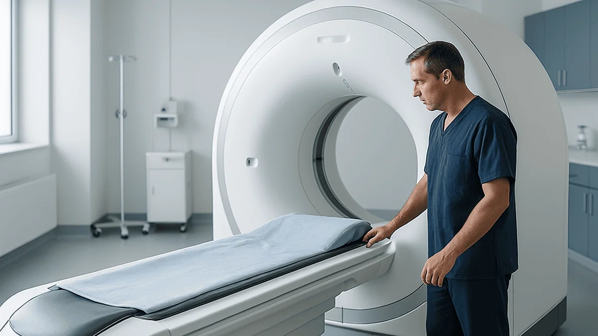
Key Takeaways
- In the PRECISION trial, MRI-first diagnosis found clinically significant cancer in 38% of men versus 26% with standard biopsy, while 28% of the MRI group avoided biopsy entirely.
- A clear prostate MRI carried an 89% negative predictive value in PROMIS, so about one clear scan in ten still hid a significant tumor.
- PI-RADS scores run from 1 to 5; a 15 mm size threshold separates 4 from 5, and PI-RADS 3 is where PSA density most often decides the next step.
- In radiology, a lesion means only an area that looks different; inflammation, benign nodules and old scars all qualify, and the score, not the word, carries the probability.
- Caffeine is avoided before the scan because it stimulates bowel movement and bladder filling, which blur the diffusion images; it is an image-quality rule, not a safety one.
- A 2025 randomized trial found the contrast-free two-sequence scan non-inferior to the full three-sequence version for detecting significant cancer in experienced centers.
Prostate MRI, a detailed magnetic scan of the prostate gland, is now widely used before biopsy when a PSA test or examination raises concern. Randomized trials show it finds more clinically significant cancers, detects fewer harmless ones, and lets roughly a quarter of men avoid a biopsy. It cannot confirm cancer on its own; a tissue biopsy is still needed for diagnosis, and results are interpreted by your clinician.
A man in his late fifties sits in a parking lot reading a two-line message on his phone: PSA slightly raised, please book a follow-up. Ten years ago the next sentence would almost certainly have been “we need to do a biopsy.” Today it is just as likely to be “we need to get a prostate MRI first.”
That shift is why the scan is trending again as of March 2026. A large international trial published in 2025 reported that a shorter, contrast-free version of the prostate MRI found clinically significant cancers about as well as the full scan, and several European health systems are now testing MRI as a formal step in screening pilots. Social feeds have run ahead of the data, with posts claiming the scan makes biopsies obsolete or, at the other extreme, that it misses most tumors.
Neither claim survives contact with the trials. Here is what the technology actually does, what it changed, and where its limits sit.
What changed recently: prostate MRI moved ahead of the biopsy needle
For roughly thirty years the pathway was fixed. A raised PSA, a blood test that measures prostate-specific antigen, a protein made by the prostate gland, led straight to a transrectal ultrasound biopsy: a dozen or so needle samples taken in a grid pattern, essentially blind to where any tumor sat. Ultrasound can show the gland’s outline but cannot reliably tell cancer from normal tissue.
Three dated results rewrote that pathway.
- January 2017, PROMIS. A UK study of 576 men gave every participant a multiparametric MRI and then a highly detailed mapping biopsy as the reference standard. The scan detected 93 percent of clinically significant cancers; the standard ultrasound biopsy detected 48 percent.
- March 2018, PRECISION. A randomized trial across 25 centers in 11 countries assigned 500 men either to standard biopsy or to MRI first, with biopsy only of suspicious areas. The MRI arm found more significant cancers (38 versus 26 percent), fewer insignificant ones (9 versus 22 percent), and 28 percent of men in that arm avoided a biopsy altogether.
- 2019 onward. National guidance in England made MRI the recommended first test for men whose PSA or examination raises suspicion, and the NHS now describes the scan as a routine step before deciding on biopsy. US guideline bodies followed, and a 2022 Swedish screening trial reported that restricting biopsy to MRI-visible lesions roughly halved the detection of low-grade cancers that would never have caused harm.
The 2025 result on contrast-free scanning is the latest piece. It matters because a scan that needs no injection is quicker and easier to offer at scale, which is exactly what a screening program would require.
What is a prostate MRI and how does the scan work?
Magnetic resonance imaging uses a strong magnet and radio waves, not X-rays, to map how water molecules behave inside tissue. For the prostate, a walnut-sized gland sitting just below the bladder and in front of the rectum, radiologists combine three kinds of picture, which is why the full exam is called multiparametric MRI, often shortened to mpMRI.
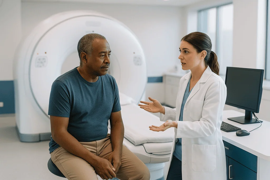
The first is T2-weighted imaging, the anatomical view. It shows the gland’s zones: the peripheral zone at the back, where about 70 percent of cancers begin, and the transition zone near the urethra, where benign enlargement happens. Healthy peripheral tissue looks bright because it holds a lot of water; dense tumor tissue looks darker.
The second is diffusion-weighted imaging. Cancer cells pack tightly, so water molecules cannot drift freely between them. The scanner measures that restriction and converts it into a map; areas where water is hemmed in stand out. Of the three sequences, this one carries most of the diagnostic weight.
The third, dynamic contrast-enhanced imaging, follows a gadolinium-based dye injected into an arm vein. Tumors grow leaky new blood vessels, so dye floods in and washes out faster than in normal tissue. This sequence helps most when the first two are ambiguous.
Most modern scans use a surface coil, a padded receiver laid over the pelvis. The endorectal coil, a probe placed inside the rectum to sharpen the signal, was common a decade ago but is now rarely required on higher-field (3 Tesla) machines. The scan takes 20 to 40 minutes, longer if contrast is given, and involves no ionizing radiation.
Can you tell from an MRI if you have prostate cancer?
The honest answer: it can tell you how likely cancer is, not whether it is there. A prostate MRI is a probability tool. The radiologist is not seeing cancer cells; they are seeing patterns of water restriction, signal intensity and blood flow that cancers tend to produce. Inflammation, scarring from past infection and ordinary benign enlargement can produce overlapping patterns.
That is why no mainstream guideline allows a diagnosis of prostate cancer to be made from imaging alone. Diagnosis requires a biopsy, meaning thin needle samples of tissue examined under a microscope by a pathologist, who assigns a Gleason grade group from 1 (least aggressive) to 5 (most aggressive). The scan decides whether a biopsy is needed and where to point it; the microscope decides what it is.
Where the scan performs best is in ruling out the cancers that matter. In PROMIS, the negative predictive value of MRI for clinically significant disease, meaning the chance that a man with a clear scan truly had no significant cancer, was 89 percent. Put the other way, roughly one clear scan in ten still hid a significant tumor. That is a strong result for a non-invasive test, but it is not zero, which is why PSA level, prostate size and family history are weighed alongside the pictures.
The reverse error also happens. The scan flags areas that turn out benign; specificity in PROMIS was only 41 percent. A suspicious MRI is a reason to look closer, not a verdict. Clinicians describe the pathway as “MRI to decide, biopsy to confirm,” and that phrasing is worth holding onto.
PI-RADS score meaning: how radiologists grade what they see
PI-RADS stands for Prostate Imaging Reporting and Data System, a standardized 1-to-5 scale, now in version 2.1, that radiologists use to express how likely a visible area is to be clinically significant cancer. “Clinically significant” is itself a defined term: in most studies it means Gleason grade group 2 or higher, the level at which treatment rather than monitoring starts to be considered.
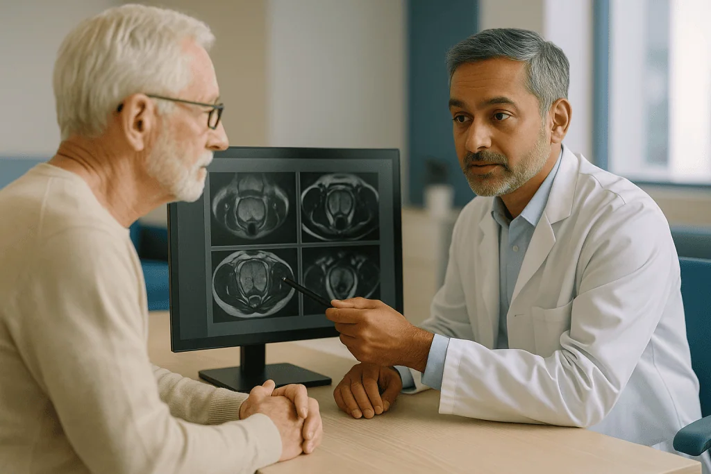
Each lesion receives its own score, and the report usually also records its size, which zone it sits in, and whether it appears to touch or breach the gland’s capsule.
| PI-RADS score | What the radiologist is saying | Approximate chance of significant cancer on biopsy | Usual next step |
|---|---|---|---|
| 1 | Very low suspicion; nothing of concern seen | Low single digits | Biopsy usually not needed |
| 2 | Low suspicion | Low single digits | Biopsy usually not needed; PSA follow-up |
| 3 | Equivocal; features present but inconclusive | Roughly 1 in 10 to 1 in 5 | Depends on PSA density and risk factors |
| 4 | High suspicion, lesion under 15 mm | Roughly 1 in 3 to 1 in 2 | Targeted biopsy recommended |
| 5 | Very high suspicion, lesion 15 mm or larger, or capsule involvement | Most cases | Targeted biopsy recommended |
The percentages are pooled estimates from published series and vary between hospitals, scanners and radiologists; your own report’s number should be read against your local team’s track record. The pivotal zone is PI-RADS 3. Here many teams use PSA density, your PSA divided by prostate volume in milliliters, as a tiebreaker: a low density nudges toward monitoring, a high density toward biopsy. That decision is a conversation, not an algorithm.
What does it mean if there are lesions on my prostate?
“Lesion” is a word that lands hard in a report, but in radiology it simply means an area of tissue that looks different from its surroundings. It carries no judgment about cause. A prostate lesion on MRI may be a tumor; it may equally be a cluster of inflamed glands (prostatitis), a benign nodule of enlarged tissue, a small cyst, or scar from an old infection or a previous biopsy.
What matters is the context the radiologist attaches to it. Three details do most of the work.
Location comes first. Most cancers arise in the peripheral zone, and a lesion there is read with more suspicion than an identical-looking nodule in the transition zone, where benign enlargement creates a chaotic, nodular background that mimics tumors. Size follows: the 15 mm threshold that separates PI-RADS 4 from 5 exists because larger lesions are more often higher grade. The third is behavior across sequences. A lesion that is dark on T2, strongly restricted on diffusion imaging and shows early contrast enhancement ticks every box; one visible on only a single sequence is far less convincing.
Many men have more than one lesion. Prostate cancer is frequently multifocal, and reports list each area with its own score. The highest-scoring lesion, called the index lesion, drives the decision.
A lesion is not a diagnosis, and it is not a treatment plan. Even when biopsy confirms cancer, a good proportion turns out to be grade group 1 disease, which guidelines increasingly manage with active surveillance, a structured program of PSA tests, repeat MRI and occasional repeat biopsy rather than immediate surgery or radiation. The scan gives your urologist a map. Deciding what to do with it is a shared decision.
MRI before prostate biopsy: how the needle learned to aim
The quieter revolution is in the biopsy room. Once a prostate MRI has marked a suspicious area, the urologist can aim at it instead of sampling the gland in a blind grid. Three methods exist.
Cognitive fusion is the simplest: the clinician studies the MRI, then steers the ultrasound probe to the matching spot by eye and anatomy. Software fusion overlays the stored MRI onto the live ultrasound image so the target appears as a highlighted zone on screen; this is what most centers mean by “MRI-ultrasound fusion biopsy.” In-bore biopsy is performed inside the MRI scanner itself and is reserved for hard-to-reach or very small lesions.
The route of the needle has shifted too. The traditional transrectal approach passes needles through the rectal wall and carries a small but real risk of infection, because the rectum is not sterile. The transperineal approach passes needles through the skin between the scrotum and anus, bypassing the bowel. Observational data from multiple centers show infection rates close to zero with the transperineal route, and several national bodies now favor it. Randomized comparisons remain limited, so the strength of that recommendation is moderate rather than definitive.
Most protocols combine the two logics: targeted samples from each MRI lesion plus a reduced set of systematic samples from the rest of the gland, because a handful of significant cancers are invisible on imaging. In PRECISION, targeted-only biopsy still found more significant disease than the systematic approach, which tells us how much of the yield comes from aiming. Whether to add systematic cores is a judgment your urologist makes based on scan quality, PSA density and prior history.
What the evidence actually says, and how strong it is
It helps to sort the claims by the kind of evidence behind them.
Strong, from randomized trials. An MRI-first pathway detects more clinically significant cancers and fewer insignificant ones than standard biopsy alone. PRECISION established this across 25 centers, and the finding has been reproduced in other randomized trials, including population-based screening studies in Sweden. The 2025 trial on contrast-free scanning adds randomized evidence that the two-sequence version is non-inferior for detecting significant disease in experienced centers.
Strong, from prospective diagnostic studies. The sensitivity and negative predictive value figures (93 percent and 89 percent in PROMIS) come from a design where every participant had both the MRI and a reference-standard biopsy, which is the right way to measure test accuracy. They are robust for men with raised PSA referred to specialist centers; accuracy in lower-risk populations is less certain.
Moderate, from observational data. Lower infection rates with transperineal biopsy, the value of PSA density in sorting PI-RADS 3 cases, and the usefulness of repeat MRI during active surveillance rest largely on cohort studies. Consistent direction, no large randomized confirmation yet.
Not yet shown. No trial has demonstrated that an MRI-first pathway reduces deaths from prostate cancer. That is the question the current European screening trials are designed to answer, and results will take years because prostate cancer progresses slowly. Finding more significant cancer earlier is a reasonable surrogate, but it is a surrogate.
Expert opinion. Exact PI-RADS thresholds for biopsy, how often to repeat scans in surveillance, and whether community scanners match trial-center performance are guided by consensus rather than hard data. Reports often read as if they were certainties. They are informed estimates.
Multiparametric or biparametric: is the contrast dye still needed?
Biparametric MRI, usually written bpMRI, drops the contrast injection and keeps the two sequences that do most of the work: T2-weighted and diffusion-weighted imaging. The scan is roughly a third shorter, needs no intravenous line, and removes the small risk of a gadolinium reaction along with the practical hurdle of checking kidney function first.
The question has always been whether the missing sequence costs accuracy. Dynamic contrast imaging was designed to rescue equivocal peripheral-zone lesions, upgrading some PI-RADS 3 findings to 4 when enhancement is early and focal. Lose it and, in theory, a few borderline cancers slip into the “probably fine” category.
The 2025 randomized trial tested exactly that in men referred with suspected cancer at centers with experienced readers. Detection of clinically significant cancer with the two-sequence scan was non-inferior to the full three-sequence version. Earlier observational comparisons had pointed the same way, but this was the first large head-to-head randomized answer.
Two caveats come with it. The result was obtained with high-quality scanning and expert radiologists; smaller centers with older machines or less experienced readers may not reproduce it, and image quality matters more when there is no contrast safety net. Second, the trial addressed first-time diagnosis, not surveillance of known cancer or assessment after treatment, where contrast still has a role.
What this means for you is practical. If your center offers a contrast-free scan, that is now an evidence-backed choice rather than a shortcut. If your radiologist wants contrast because your prostate is large, you have had a previous biopsy, or the first images were unclear, that is also reasonable. The choice is theirs to make with you, not a measure of quality in either direction.
Prostate MRI preparation: how do I get ready for the scan?
Preparation for a prostate MRI is lighter than most people expect, and instructions vary between centers, so the sheet your imaging department sends you overrides anything written here.
The common requests share one logic: keep the pelvis still and the picture clear. Gas and stool in the rectum distort the diffusion images, so some centers ask for a light meal the day before and a small enema a few hours ahead; others ask only that you empty your bowels that morning. Many ask you to arrive with a comfortably full bladder rather than a very full one, because a tense bladder pushes on the gland and makes lying still harder.
Ejaculation is often discouraged for one to three days beforehand. The seminal vesicles, two small sacs behind the prostate that store fluid, collapse after ejaculation and are easier to assess when full, and there is some evidence that the gland itself looks slightly different in the hours after.
Safety screening is universal. You will be asked about pacemakers, cochlear implants, aneurysm clips, metal fragments and any work history involving metal grinding. Most modern joint replacements are MRI-safe, but a hip prosthesis can throw a shadow of distortion across the pelvis that degrades image quality; tell the department in advance so they can adjust the protocol. If contrast is planned, a recent kidney function blood test is usually required.
Medicines are generally continued as normal. Do not stop or alter any prescription for the scan unless the prescribing clinician specifically tells you to. Bring a list of what you take, including blood thinners, because that information matters more for any biopsy that follows than for the scan itself.
Why no caffeine before a prostate MRI, and what the scan feels like
The caffeine instruction puzzles people because the scan involves no fasting and nothing enters the gut. The reason is movement. Caffeine stimulates bowel contractions and increases urine production. Peristalsis, the rhythmic squeezing of the intestine, shifts the rectum against the back of the prostate during the several minutes each sequence takes, smearing the diffusion images that carry the most diagnostic weight. A filling bladder adds pressure and the urge to move. Skipping coffee, tea, energy drinks and some sodas for a few hours beforehand is a low-effort way to protect image quality; it is not a safety rule.
For the same reason, some centers give a short-acting muscle-relaxing injection before scanning to quiet the bowel. If you have glaucoma, a heart rhythm condition or difficulty passing urine, mention it, because those conditions affect whether that injection is appropriate. The radiology team decides.
The scan itself is undramatic. You lie on your back on a sliding table, a padded coil is strapped over your lower abdomen, and you are moved into the bore of the magnet feet first. The machine is loud, a mix of knocking and buzzing, so you are given earplugs or headphones. Each sequence lasts two to six minutes; the technologist speaks to you between them. If contrast is used, you may notice a cool feeling in the arm and a brief metallic taste.
Claustrophobia is the most common difficulty. Wider-bore scanners help, as does knowing that your head is usually near the opening. A radiologist typically reads the images within days, and the report goes to the clinician who requested it, who explains it to you in context.
What a prostate MRI can miss
A test that catches nine in ten significant cancers misses one in ten, and it pays to understand which ones.
Small tumors are the obvious gap. Lesions under about 5 mm sit at the edge of what diffusion imaging resolves, and some aggressive cancers grow in a diffuse, sheet-like pattern rather than forming a discrete nodule. Cribriform and intraductal patterns, two microscopic growth styles associated with worse outcomes, can be surprisingly inconspicuous on imaging.
The transition zone is the second blind spot. Benign enlargement fills this area with nodules of varying signal, and a cancer hiding among them can look like just another nodule. Readers are trained to look for a lens-shaped, “smudged charcoal” appearance, but the false-negative rate here is higher than in the peripheral zone.
Technical factors matter as much as biology. Rectal gas, hip implants, patient motion and lower-field scanners all reduce sensitivity. Reader experience is the largest single variable; studies comparing radiologists show meaningful disagreement at PI-RADS 3, and centers that read high volumes outperform those that read few. Many health systems now require double reading or a quality checklist for exactly this reason.
What follows is not alarm but a sensible safety net. A clear MRI with a PSA that keeps rising, a high PSA density, a strong family history or a known high-risk gene variant is not the end of the story; most guidelines advise continued PSA monitoring and a low threshold for repeat imaging or systematic biopsy in that situation. The scan shifts the odds. It does not close the file, and a good clinician will say so plainly.
Who should think about screening, and at what age?
MRI is not a screening test in the strict sense. Nobody is scanned off the street; the scan sits one step downstream, after a PSA blood test or a clinical examination has raised a question. Understanding who gets to that first step is therefore the real screening question.
Major US bodies frame PSA testing as an individual decision made with a clinician, because the test finds many cancers that would never have caused harm, and treatment carries risks to urinary and sexual function. The conversation is generally recommended from around age 50 for men at average risk and from 40 to 45 for men at higher risk: those of African ancestry, those with a father or brother diagnosed before 65, and those who carry inherited variants such as BRCA2. Screening is generally not recommended for men over 70 or those with a life expectancy under 10 to 15 years, because the slow course of most prostate cancers means the harms tend to outweigh the benefits.
What prostate MRI has done is change the arithmetic of that conversation. The main objection to PSA screening was always overdiagnosis: find a borderline PSA, biopsy blind, find a grade group 1 cancer, treat it, live with the side effects, gain nothing. An MRI step lets roughly a quarter of men with a raised PSA skip biopsy and roughly halves the detection of harmless cancers in screened populations. That is why European screening trials now build MRI into their design.
The evidence for population screening with an MRI step remains incomplete; mortality data are pending. For an individual man the practical point is simpler. If a PSA test is on the table, ask what happens next if it is raised, and whether MRI is part of that pathway.
Common myths about prostate MRI
“A clear MRI means I do not have cancer.” A clear scan means significant cancer is unlikely, about one chance in ten in the best studies. Low-grade cancer is often invisible on MRI by design, which is the point. Ongoing PSA monitoring stays sensible.
“MRI has made biopsy obsolete.” It has made blind biopsy obsolete in many centers. Diagnosis still requires tissue under a microscope, and no guideline supports treating prostate cancer on imaging alone.
“A lesion on the report means cancer.” A lesion is an area that looks different. Inflammation, benign nodules and scars all qualify. The PI-RADS score, not the word lesion, carries the probability.
“MRI can replace the PSA test.” The two answer different questions. PSA is a simple, repeatable blood test that decides who needs imaging; MRI is a detailed scan that decides who needs biopsy. Scanning every man regardless of PSA has not been tested at population scale and would generate large numbers of equivocal findings.
“The scan exposes you to radiation.” MRI uses magnets and radio waves. There is no ionizing radiation, which is one reason it suits repeat use in surveillance.
“You need a probe inside you.” Endorectal coils are now rarely used. Most modern scans use an external pad over the pelvis.
“Contrast dye is dangerous.” Serious reactions to gadolinium-based contrast are rare. Tiny deposits have been detected in the brain after repeated doses, without demonstrated harm so far, and regulators continue to monitor. Men with significantly reduced kidney function are screened beforehand.
“MRI results are objective.” Reading a prostate MRI is a skill. Agreement between radiologists is good at scores 1, 2 and 5, and weaker at 3. Ask whether your center uses structured reporting and quality review.
When to see a doctor
Most prostate MRI appointments end with a report and a scheduled conversation, and that conversation with the clinician who ordered the scan is where every decision belongs. A few situations warrant contact sooner.
After the scan itself, problems are uncommon. If you received contrast and develop hives, swelling of the face or lips, wheezing or difficulty breathing within hours, seek urgent care; these are signs of an allergic reaction. Pain, redness or swelling at the injection site that worsens after a day should be checked.
After a biopsy, the red flags are clearer and more important. Fever or shaking chills in the days that follow can signal infection spreading to the bloodstream, which needs same-day assessment. Being unable to pass urine, passing large clots, or rectal bleeding that does not settle within hours also needs prompt attention. Small amounts of blood in urine, stool or semen for a few weeks are expected and not alarming.
Independently of any scan, see a doctor if you notice persistent new changes in urination, blood in urine or semen, or unexplained bone or pelvic pain lasting more than a few weeks. These usually have benign explanations, but they deserve assessment rather than an internet search.
Finally, see your doctor to talk, not only when something is wrong. If you are approaching 50, or 40 to 45 with a family history, African ancestry or a known high-risk gene variant, the right time to discuss PSA testing and what an MRI pathway would mean for you is before any result exists. Bring your questions about the report, the score and the options. Do not change any medicine, including blood thinners, ahead of a scan or biopsy unless the prescribing clinician tells you to.
Frequently asked questions
Can you tell from an MRI if you have prostate cancer?
No, an MRI estimates probability; it does not diagnose. The scan grades suspicious areas on the PI-RADS 1-to-5 scale, and the higher the score the more likely a biopsy will find significant cancer. Only tissue examined by a pathologist confirms cancer and its grade. A clear MRI makes significant cancer unlikely, around a one-in-ten residual chance in the best studies, but not impossible.
How do I prepare for a prostate MRI?
Follow your imaging center’s sheet, since instructions vary. Typical prostate MRI preparation includes emptying your bowels that morning, sometimes a small enema, avoiding ejaculation for one to three days, arriving with a comfortably full bladder, and skipping caffeine for a few hours. You will be screened for metal implants, and a kidney blood test is usually needed if contrast is planned. Continue your medicines unless your prescriber says otherwise.
Why no caffeine before a prostate MRI?
Caffeine stimulates bowel contractions and urine production, and both create movement in the pelvis during the scan. The rectum sits directly behind the prostate, so even small shifts blur the diffusion-weighted images that radiologists rely on most. Avoiding coffee, tea, energy drinks and caffeinated sodas for a few hours beforehand simply improves picture quality. It is not a safety requirement.
What does a prostate lesion on MRI actually mean?
A lesion is an area that looks different from surrounding tissue, nothing more specific. It can be cancer, but it can also be inflammation, a benign nodule from prostate enlargement, a cyst or scar from a previous biopsy. The PI-RADS score, size, zone and appearance across sequences tell your clinician how suspicious it is. A lesion is a reason to discuss next steps, not a diagnosis.
What does a PI-RADS 3 score mean?
PI-RADS 3 means equivocal: features are present but inconclusive. In pooled studies roughly one in ten to one in five such lesions turns out to be clinically significant cancer on biopsy. Many teams use PSA density, your PSA divided by prostate volume, plus family history and prior results to decide between biopsy and monitoring. The decision is a shared one with your urologist.
Is an MRI before prostate biopsy always necessary?
Not always, but it is now the recommended first step in many guidelines when PSA or examination raises concern. Randomized trials show MRI before prostate biopsy finds more significant cancers, fewer harmless ones, and spares about a quarter of men a biopsy. Some men proceed to biopsy without MRI when scanning is unavailable or contraindicated, for example with certain implants; your clinician weighs that.
Does a prostate MRI need contrast dye?
Increasingly, no. A 2025 randomized trial found that the two-sequence contrast-free scan detected clinically significant cancer as well as the full three-sequence version in experienced centers. Contrast still helps when images are unclear, the prostate is very large, or after a previous biopsy or treatment. Whether to include it is the radiologist’s judgment based on your situation and the scanner available.
How long does a prostate MRI take, and does it hurt?
The scan takes roughly 20 to 40 minutes, longer if contrast is used. It is painless; you lie still on your back with a padded coil over the pelvis while the machine makes loud knocking sounds, so earplugs or headphones are provided. Most centers no longer use a rectal coil. The main discomfort is lying still in a confined space, which wider-bore scanners ease.
Can a prostate MRI miss cancer?
Yes. In the PROMIS study the scan detected 93 percent of clinically significant cancers, so about 7 percent were missed, and roughly one clear scan in ten hid a significant tumor. Small lesions, diffuse growth patterns, transition-zone tumors and poor image quality are the usual reasons. A rising PSA after a clear scan is a reason for continued follow-up, not reassurance.
Can I have a prostate MRI with a hip replacement or pacemaker?
Usually yes with a hip replacement; most modern prostheses are MRI-safe, though the metal can distort pelvic images, so tell the department in advance. Pacemakers and other electronic implants are more complex: many newer devices are MRI-conditional under specific settings, while some older ones are not. Your imaging team checks the device details and decides what is safe.
References
- NHS: Prostate cancer, diagnosis
- MedlinePlus: Prostate Cancer Screening
- CDC: Screening for Prostate Cancer
This article is for general information only and is not a substitute for professional medical advice. Please consult a qualified doctor about your individual situation.

More from the Blog
Ovarian Cancer Screening and CA-125: Why There Is Still No Routine Test, and Who Is Monitored
No routine ovarian cancer screening test is recommended for women at average risk. Two large randomized trials, PLCO and UKCTOCS, found that annual CA-125…
3D Mammograms and Breast Ultrasound: How Modern Breast Screening Layers Up
A 3D mammogram, or digital breast tomosynthesis, captures X-ray images in thin slices so radiologists can see through overlapping tissue. Studies suggest it detects…

APOE4: What Carrying the Alzheimer’s Risk Gene Means, and Does Not Mean
APOE4 is the most common gene variant linked to late-onset Alzheimer's disease. One copy roughly doubles to triples lifetime risk compared with the most…

Vitamin D Test: What Level Is Actually Deficient and Who Needs Testing
A vitamin D test measures 25-hydroxyvitamin D in blood. Most US laboratories and the NIH call a level below 12 ng/mL (30 nmol/L) deficient…
Preventive Care Checklist: The Screenings and Vaccines Recommended by Age
Preventive care is the set of screenings, vaccines and counseling that find or prevent disease before symptoms appear. For most adults that means blood…

Whole Genome Sequencing and Polygenic Risk Scores: What Consumer Genomics Can Predict
Whole genome sequencing reads nearly all 3 billion letters of a person's DNA and can reliably identify rare single-gene variants linked to conditions such…