

Seborrheic Keratosis and Cherry Angiomas: The Harmless Growths of Adult Skin

Key Takeaways
- Essentially every adult over 50 has at least one seborrheic keratosis, and more than three-quarters of people over 75 have cherry angiomas, so neither growth is a sign that anything is wrong.
- Both growths are driven by acquired, non-inherited mutations in growth-signaling genes (FGFR3 and PIK3CA in keratoses; GNAQ and GNA11 in angiomas), not by viruses, toxins, or diet.
- The only FDA-approved topical treatment for seborrheic keratosis was withdrawn from the market in 2019, so no approved cream for home use exists as of spring 2025.
- Scraping off a keratosis at home risks infection, long-lasting discoloration, and regrowth, and it destroys the tissue a pathologist would need if the spot were actually a melanoma.
- In-office cryotherapy and curettage clear similar proportions of keratoses in small trials, but freezing carries a higher risk of pale patches on deeper skin tones and on the face.
- A treated cherry angioma rarely returns in the same spot, yet new ones keep appearing with age because the underlying tendency remains.
Seborrheic keratosis and cherry angiomas are two of the most common benign skin growths in adults, and neither is cancer nor a sign of poor health. Seborrheic keratoses are waxy, stuck-on brown or tan patches; cherry angiomas are small, bright-red bumps made of clustered blood vessels. Both usually need no treatment, but a clinician should examine any growth that changes, bleeds, or looks different from the others.
The video has millions of views. A hand, a pair of tweezers, and a crusty brown patch on someone’s back that lifts away in one satisfying piece. The caption promises you can do the same at home. Scroll a little further and a second creator swears that dabbing apple cider vinegar on the little red dots along her collarbone made them vanish in a week. As of spring 2025, searches for seborrheic keratosis and cherry angioma are climbing alongside those clips, and dermatology clinics report the same question from patients: can I just take these off myself?
It is a fair question. By your fifties, the odds are strong that you carry at least one of these growths, and by your seventies you may have dozens. They arrive uninvited, they are not pretty, and the internet is full of confident advice.
So let’s separate what medical evidence supports from what simply looks convincing on camera.
What is seborrheic keratosis, and why do doctors call them the barnacles of aging?
A seborrheic keratosis (plural: keratoses) is a benign overgrowth of keratinocytes, the cells that make up the outer layer of skin. It is not a mole, not a wart, and not a form of skin cancer. Dermatologists have a fond nickname for them, “the barnacles of aging,” because they seem to attach themselves to the skin’s surface the way barnacles cling to a hull.
That image is more accurate than it sounds. The classic seborrheic keratosis looks as if it has been pressed onto the skin rather than grown from within: a slightly raised, waxy or warty patch with a sharp border, ranging from pale tan to nearly black. Sizes run from a few millimeters to several centimeters. Some feel greasy, others feel like a rough scab. Many carry tiny plugs of keratin, the tough protein in nails and hair, visible as small pits under magnification.
They favor the chest, back, scalp, face, and neck, though they can appear almost anywhere except the palms, soles, and mucous membranes. Numbers vary widely. One person may have a single patch on the temple; another may have a hundred scattered across the torso. In an Australian survey cited by StatPearls, essentially every participant over 50 had at least one, and estimates suggest tens of millions of American adults carry them.
Two variants deserve a mention because they confuse people. Dermatosis papulosa nigra describes small, dark, smooth seborrheic keratoses that cluster on the cheeks and temples, most common in people with darker skin tones; they often appear in the twenties and thirties. Stucco keratoses are pale, chalky bumps that gather around the ankles and lower legs. Both are simply regional dialects of the same benign growth.
Cherry angioma: the tiny red dots that show up in your thirties and never stop
A cherry angioma is a small, benign cluster of dilated blood vessels sitting just beneath the skin’s surface. The name is descriptive: most are the color of a ripe cherry, though some lean purple. They range from a pinpoint to about a quarter inch across, and they may sit flat or rise into a soft dome.

If you have one, you probably have several. They begin appearing in early adulthood, often noticed first as a single red speck on the chest or upper arm, and then accumulate steadily. MedlinePlus notes they are most common after age 30 and increase in number with age. Surveys of older adults routinely find them in three-quarters or more of people past 75. The trunk is the favorite location, followed by the arms, shoulders, and scalp; they rarely appear on the hands or feet.
Because they are made of blood vessels, cherry angiomas behave differently from a seborrheic keratosis. Press one gently with a clear glass and it may pale slightly. Nick one while shaving or scratching and it bleeds more freely than you would expect from something so small, sometimes for several minutes. That bleeding is alarming in the moment but stops with steady pressure and does not signal anything dangerous.
A short burst of new angiomas is common during pregnancy and sometimes in people with liver disease, which has led to speculation about hormonal and chemical triggers. The honest position is that the evidence there is observational and inconsistent. What is well established is simpler: cherry angiomas track with age and with your genes, and having many of them says nothing about your heart, your circulation, or your overall health.
What changed recently in how these growths are understood and treated
Three developments explain why a pair of decades-old skin growths keeps resurfacing in the news and on social feeds.
The first is genetic. Researchers have identified that most seborrheic keratoses carry acquired mutations, changes that occur in a single patch of skin cells during life rather than being inherited, in growth-signaling genes such as FGFR3 and PIK3CA. StatPearls, updated in the NIH Bookshelf, summarizes this evidence. In 2019, a separate research group reported that cherry angiomas frequently carry acquired mutations in the GNAQ and GNA11 genes, which regulate signaling inside blood-vessel cells. These findings reframe both growths as tiny, self-limited clones of cells with a stuck accelerator, not as infections or reactions to toxins.
The second is pharmaceutical, and it is a story of a door opening and closing. In December 2017, the U.S. Food and Drug Administration approved Eskata, a hydrogen peroxide solution applied in a clinician’s office, as the first prescription topical treatment for raised seborrheic keratoses. Its maker withdrew it from the market in 2019 for commercial reasons, not safety recalls. As of spring 2025, no FDA-approved topical treatment for seborrheic keratosis remains on sale in the United States, a fact that matters when you see a cream advertised online.
The third is cultural. Short-form video has turned at-home removal into content. Clips of scraping, freezing, or acid-dabbing draw enormous audiences, and the growths in those videos are rarely confirmed by biopsy. The Mayo Clinic and Cleveland Clinic patient pages, both revised in recent years, now explicitly warn against home removal, a reflection of how often clinicians are seeing the aftermath.
Why do seborrheic keratoses and cherry angiomas appear with age?
Nobody has a complete answer, but the outline is clearer than it was twenty years ago.

Start with the cells. Every time a skin cell divides, it copies its DNA, and every copy carries a small risk of error. Over decades, some errors land in genes that tell cells to keep growing. In a seborrheic keratosis, the affected keratinocytes pile up into a thick, well-organized plaque that never invades deeper tissue, which is why these growths stay benign despite carrying mutations. In a cherry angioma, the affected cells line small blood vessels, which balloon and multiply into a compact red knot. Age is the main risk factor simply because time allows more cell divisions and more chances for an error.
Genetics tilt the odds. Seborrheic keratoses run strongly in families, and people with a parent who developed dozens tend to follow the same path. Cherry angiomas show a similar familial pattern, though it is less well quantified.
Sunlight plays a supporting role rather than a starring one. Seborrheic keratoses do appear more often on sun-exposed areas such as the face and upper back, and some studies link cumulative ultraviolet exposure to higher counts. Yet they also grow on skin that never sees daylight, so sun is not required. For cherry angiomas, the evidence tying them to sun exposure is weak.
What the evidence does not support is equally useful. Neither growth is caused by a virus, unlike common warts. Neither reflects poor diet, a struggling liver, a toxin buildup, or a lack of any vitamin. They are not contagious. And developing one does not raise your risk of skin cancer; it is simply a very common consequence of skin that has been in use for a long time.
Seborrheic keratosis vs. cherry angioma vs. other common growths: how they differ
Adults collect a small catalog of benign skin marks, and the labels blur. This table sets four of the most common side by side. It is meant to help you understand what a clinician is looking at, not to replace an examination; the same person should evaluate any spot that stands out from the rest.
| Growth | Made of | Typical look | Usual sites | Typical onset |
|---|---|---|---|---|
| Seborrheic keratosis | Surface skin cells (keratinocytes) | Waxy or warty, stuck-on, tan to black, sharp edge | Trunk, face, scalp, neck | 40s onward; earlier in some families |
| Cherry angioma | Clustered small blood vessels | Bright red to purple dome or dot, may bleed if nicked | Trunk, arms, shoulders | 30s onward, increasing with age |
| Skin tag (acrochordon) | Loose skin and connective tissue | Soft, skin-colored, on a narrow stalk | Neck, armpits, groin, eyelids | Adulthood; more with friction and weight gain |
| Solar lentigo (age spot) | Pigment-rich surface cells | Flat, uniform tan or brown, no texture | Sun-exposed face, hands, shoulders | Middle age, after years of sun |
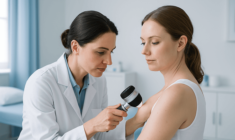
The hardest distinction is not among these four. It is between a dark, irregular seborrheic keratosis and melanoma, the most serious skin cancer, which can occasionally mimic one. Dermatologists use a handheld magnifier called a dermatoscope to look for features such as keratin plugs and a “brain-like” surface pattern that point toward a keratosis. When any doubt remains, they remove the growth and send it to a pathologist. That is the safety net, and it is why the right response to an unusual spot is an appointment rather than a guess.
What the evidence actually says, and how strong it is
Medical claims come in grades, and it helps to know which grade you are reading.
Strong evidence (large observational studies and pathology series). Both growths are benign. Decades of biopsy data show that seborrheic keratoses and cherry angiomas do not transform into cancer. Their prevalence rises steeply with age, and seborrheic keratoses cluster in families. These points are as settled as anything in dermatology.
Moderate evidence (molecular studies, smaller cohorts). Acquired mutations in FGFR3 and PIK3CA drive many seborrheic keratoses; acquired mutations in GNAQ and GNA11 appear in a majority of cherry angiomas studied. Sun exposure modestly increases seborrheic keratosis counts on exposed skin. These findings come from laboratory analyses of removed tissue rather than trials, so the causal chain is convincing but not complete.
Randomized trial evidence (limited). The clinic-applied hydrogen peroxide solution approved in 2017 was tested in two randomized, controlled trials against an inactive solution and cleared a meaningful share of treated lesions, with local irritation as the main side effect. That product is no longer marketed. Small trials have compared cryotherapy with curettage, generally finding similar clearance with different cosmetic trade-offs. No randomized trial supports any over-the-counter cream or home remedy for either growth.
Expert opinion and case reports. The association between a sudden eruption of seborrheic keratoses and internal cancer, known as the sign of Leser-Trélat, rests on case reports and remains debated. Links between cherry angiomas and pregnancy hormones or liver disease are observational and inconsistent.
The practical reading: you can trust that these growths are harmless, you can trust that in-office removal works, and you should be skeptical of anything sold to you for home use.
What happens if seborrheic keratosis is left untreated?
Usually nothing. That is the honest and slightly anticlimactic answer, and it is backed by the largest body of evidence on the topic.
A seborrheic keratosis that is left alone typically does one of three things. Most stay exactly as they are for years. Some thicken slowly and darken, gaining a more warty surface. A minority fall off on their own, often after minor trauma, and then either stay away or regrow a paler version in the same spot. None of these paths lead to cancer, because the cells involved lack the machinery to invade deeper tissue or spread.
The realistic downsides are practical rather than medical. Thick keratoses on the back can catch on clothing and bra straps. Ones on the scalp snag combs. When a growth is rubbed repeatedly it can become inflamed, red at the edges, itchy, or crusted. An irritated seborrheic keratosis can look worrying, and that appearance, not any underlying danger, is what most often prompts a visit to the doctor. The inflammation settles once the friction stops or the growth is removed.
There is one genuine reason not to ignore a growth entirely: certainty. Leaving a spot untreated is perfectly reasonable once a clinician has confirmed what it is. Leaving a spot unexamined because you assume it is a harmless keratosis is a different decision. Melanoma occasionally hides in plain sight among a crop of keratoses, and the two can be difficult to tell apart without a trained eye. A single baseline skin check, with a note about which spots are known keratoses, converts “probably fine” into “confirmed fine” and gives you a reference point for the years ahead.
Is it okay to scratch off seborrheic keratosis at home?
No, and the reason is not squeamishness.
The viral clips make it look easy because a mature seborrheic keratosis is loosely attached. Its base sits above the deeper skin, so it can be levered off with a fingernail or tweezers. What the camera does not show is what follows. The raw base weeps and scabs. Bacteria on skin and under nails move into an open wound. Healing frequently leaves a pale or dark patch that lasts longer than the original growth would have, and in people with deeper skin tones the discoloration can persist for a year or more. Regrowth is common because fragments of the abnormal cells remain.
The larger problem is one of identification. Self-removal assumes you know what you are removing. Dermatologists with a dermatoscope and years of pattern recognition still send a percentage of suspected keratoses to the lab because they cannot be sure. If a spot that was actually a melanoma is scraped off at home, the tissue that would have allowed a pathologist to diagnose it is gone, the wound heals over, and the cancer continues beneath the scar unseen. That scenario is uncommon, but it is precisely the kind of rare, severe outcome that medical advice is built to prevent.
The same caution applies to picking at a cherry angioma. Because it is a knot of blood vessels, tearing it produces brisk bleeding that can be difficult to stop without pressure, and the angioma returns because the vessels beneath remain.
If a keratosis catches, itches, or bothers you enough to want it gone, that is a legitimate reason to ask a clinician to remove it. The procedure takes minutes and, unlike a bathroom-mirror attempt, includes the option of examining what came off.
Is there a cream that gets rid of seborrheic keratosis?
Not one you can buy today, and none you can use at home.
The only topical product ever approved by the FDA for seborrheic keratosis was Eskata, a concentrated hydrogen peroxide solution. It was designed for application by a clinician, not for patient use, and it was tested in randomized trials that showed clearance of a meaningful proportion of treated growths. It has been off the market since 2019. Whatever is currently advertised online as “the seborrheic keratosis cream” is not that product.
Over-the-counter creams fall into a few categories, and the evidence is thin across all of them. Exfoliating acids such as glycolic or salicylic acid can soften the surface of a thin keratosis and make it look slightly flatter, but they do not remove it, and repeated use irritates the surrounding skin. Retinoid creams, which speed up skin cell turnover, have been proposed and studied only in small, uncontrolled reports; they are not approved for this purpose, and using a prescription retinoid on a growth is an off-label decision that belongs to the prescribing clinician. Wart removers containing high-strength salicylic acid are formulated for a viral infection that seborrheic keratosis does not have, and they can burn healthy skin around the growth.
Then there is the home remedy aisle: apple cider vinegar, tea tree oil, hydrogen peroxide from the pharmacy shelf, iodine, castor oil. No randomized or controlled study supports any of these. Vinegar and drugstore hydrogen peroxide are mild acids and oxidizers; applied often enough they cause a chemical burn, which lifts the surface of the growth along with the skin underneath. The result is a wound, a scar, and, frequently, a keratosis that returns.
A cream that safely dissolves these growths at home would be welcome. The evidence says it does not yet exist.
What is the best way to get rid of seborrheic keratosis?
The best method is the one a clinician chooses for your particular growth, your skin tone, and the location, because each technique trades speed, scarring, and pigmentation risk differently.
Cryotherapy uses liquid nitrogen sprayed or applied to freeze the growth, which blisters and falls away over one to two weeks. It is quick and needs no anesthetic. Its main drawback is pigment change: freezing damages melanocytes, the pigment-producing cells, so it can leave a lighter patch, a risk that rises with deeper skin tones and on the face.
Shave removal uses a small blade to slice the growth off flush with the skin after a numbing injection. Because the tissue is removed intact, it can be sent to a pathologist, which makes this the preferred approach whenever there is any doubt about the diagnosis. Healing leaves a flat, sometimes slightly pale mark.
Curettage scrapes the growth away with a spoon-shaped instrument, often paired with light electrocautery to seal the base. It suits thicker lesions.
Electrodesiccation uses a fine electric current to dry out the growth, then it is wiped away. Dermatologists often favor it for the small, numerous facial lesions of dermatosis papulosa nigra because it can be controlled precisely.
Laser treatment, using devices that target water or pigment in the growth, is an option for cosmetic clearance of many lesions at once.
Small comparative trials suggest cryotherapy and curettage clear similar proportions of lesions, with patients often preferring the cosmetic result of curettage. Every method carries a chance of temporary redness, a small scar, or a change in skin color, and every method occasionally sees regrowth. Removal for purely cosmetic reasons is a reasonable choice; the decision about whether and how belongs in a conversation with the person holding the instrument.
Can cherry angiomas be removed, and do they come back?
Yes to the first question, and the answer to the second is nuanced: the one removed rarely returns, but new ones keep arriving.
Because a cherry angioma is a bundle of blood vessels, the goal of treatment is to seal or destroy those vessels rather than to scrape a surface. Three approaches dominate.
Electrocautery touches the angioma with a fine heated or electrified tip that coagulates the vessels. It is fast, works for lesions of almost any size, and typically leaves a small scab that heals within a week or two. A tiny pale or slightly indented mark can remain.
Vascular lasers, including pulsed-dye and certain green-wavelength lasers, deliver energy absorbed specifically by hemoglobin, the red pigment in blood. The vessel heats and collapses while the surrounding skin is spared. Lasers are the usual choice on the face and for people who want to treat many angiomas in one session. Temporary bruising and darkening are common; permanent scarring is unusual.
Shave removal is reserved for raised angiomas that bleed repeatedly or whenever the clinician wants tissue for the pathologist. Bleeding during the procedure is expected and controlled with cautery.
Cryotherapy is used less often for cherry angiomas because blood flow carries heat back into the tissue and makes freezing less reliable.
On recurrence, the evidence is largely observational but consistent: a properly treated angioma usually does not regrow at the same spot, yet the underlying tendency remains, and most people continue to develop new ones over the years. Removal is almost always cosmetic. Medical reasons include an angioma that bleeds often because of its position, or a red bump whose appearance the clinician cannot confidently confirm by eye, since a rare pigmented melanoma can occasionally look reddish.
Can a sudden crop of seborrheic keratoses mean something else is going on?
This is the one corner of the topic where the reassuring message needs a careful footnote.
Since the late 1800s, physicians have described patients who developed a large number of seborrheic keratoses very rapidly, sometimes over weeks, and were subsequently found to have a cancer elsewhere in the body, most often in the digestive tract. The pattern is called the sign of Leser-Trélat, after the two surgeons credited with describing it. The proposed mechanism is that certain tumors release growth factors into the bloodstream that stimulate skin cells to proliferate.
It is important to be honest about the quality of this evidence. It consists almost entirely of case reports and small series. There has never been a controlled study showing that people with a sudden eruption of keratoses are more likely to have cancer than people without one, and skeptics point out that because seborrheic keratoses are so common in the age group where cancers also occur, coincidence alone would produce many such stories. Several reviews have concluded that the sign is unproven, and leading dermatology references classify it as a possible but debated association.
What this means for you: the slow, steady accumulation of keratoses over years, which is how nearly everyone acquires them, is not the pattern described in these reports and carries no such implication. The specific scenario in the literature is an abrupt appearance of many new growths in a short time, often with itching, in a person who may also feel unwell in other ways. If that describes you, the sensible step is not alarm but a routine appointment where a clinician can take a history, examine the skin, and decide whether any further evaluation is warranted. That decision is theirs to make, based on the whole picture rather than the skin alone.
Common myths about seborrheic keratosis and cherry angiomas, corrected
Viral health content thrives on a kernel of truth wrapped in a confident claim. Here are the claims circulating most widely, and what the evidence says about each.
“They are a sign of toxins or a sluggish liver.” There is no evidence that seborrheic keratoses reflect anything about internal organ function. Cherry angiomas do appear somewhat more often in people with advanced liver disease, but that is an observational association in a small subgroup, not a diagnostic sign, and the overwhelming majority of people with angiomas have healthy livers.
“They turn into skin cancer if you leave them.” Decades of pathology data show they do not. The concern with a dark growth is that it might have been a cancer all along and misidentified, which is why examination matters and why the growth itself is not the threat.
“Apple cider vinegar dissolves them.” Vinegar is a weak acid. Applied repeatedly it causes a chemical burn that can lift the surface of a keratosis, along with healthy skin. What the before-and-after photos do not show is the scar, the discoloration, or the regrowth months later. No controlled study supports the practice.
“Cherry angiomas mean you have too much bromine in your body.” This claim traces to older, isolated case reports involving occupational chemical exposure. It has never been confirmed in a broader population and does not apply to ordinary life.
“If it falls off on its own, it is gone for good.” Sometimes. Just as often a paler or flatter version regrows in the same spot because the underlying cells remain.
“You can freeze them off with wart spray from the pharmacy.” Home freezing kits are designed for warts and reach shallower temperatures than clinic liquid nitrogen. Results are inconsistent, blistering is common, and the identification problem remains: you are treating a spot no one has confirmed.
“They are contagious.” Neither growth involves a virus or bacterium. Sharing towels or touching them spreads nothing.
When to see a doctor about a seborrheic keratosis or cherry angioma
Most of these growths never need a medical visit beyond a routine skin check. Certain patterns, however, warrant an appointment, and a few call for prompt attention.
Schedule a visit when:
- A growth is new and you are not sure what it is, particularly if it is dark, irregular in outline, or has more than one color.
- An existing spot changes noticeably in size, shape, color, or texture over weeks to months.
- One growth looks different from all your others, the so-called “ugly duckling.”
- A spot bleeds without being scratched or bumped, or a small wound in that area will not heal within a few weeks.
- A growth becomes persistently itchy, painful, or inflamed.
- Many new keratoses appear rapidly over a short period.
- You want a growth removed because it catches on clothing, bleeds when shaved, or bothers you cosmetically.
Seek care promptly, rather than waiting for a routine slot, if a growth develops a spreading red border with warmth and swelling, which can indicate infection, or if a cherry angioma bleeds heavily and does not stop after ten minutes of firm, continuous pressure.
What to expect at the visit is reassuringly simple. A clinician will look at the spot, usually with a dermatoscope, and compare it to the rest of your skin. In most cases they will confirm the diagnosis by eye and tell you no treatment is needed. If they have any doubt, they will remove the growth and send it to a pathologist, which is the only way to be certain. If you take a blood thinner, mention it before any removal, since it affects bleeding during the procedure.
Every decision about whether to remove a growth, which technique to use, and whether any further evaluation is warranted rests with the clinician who examines you. This article can explain the options; it cannot see your skin.
Frequently asked questions
What is the best way to get rid of seborrheic keratosis?
The best method depends on the growth’s thickness, its location, and your skin tone, so the choice belongs to the clinician examining you. Common in-office options include cryotherapy (freezing), shave removal, curettage (scraping), electrodesiccation, and laser. Shave removal is preferred when there is any doubt about the diagnosis because the tissue can be examined by a pathologist. All methods carry small risks of scarring, color change, or regrowth.
What happens if seborrheic keratosis is left untreated?
Usually nothing harmful. Most seborrheic keratoses stay the same for years, some slowly thicken or darken, and a few fall off on their own. They do not turn into cancer. The practical downsides are irritation from clothing or jewelry and occasional inflammation. The one reason not to ignore a growth entirely is to have a clinician confirm it is a keratosis rather than something that merely resembles one.
Is there a cream that gets rid of seborrheic keratosis?
Not one available today. The only FDA-approved topical treatment, a clinic-applied hydrogen peroxide solution approved in 2017, was withdrawn in 2019. Over-the-counter exfoliating acids may soften the surface but do not remove the growth, and no controlled study supports vinegar, tea tree oil, or similar home remedies. Any prescription cream used for this purpose would be off-label, a decision that rests with the prescribing clinician.
Is it okay to scratch off seborrheic keratosis?
No. Although a mature keratosis is loosely attached and can be pried off, doing so risks infection, prolonged discoloration, and regrowth from cells left behind. More importantly, a dark growth that looks like a keratosis can occasionally be a melanoma, and scraping it off destroys the tissue needed to diagnose it. If a growth bothers you, ask a clinician to remove it; the procedure takes minutes.
What causes cherry angiomas to suddenly appear?
Cherry angiomas arise from acquired mutations in genes such as GNAQ and GNA11 inside blood-vessel cells, and their number rises steadily with age. A temporary burst of new angiomas is common in pregnancy and has been observed in some people with liver disease, though those associations are observational and inconsistent. Family history plays a role. Sudden appearance of several red dots is normal and not a sign of internal illness.
Can a cherry angioma be removed permanently?
A properly treated cherry angioma rarely regrows in the same spot. Electrocautery, vascular laser, and shave removal all seal or remove the abnormal vessels. What cannot be prevented is the tendency to form new angiomas elsewhere, which continues with age. Removal is almost always cosmetic; medical reasons include an angioma that bleeds repeatedly because of its position or one whose appearance a clinician cannot confirm by eye.
How can I tell a seborrheic keratosis from melanoma?
You cannot do so reliably on your own, and neither can many clinicians without a dermatoscope. Seborrheic keratoses typically look stuck-on, waxy, and sharply bordered, but dark or irregular ones can mimic melanoma closely. A clinician looks for features such as keratin plugs and a brain-like surface pattern, and removes anything uncertain for pathology. Any spot that changes, bleeds, or looks unlike your others deserves an examination.
Do seborrheic keratoses spread or multiply?
They multiply in the sense that most people develop more over time, sometimes dozens, but they do not spread from one spot to another and are not contagious. Each growth is a separate cluster of cells that acquired its own mutation. The tendency to form many keratoses runs in families and increases with age, and sun exposure modestly raises counts on exposed skin.
Why does a cherry angioma bleed so much when scratched?
Because it is a compact knot of dilated blood vessels sitting just under the surface, so even a small tear opens several vessels at once. The bleeding can look dramatic but stops with firm, continuous pressure for several minutes. Seek care if bleeding continues beyond ten minutes of pressure. Repeated bleeding from an angioma in a spot you shave or rub is a reasonable reason to have it treated.
Does a sudden crop of seborrheic keratoses signal cancer?
Rarely, and the evidence is weak. The sign of Leser-Trélat describes an abrupt eruption of many keratoses in someone later found to have an internal cancer, but it rests on case reports rather than controlled studies, and many experts consider it coincidental given how common keratoses are. The slow accumulation most people experience does not fit this pattern. A rapid outbreak is worth a routine visit, not alarm.
References
- Cleveland Clinic: Seborrheic Keratosis
- MedlinePlus Medical Encyclopedia: Seborrheic keratosis
- MedlinePlus Medical Encyclopedia: Cherry angioma
- NIH National Library of Medicine, StatPearls: Seborrheic Keratosis
This article is for general information only and is not a substitute for professional medical advice. Please consult a qualified doctor about your individual situation.

More from the Blog

Acute vs Chronic Urticaria: How the Six-Week Line Changes the Way Hives Are Investigated
Acute urticaria is hives that settle within six weeks; chronic urticaria is hives that keep appearing, on most days, for longer than six weeks.…

Seborrheic Dermatitis in Babies and Adults: How Cradle Cap and Adult Flares Are Approached
Cradle cap and adult seborrheic dermatitis are the same inflammatory skin condition seen at two different ages. In babies it shows as greasy, yellowish…
Glycolic vs Lactic vs Salicylic Acid: Choosing an Exfoliating Acid by Skin Type
Glycolic, lactic and salicylic acid all loosen the glue between dead skin cells, but they suit different skin. Glycolic acid, the smallest alpha-hydroxy acid,…

Microneedling at Home vs in Clinic: The Depth Difference and the Infection Risk
Microneedling at home typically uses needles 0.25–0.5 mm long that reach only the outer skin layers, while clinic devices reach 1–2.5 mm into the…

Which Folliculitis Type Is It? Why Bacterial, Fungal and Razor Causes Are Treated Differently
Folliculitis is inflammation of hair follicles, and the main types are bacterial (usually staphylococcal, or Pseudomonas after hot tubs), fungal (a yeast called Malassezia),…

Milia: The Tiny White Bumps That Are Not Pimples, and Why Squeezing Fails
Milia are tiny, firm, white or yellowish cysts, usually 1 to 2 millimeters wide, that form when the skin protein keratin gets trapped just…