

Seroma After Surgery: Why Fluid Collects and How It Is Managed

Key Takeaways
- A seroma usually appears 7 to 14 days after surgery, often within a day or two of a drain being removed, and most small ones are reabsorbed within a month.
- Mastectomy with axillary lymph node removal and abdominoplasty carry the highest seroma rates because both leave large raw surfaces across lymph-rich, mobile tissue.
- Quilting or progressive tension sutures that close dead space from inside are the best-supported preventive measure, backed by multiple randomised trials.
- Fibrin sealants have been tested in many trials and most pooled analyses show no reliable reduction in seroma formation.
- Serous fluid is sterile, so an uninfected seroma does not need antibiotics; redness, warmth, fever, or cloudy fluid are the signs that change that.
- Needle aspiration in clinic under sterile conditions is the standard treatment for larger collections; home drainage, heat, and vigorous massage add risk without evidence of benefit.
A seroma is a pocket of clear or straw-colored fluid that collects under the skin after surgery, most often where tissue was lifted or lymph nodes were removed. Many small seromas are absorbed by the body over weeks; larger or uncomfortable ones are usually drained with a needle in clinic. Fast swelling, redness, fever, or cloudy fluid should be checked promptly by your surgical team.
Scroll through any recovery forum this season and you will find the same photograph, taken from above: a soft, sloshing bulge below a healing incision, captioned with a question mark. Body-contouring operations, especially abdominoplasty and liposuction, have climbed steadily, and with them a wave of videos in which patients press a fingertip into the swelling and watch it ripple like a waterbed. Search interest in the word seroma has followed. As of mid-2026, it sits among the most-queried surgical complications, alongside “is this normal” and “should I be worried.”
The honest answer is: usually normal, occasionally worth a phone call, rarely dangerous. A seroma is one of the oldest and best-understood problems in surgery, yet it still unsettles people because it appears days after they thought the hard part was over.
This explainer walks through why that fluid gathers, which operations invite it, what the trials genuinely show about preventing and treating it, and the handful of signs that should move you from watchful patience to a same-day clinic visit.
What is a seroma, and why does it feel like a water balloon?
Picture the inside of a healing wound not as a sealed seam but as a shallow cave. When a surgeon separates skin and fat from the muscle beneath, or removes a lump, a lymph node, or a stretch of tissue, a space is left behind. The body wants to fill it. In the first days it floods the area with plasma-like fluid, the same watery component of blood that leaks from capillaries during any inflammation. A seroma (pronounced seh-ROH-muh) is simply that fluid pooling faster than the surrounding tissue can absorb it.
Serous fluid is pale yellow, thin, and sterile. It is not pus, and it is not blood, although it may be tinged pink early on. Because it sits in a soft pocket without rigid walls, it moves when you touch it, producing the signature slosh or fluid wave that patients describe. Doctors call this fluctuance: the sense that a lump shifts under pressure rather than staying firm.
Most seromas appear 7 to 14 days after an operation, often right after a surgical drain has been removed. Some are the size of a grape and go unnoticed until a follow-up visit. Others stretch the skin visibly and feel heavy or tight when you stand. Patient guidance from the NHS and Mayo Clinic lists seroma among the expected possibilities after mastectomy, lymph node removal, and abdominoplasty, which tells you how routine it is.
Here is the reassuring part. A seroma is a mechanical problem, not an infection and not a sign that something went wrong with the operation itself. The surrounding tissue, called the capsule, gradually knits down, the lymphatic channels reconnect, and the fluid is reabsorbed. That process can be slow, and occasionally it needs help, but the direction of travel is almost always toward resolution.
Why does fluid collect after surgery in the first place?
Three forces work together, and understanding them makes the management choices feel logical rather than arbitrary.

The first is dead space. Surgeons use that stark phrase for any gap between tissue layers that no longer touch. Every operation that lifts a flap of skin, removes bulk, or dissects widely leaves some. Nature abhors a vacuum, and the body fills the gap with the fluid that is already seeping from freshly cut capillaries.
The second is lymphatic disruption. The lymphatic system is a network of fine vessels that returns excess tissue fluid to the bloodstream. Cutting through fat and tissue severs thousands of these microscopic channels. Unlike blood vessels, they do not clot shut; they simply weep lymph into the wound until they scar over or find new routes. This is why operations in lymph-rich zones, such as the armpit (axilla) and groin, are notorious for persistent seromas.
The third is inflammation. Surgery triggers the same cascade as any injury: blood vessels become leakier, white cells arrive, and the fluid that bathes the area becomes richer in protein. Protein-rich fluid is harder to reabsorb, so the pocket persists.
Other factors tip the balance. Motion matters: tissue that slides against the layer beneath it with every step or arm swing cannot seal. Larger body surface area means more raw tissue and more leakage. Energy devices used to cut and coagulate tissue, including electrocautery, leave a thin zone of heat-injured cells that may weep more than a scalpel cut. None of these are failures of technique. They are the ordinary physics of healing, and surgeons spend considerable effort trying to counter them with drains, compression, and suturing methods that close dead space from within.
Which operations carry the highest seroma risk?
Not all surgeries are equal here. Risk rises with how much tissue is lifted, how many lymphatic channels are cut, and how mobile the area is afterward.
Breast surgery sits near the top. After mastectomy, the removal of a breast, a flat raw surface the size of a dinner plate is left under the skin, and reported seroma rates in observational series span a wide range, commonly cited between roughly 15 and 50 percent depending on how the condition is defined. Add axillary lymph node dissection, the removal of lymph nodes under the arm, and the rate climbs further because the lymphatic highway to the arm has been interrupted. The NHS mastectomy guidance names seroma as a recognized after-effect and explains that it is typically drained in clinic.
Abdominoplasty, the surgical tightening of the abdominal wall with removal of excess skin and fat, is the other headline procedure. The skin flap is elevated across most of the abdomen, so the dead space is enormous, and the area moves with every breath, cough, and step. Mayo Clinic lists fluid accumulation (seroma) first among its stated risks for this operation.
Other common settings include:
- Liposuction, particularly when large volumes are removed or when it is combined with skin excision.
- Hernia repair, especially large incisional or inguinal hernias repaired with mesh, where fluid can gather between the mesh and the tissue.
- Groin and pelvic lymph node surgery for melanoma or gynecologic cancers.
- Orthopedic and spinal procedures with wide soft-tissue exposure.
- Breast augmentation, breast lift, and reduction, where seromas are less common but can form around an implant or along a long incision.
Personal factors matter too. Higher body weight, larger resection volumes, smoking, poorly controlled diabetes, and the use of certain blood-thinning medicines are repeatedly associated with higher seroma rates in observational studies, although association is not the same as cause, and no single factor predicts who will develop one.
Seroma after tummy tuck: why body contouring is the current flashpoint
The phrase seroma after tummy tuck has become the single most common way people arrive at this topic, and the reason is partly demographic. Body-contouring surgery has grown as more adults seek skin removal after significant weight loss, whether through lifestyle change, bariatric surgery, or newer weight-management medicines. Larger skin excisions mean larger raw surfaces.
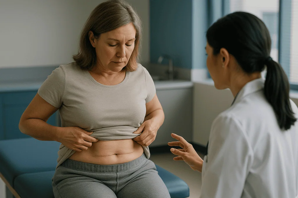
There is also a social-media effect. Recovery diaries are filmed daily, and the second week, when drains come out and swelling can shift into a defined fluid pocket, makes for alarming footage. A lower-abdominal seroma often appears as a soft, jiggling mound just above the pubic area or over the hips, where gravity pulls fluid when the patient stands. Viewers who have had similar surgery recognize it instantly, and the comment threads fill with home remedies of wildly varying quality.
Surgeons have responded to the underlying problem with technique rather than slogans. Many now use progressive tension sutures, a method of stitching the lifted flap back down to the muscle layer at multiple points so that dead space is closed from the inside. This has allowed some teams to offer drainless abdominoplasty. Others keep drains in longer, add abdominal compression garments for several weeks, or use liposuction more conservatively over the flap.
Patients are told to expect swelling, and swelling is not a seroma. The distinction is texture and timing. Generalized firm puffiness across the whole abdomen in week one is inflammation. A discrete, soft, shifting bulge that appears or grows after the drains are out is more likely fluid. Only an examination, sometimes with ultrasound, settles it.
The key message for anyone considering this surgery: ask your surgeon beforehand how they manage dead space, what their own seroma rate looks like, and what you should do if a fluid pocket appears after you go home. A clear plan turns a frightening bulge into a manageable follow-up visit.
What changed recently in how seroma is understood and managed
Seroma is old news in surgical textbooks, but the way it is handled has shifted over the past several years, and the current patient-facing guidance reflects that.
First, dead-space closure has moved from niche to mainstream. Quilting or progressive tension sutures, once associated mainly with abdominoplasty, are now widely studied after mastectomy and lymph node surgery. Randomised trials published through the early 2020s have generally found lower seroma rates and earlier drain removal when the flap is sutured down, which is why this appears increasingly in surgical protocols rather than only in research papers.
Second, the pendulum on drains has swung toward shorter use. Older practice left suction drains in for one to two weeks. Trials over the past decade suggest that removing them earlier, once output has fallen, does not substantially raise seroma rates in most settings, and it reduces discomfort and infection risk. Current NHS and Mayo Clinic mastectomy information describes drains as a short-term measure, typically removed within days to about a week, with clinic aspiration available if fluid later collects.
Third, infection control guidance has been refreshed. The CDC’s patient-facing material on surgical site infections, updated in 2024, emphasizes the warning signs, redness, warmth, drainage, and fever, that separate a benign fluid collection from an infected one. That distinction matters because a sterile seroma rarely needs antibiotics, and unnecessary antibiotics carry their own harms.
Fourth, imaging has become more routine. Point-of-care ultrasound in clinic lets a surgeon confirm fluid, estimate its volume, and guide a needle safely, rather than relying on touch alone.
What has not changed is the fundamental approach: observe small collections, aspirate large or symptomatic ones, and reserve more invasive options for the persistent minority. The tools are sharper; the philosophy is the same.
Seroma vs hematoma vs infection: how clinicians tell them apart
Three different things can swell a surgical site, and they call for three different responses. A hematoma is a collection of blood rather than serous fluid. An abscess is a collection of pus caused by infection. Telling them apart is the first job at any follow-up visit, and the clues are consistent enough to summarize.
| Feature | Seroma | Hematoma | Abscess (infection) |
|---|---|---|---|
| Typical timing | Days 7–14, often after drain removal | First 24–72 hours | Days 5–14 or later |
| Feel | Soft, shifting fluid wave | Firm, tense, may feel like a bruise under tension | Hot, tender, may be firm or fluctuant |
| Skin color | Normal or slightly stretched | Purple, blue, or dark bruising | Red, spreading warmth |
| Pain | Mild heaviness or tightness | Often significant, sudden | Throbbing, worsening |
| Fever | Absent | Usually absent | Common |
| Fluid if drained | Clear or straw-colored | Dark blood or clot | Cloudy, thick, foul-smelling |
| Usual management | Observe or aspirate | Pressure, sometimes surgical evacuation | Drainage plus antibiotics |
A fourth entity, the lymphocele, is a seroma made predominantly of lymph rather than plasma. It behaves like a seroma but can be more stubborn, particularly after groin or pelvic lymph node surgery, because the lymphatic leak keeps refilling the pocket.
The categories blur at the edges. A seroma can become infected if bacteria reach it, often through a needle or a wound that has not fully closed. An old hematoma liquefies over weeks into something that looks and feels like a seroma. This is why surgeons examine, and sometimes image or sample, rather than guess, and why a seroma that suddenly becomes warm, red, or painful is treated as a possible infection until proven otherwise.
How is a seroma diagnosed?
Most seromas are diagnosed by hand. A surgeon or nurse presses gently on the swelling with two fingers and feels for the fluid wave. They check the overlying skin for redness and warmth, ask about fever, and look at the incision for gaps or drainage. In the majority of cases, that is enough.
Ultrasound is the next step when the picture is unclear or when the surgeon wants to drain the pocket safely. Ultrasound is a painless scan that uses sound waves to show fluid as a dark, well-defined area beneath the skin. It distinguishes fluid from solid swelling, estimates volume, and reveals whether the collection has septations, which are thin internal walls that divide it into chambers. Septated collections drain less completely with a single needle pass.
Aspiration itself is diagnostic. If the fluid that comes back is clear or pale yellow, the question is answered. If it is bloody, the collection may be a liquefying hematoma. If it is cloudy, thick, or smells, the sample is sent to the laboratory for culture, a test that grows any bacteria present so the right antibiotic can be chosen.
CT or MRI scans are rarely needed for a simple seroma. They are reserved for deep collections after abdominal or spinal surgery, for suspected involvement of mesh or an implant, or when a surgeon needs to rule out other causes of swelling such as a recurrent hernia or, after cancer surgery, a recurrence of disease.
One point patients often miss: a seroma is not usually visible on routine blood tests, and a normal white cell count does not exclude infection in the early stages. Clinical examination remains the foundation, and that is why a quick in-person check is worth far more than a photograph sent to a friend who has had the same operation.
How long does a seroma last?
The question how long does a seroma last has no single answer, but the pattern is predictable enough to set expectations.
Small seromas, roughly the size of a walnut or smaller, are often reabsorbed within two to four weeks without any intervention. The body gradually balances production and absorption as the lymphatics heal and inflammation settles. These may never be noticed at all.
Medium collections, those that cause a visible bulge or a sensation of heaviness, commonly persist for four to eight weeks. Many are drained once or twice in clinic during that period, each aspiration temporarily emptying the pocket while the tissue continues to seal.
A minority become chronic, meaning they persist beyond about six to eight weeks and keep refilling after repeated drainage. These are more common after axillary or groin lymph node surgery, large abdominoplasty flaps, and hernia repairs with mesh. The pocket develops a smooth lining, sometimes called a pseudocapsule, which prevents the walls from sticking together. Chronic seromas account for a small fraction of all cases but a large share of the frustration in online forums.
Several things influence the timeline:
- Location: lymph-rich zones heal slower than areas with sparse lymphatics.
- Movement: the arm after breast surgery or the trunk after abdominoplasty keeps shearing the healing surfaces.
- Volume of surgery: more tissue removed, more surface to seal.
- Individual healing: age, nutrition, smoking status, and medical conditions all play a role.
The pattern to watch is trend, not size on any single day. A seroma that is slowly shrinking, even over weeks, is doing what it should. One that is enlarging, becoming painful, or stretching the skin to the point of shine or redness has stopped behaving, and that is when your team needs to see it.
How is a seroma managed: watchful waiting and needle aspiration
Management follows a ladder, and most people never climb past the second rung.
The first rung is observation. A small, painless, stable seroma is left alone. Surgeons may ask patients to wear a compression garment, keep the area supported, and avoid heavy lifting or vigorous repetitive movement for a few weeks. The reasoning is mechanical: less motion means less shearing, which gives the tissue layers a chance to adhere. Patients are usually asked to monitor the swelling and the skin over it, and to report changes.
The second rung is needle aspiration. In clinic, the surgeon cleans the skin, inserts a fine needle into the fluid pocket, and draws the fluid into a syringe. The procedure typically takes a few minutes and is usually described as mildly uncomfortable rather than painful, because the overlying skin is often partly numb after surgery. Aspiration relieves pressure and lets the pocket walls touch. Some seromas resolve after a single drainage; others refill and need the procedure repeated at weekly or fortnightly intervals.
Aspiration is not without small risks. Every needle pass carries a chance of introducing bacteria into a sterile space, which is why surgeons use careful sterile technique and avoid draining collections unnecessarily. There is also a small risk of injuring a blood vessel or, after reconstruction, an implant, which is why ultrasound guidance is used in tricky locations.
What patients should not do is attempt drainage at home. Kitchen-grade hygiene is not sterile, needle depth is unpredictable, and an infected seroma is a far larger problem than an uninfected one. The same applies to squeezing or massaging the pocket toward the incision in the hope it will leak out; this can open the wound.
If aspiration repeatedly fails, the surgeon moves up the ladder, and that is where the options become more varied and the evidence more mixed.
Drains, compression and sclerotherapy: what helps a stubborn seroma?
For the persistent minority, several further options exist. Their evidence base ranges from solid to thin, and the choice belongs to the treating surgeon, who knows the anatomy of the specific wound.
Reinsertion of a drain is the most common next step. A thin tube is placed into the pocket under sterile conditions and connected to a suction bulb, allowing continuous emptying for days to weeks while the cavity collapses. Drains are uncomfortable and need care, but they avoid repeated needle passes.
Sclerotherapy involves instilling an irritant into the emptied cavity to inflame the lining so that the walls stick together. Agents used in published series include doxycycline, an antibiotic repurposed for its irritant effect, and talc. Reports describe variable success, mostly from small observational series rather than large randomised trials, and the procedure can be painful. Any use is a specialist decision made by the surgeon, and this article does not describe amounts or technique.
Fibrin sealants and tissue glues, sprayed onto the raw surface during the original operation or injected later, have been tested in multiple randomised trials, chiefly in breast and axillary surgery. The pooled results are inconsistent, with some trials showing modest reductions in fluid volume and others showing no difference. Most reviews conclude that routine use is not supported.
Compression garments are near-universal after abdominoplasty and liposuction. Their logic is sound and surgeons strongly endorse them, but high-quality trial evidence that they reduce seroma incidence is limited; the recommendation rests largely on expert consensus.
Surgical revision is the final rung. The surgeon reopens the area, removes the smooth pseudocapsule that lines a chronic seroma, and closes the dead space with quilting sutures. It is rarely needed, but for a seroma that has persisted for months despite everything else, it is often definitive.
What the evidence actually says about seroma prevention and treatment
Grading the evidence honestly means separating what has been tested in randomised trials from what is practiced on the strength of experience.
Strong to moderate evidence (randomised trials, several meta-analyses):
- Quilting or progressive tension sutures after mastectomy and abdominoplasty reduce seroma rates and drain duration. Multiple trials point the same direction, although studies vary in how they define a seroma.
- Early drain removal based on output does not substantially increase clinically significant seromas in most breast surgery trials and reduces patient discomfort.
- Fibrin sealants have been tested extensively; results are inconsistent and most pooled analyses show no reliable benefit.
Moderate to weak evidence (smaller trials, observational cohorts):
- Shoulder immobilization or delayed arm exercises after axillary surgery may modestly lower seroma rates but risk stiffness; the trade-off is debated.
- Energy-device choice, such as ultrasonic scalpel versus electrocautery, shows small differences in some trials and none in others.
- Sclerotherapy for chronic seroma is supported mainly by case series.
Expert opinion and physiological reasoning (little trial data):
- Compression garments after body contouring.
- Activity restriction beyond the first couple of weeks.
- Dietary measures, including high-protein intake, to speed resolution.
Two caveats run through the whole literature. First, definitions differ: some studies count any fluid seen on ultrasound, others only collections needing aspiration, so headline percentages are not comparable across papers. Second, most trials come from breast cancer surgery, and findings do not automatically transfer to abdominoplasty or hernia repair.
Taken together, the evidence favors prevention at the operating table through dead-space closure, supports a conservative, stepwise approach afterward, and offers no single intervention that reliably speeds resolution once a seroma has formed. That is less satisfying than a viral fix, but it is what the data show.
Can you lower your seroma risk before surgery?
You cannot eliminate the risk, because dead space and cut lymphatics are built into the operation. You can shift the odds, and most of the levers are the same ones that improve healing generally.
Stop smoking well in advance. Nicotine narrows blood vessels and slows tissue repair, and smokers appear in the higher-risk group in nearly every observational series on wound complications. Surgeons commonly ask for several weeks of abstinence before elective procedures, and the benefit extends far beyond seroma.
Bring blood sugar under control if you live with diabetes. Elevated glucose impairs white cell function and collagen formation. Your surgical team may check a long-term glucose marker before scheduling.
Discuss every medicine and supplement with your surgeon and prescribing clinician. Anticoagulants, antiplatelet drugs, and some supplements affect bleeding and fluid dynamics. Never stop or adjust a prescribed medicine on your own; the decision to pause anything belongs to the clinician who prescribed it, weighing the reason it was started against the surgical risk.
Ask about technique. Reasonable questions include whether the surgeon uses quilting or progressive tension sutures, how they decide when to remove drains, and what their follow-up plan is if fluid collects. A surgeon who answers readily is giving you a window into how they think.
Plan for rest. The first two weeks after abdominoplasty or breast surgery are not the time to return to a physically demanding job. Arrange help with lifting, childcare, and household tasks before the operation rather than after.
Finally, follow the specific instructions you are given on garments, arm movement, and showering, even when they feel overcautious. Protocols differ between surgeons for good reasons tied to their technique, and the forum consensus is no substitute for the plan written for your wound.
Common myths about seroma, corrected
The comment threads beneath recovery videos are full of confident advice. Some of it is harmless; some is not.
Myth: A seroma means the surgeon made a mistake. Fluid collection is a known, expected risk of any operation that creates dead space. Experienced surgeons with excellent technique still see seromas. Rates vary by procedure and patient far more than by surgeon error.
Myth: You can drain it yourself with a clean needle. Household cleanliness is not sterility. Home aspiration risks infection, bleeding, and damage to deeper structures or implants. Infected seromas require antibiotics and sometimes surgery; sterile ones usually do not.
Myth: Massage will push the fluid out. Vigorous massage over a fresh wound can open the incision, worsen inflammation, and shear the healing layers apart. Gentle lymphatic drainage techniques are sometimes recommended by surgeons in specific situations, but only on their instruction.
Myth: A seroma always needs antibiotics. Serous fluid is sterile. Antibiotics do nothing to a sterile collection and add side effects and resistance risk. They are prescribed when examination or culture shows infection.
Myth: Heat packs will dissolve it. Heat increases blood flow and can increase fluid production in an inflamed area. There is no evidence that heat resolves seromas, and burns over partly numb post-surgical skin are a real hazard.
Myth: If it is not drained, it will turn into cancer or a permanent lump. Seromas are benign fluid. Chronic ones can develop a fibrous lining that needs surgical removal, but they do not transform into tumors.
Myth: Protein shakes make seromas disappear faster. Adequate nutrition supports healing in general. No trial shows that supplementing protein above normal dietary needs shortens seroma duration.
The thread that ties these together: seromas resolve through tissue adherence and reabsorption, both of which take time and are helped by rest and, when needed, sterile clinic drainage. Shortcuts tend to add complications rather than remove them.
When to see a doctor about a seroma
Most seromas are a follow-up-visit problem, not an emergency. A few signs change that calculation, and the CDC’s guidance on surgical site infection and the NHS’s post-operative advice converge on the same list.
Contact your surgical team the same day if you notice:
- Spreading redness, warmth, or streaking of the skin over or around the swelling.
- Fever, chills, or feeling generally unwell.
- Cloudy, thick, green, yellow, or foul-smelling fluid leaking from the incision.
- Rapid enlargement of the swelling over hours rather than days.
- Increasing pain, especially throbbing pain that wakes you or does not respond to the pain relief you were prescribed.
- The incision opening, gaping, or showing exposed deeper tissue.
- Skin over the swelling becoming shiny, thin, dusky, or developing a blister.
Seek emergency care if you have a high fever with confusion, a racing heart, difficulty breathing, or a sudden tense, painful swelling with bruising that suggests active bleeding.
Arrange a routine but prompt appointment if the swelling is soft and painless but still growing after two weeks, if it has not started shrinking by about six weeks, or if it interferes with movement, sleep, or wearing your compression garment.
After breast cancer surgery, any new swelling of the arm or hand on the operated side should also be reported, because it may signal lymphedema, a separate condition in which lymph fluid builds up in the limb, rather than a seroma at the surgical site.
Bring details with you: when the swelling appeared, whether it has changed, your temperature readings, and photographs taken in consistent lighting. Do not alter or stop any prescribed medicine, including blood thinners, before the appointment unless the prescribing clinician tells you to. Every decision about aspiration, antibiotics, imaging, or revision rests with the surgical team who performed the operation and knows exactly what was done inside the wound.
Frequently asked questions
What does a seroma after tummy tuck feel like?
It feels like a soft, shifting pocket of fluid under the skin, usually in the lower abdomen or over the hips, that ripples or sloshes when pressed. It is typically painless or mildly heavy and appears after the drains come out. General firm swelling across the whole abdomen in week one is inflammation, not a seroma. Only an examination, sometimes with ultrasound, confirms which one you have.
How long does a seroma last without drainage?
Small seromas often reabsorb within two to four weeks on their own. Medium collections commonly take four to eight weeks and may be aspirated once or twice along the way. A minority persist beyond six to eight weeks and are considered chronic; these are more likely after lymph node surgery or large abdominoplasty flaps and may need a drain or surgical revision.
What is the difference between a seroma vs hematoma?
A seroma contains clear or straw-colored serous fluid and usually forms a week or two after surgery, feeling soft and shifting. A hematoma contains blood, typically appears within the first 72 hours, feels firm and tense, and is often accompanied by purple or blue bruising and more pain. Both are examined by the surgical team; hematomas sometimes need urgent evacuation.
Is a seroma after mastectomy dangerous?
In most cases, no. Seroma is a common and expected occurrence after mastectomy, especially when lymph nodes under the arm are removed, and it is usually managed by observation or clinic aspiration. It becomes a concern if it grows rapidly, becomes red, hot, or painful, or if you develop fever, because those signs suggest infection or bleeding that needs prompt treatment.
Can a seroma go away on its own?
Yes, many do. As the lymphatic channels heal and the tissue layers stick back together, the body gradually reabsorbs the fluid. This is most reliable for small, painless collections. Larger ones may need one or more needle aspirations to help the pocket walls touch. A seroma that keeps refilling after repeated drainage is unlikely to resolve without a drain or surgical revision.
Does a seroma need antibiotics?
Not unless it is infected. Serous fluid is sterile, and antibiotics have no effect on a clean collection while adding side effects and resistance risk. Antibiotics are prescribed when examination shows spreading redness, warmth, fever, or when fluid drawn from the pocket is cloudy and grows bacteria on culture. That decision is made by the surgical team after assessing the wound.
Can I drain a seroma at home?
No. Home drainage risks introducing bacteria into a sterile space, injuring blood vessels or an implant, and reopening the wound. An infected seroma is a much larger problem than an uninfected one and may require antibiotics or surgery. Aspiration should be done in clinic under sterile conditions, often with ultrasound guidance, by the team who performed the operation.
Does a compression garment prevent seroma?
Surgeons widely recommend compression after abdominoplasty and liposuction because it limits movement between tissue layers and supports the flap, which is physiologically sensible. High-quality randomised trial evidence that garments reduce seroma incidence is limited, so the recommendation rests mostly on expert consensus. Follow your surgeon’s specific instructions on how long to wear it.
Why do seromas form after lymph node removal?
Lymph nodes sit on a network of fine lymphatic vessels that carry tissue fluid back to the bloodstream. Removing nodes cuts many of these channels, and unlike blood vessels they do not clot shut. They leak lymph into the wound until they scar over, which can take weeks. This is why axillary and groin surgery produce some of the most persistent seromas.
What is a chronic seroma and how is it treated?
A chronic seroma is a fluid collection that persists beyond roughly six to eight weeks and refills after repeated aspiration, often because a smooth lining has formed around it. Treatment options include placing a drain, sclerotherapy to inflame the lining so the walls stick, or surgical removal of the lining with quilting sutures. The surgeon chooses based on location, size, and what has already been tried.
References
- NHS: Mastectomy, including recovery and seroma
- CDC: About surgical site infections
- MedlinePlus: Surgical wound care, open
This article is for general information only and is not a substitute for professional medical advice. Please consult a qualified doctor about your individual situation.

More from the Blog

Fasting Before Surgery: The Real Rules on Food and Water, and Why They Exist
Most adults must stop eating solid food six to eight hours before anesthesia, while small amounts of clear fluids such as plain water are…

General Anaesthesia: How It Works, How Safe It Is and How You Recover
General anesthetics are medicines, given through a vein or as an inhaled gas, that temporarily quiet communication between brain cells so you feel nothing…

Tonsillectomy: How Long It Takes and What to Expect
The tonsillectomy operation itself usually takes about 20 to 30 minutes under general anesthesia, and most people go home the same day after a…

Smoking, Vaping and Surgery: Why Surgeons Insist You Stop, and for How Long
No, surgeons advise against smoking or vaping nicotine before any operation. Quitting at least four weeks ahead measurably lowers the risk of wound, heart,…

How Long Does General Anesthesia Last? Duration, Wake-Up and the Foggy Hours After
General anesthesia lasts exactly as long as the anesthesia team keeps delivering medication, whether the operation takes twenty minutes or eight hours. Once the…

Thyroidectomy: What It Is, How Long It Takes, Whether It Is Dangerous and Sleeping Afterwards
A thyroidectomy is surgery to remove all or part of the thyroid, the butterfly-shaped gland at the front of the neck. It is done…