

The Sacrum: The Keystone Bone Connecting Your Spine and Pelvis

Key Takeaways
- The sacrum begins life as five separate vertebrae (S1–S5) that fuse into a single bone, a process usually complete between the mid-teens and about age 30.
- Its name comes from the Latin os sacrum, "holy bone," translated from the ancient Greek hieron osteon, though historians still debate exactly why the ancients considered it sacred.
- The sacroiliac joints move only a few degrees and a couple of millimeters, yet research suggests they account for roughly 15 to 30 percent of chronic low back pain cases.
- Sacral insufficiency fractures in older adults with osteoporosis can occur without any fall, often hide on standard X-rays, and are frequently mistaken for ordinary back pain.
- Nerves passing through the sacral canal control bladder, bowel, and sexual function, new saddle-area numbness or loss of bladder control with back pain is a medical emergency.
- Inflammatory sacroiliitis typically causes morning stiffness that improves with movement, the opposite of mechanical strain, and often starts before age 45: a pattern worth mentioning to a doctor.
Quick Answer
The sacrum is a triangular bone at the base of the spine, formed from five vertebrae that fuse during early adulthood. Wedged between the two hip bones like a keystone, it transfers the weight of your upper body into the pelvis and legs through the sacroiliac joints. Pain near the sacrum most often comes from those joints, ligaments, or nearby muscles rather than the bone itself.
In yoga classes across the country, instructors keep telling people to “tuck the sacrum”, and most students dutifully tuck something, without being entirely sure what. Ask a room of adults to point to this bone and you will get gestures ranging from the mid-back to somewhere near the hip pocket.
That vagueness is a shame, because the sacrum quietly does one of the hardest jobs in the skeleton. Every time you stand, walk, or lift a bag of groceries, the entire weight of your head, arms, and torso funnels down the spine into this single wedge of bone, which splits the load and passes it to your legs. Roman engineers made arches stand for two thousand years using the same trick; the body got there first.
Here is what this underappreciated triangle actually is, why ancient anatomists called it sacred, and what it usually means when the area starts to ache.
Where Is the Sacrum, Exactly?
Slide your hand down the center of your lower back, past the inward curve of the waist, until the surface flattens and turns firm and slightly bony. That broad triangle sitting just above the crease of the buttocks is the sacrum. People who have two small dimples at the low back, sometimes nicknamed the dimples of Venus, can use them as landmarks: the sacrum spans the space between and just below them, where it meets the hip bones on either side.
On a skeleton, the answer to “where is the sacrum” is precise. It sits directly beneath the fifth and lowest lumbar vertebra, above the coccyx (tailbone), and wedged between the two large, wing-shaped iliac bones of the pelvis. The bone tilts forward at a noticeable angle, which is why its upper edge, a ridge called the sacral promontory, juts into the pelvic cavity and serves as a key landmark in obstetrics and pelvic measurements.
One detail surprises people: the sacrum is not just a back bone. Its front surface forms the rear wall of the pelvis, curving gently around the pelvic organs. The rectum lies directly in front of it, which is why deep pelvic conditions occasionally make themselves felt as an ache over the sacrum. You can touch its back surface through the skin; its front belongs entirely to the pelvis.
Sacrum Anatomy: Five Bones That Became One
Sacrum anatomy starts with a countable fact: you were born with five separate sacral vertebrae, labeled S1 through S5. During adolescence and early adulthood they knit together into a single solid bone, a process that typically finishes somewhere between the mid-teens and about age 30. Look closely at an adult sacrum and you can still read its history, four faint horizontal ridges mark the old boundaries between segments, like growth rings in a tree.
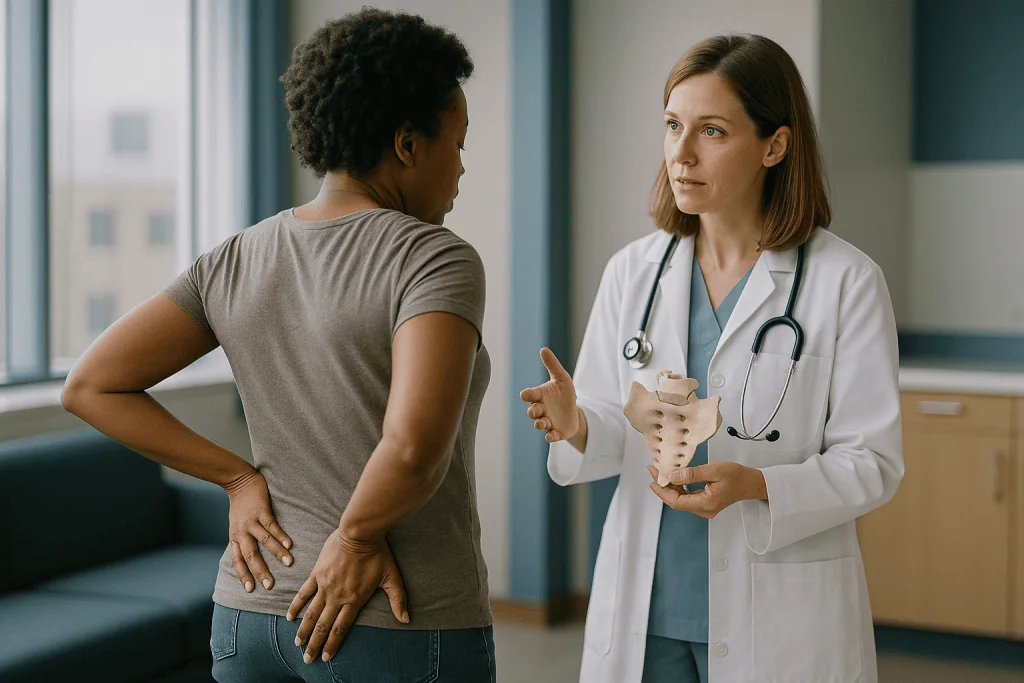
The finished bone has a distinctive architecture:
- The base, its broad top surface, carries the entire spine and joins the fifth lumbar vertebra at the lumbosacral joint.
- The apex, the narrow bottom tip, connects to the coccyx.
- The alae (Latin for “wings”) flare out on each side and meet the hip bones.
- Four pairs of sacral foraminaopenings on both the front and back surfaces, let nerves and blood vessels pass through, eight tunnels per side in total.
- The sacral canal runs down the middle, a continuation of the spinal canal, ending at a small opening near the bottom called the sacral hiatus.
Down the back of the bone runs a bumpy midline ridge, the median sacral crest: the fused remnants of what would have been individual spinous processes, the knobs you can feel along the rest of your spine. Rough ear-shaped patches on each side, called auricular surfaces, form the sacrum’s half of the sacroiliac joints.
Why Is the Sacrum Called the Sacred Bone?
The name is a genuine antique. “Sacrum” is short for the Latin os sacrumliterally “holy bone”, which itself was a translation of the ancient Greek hieron osteon, the term used by anatomists in the classical world. What nobody has settled, even after centuries of scholarly argument, is why the ancients considered it sacred in the first place.
Three theories keep circulating. The first holds that the sacrum, as a large bone surrounded by prized cuts, was part of the portion of sacrificial animals offered to the gods. The second points to old beliefs, appearing in several traditions, that this dense, slow-to-decay bone was the seat of the soul or the seed from which the body would be resurrected. A third, more prosaic explanation notes that the Greek word hieron could also carry the sense of “great” or “mighty,” so the name may simply have meant “the big bone,” and the mystical readings came later. Historians of medicine generally admit the evidence does not let us pick a winner.
As for other names: in everyday clinical language you will hear the region called the sacral spine, and its segments referred to as S1 through S5. Older anatomy texts sometimes used “vertebra magna.” Whatever the label, it is the same triangular keystone doing the same unglamorous structural work.
Is the Sacrum Part of the Back or the Buttock?
Honest answer: both, and that dual citizenship explains a lot of confusion at the doctor’s office. Structurally, the sacrum is the fifth and final section of the vertebral column, after the cervical, thoracic, and lumbar regions, so anatomists count it as spine. At the same time, it is a full member of the pelvic girdle, locked between the hip bones and forming the back wall of the pelvis.
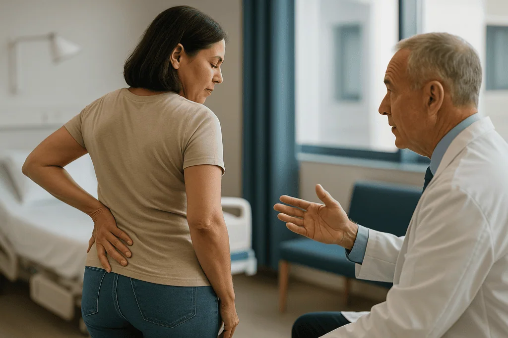
Where you feel it muddies things further. The bone’s upper portion sits at the very bottom of what most people call the low back. Its lower half lies beneath the upper buttock, and the gluteus maximus, the largest muscle in the body, anchors part of its origin directly onto the sacrum’s back surface. Piriformis, a small deep hip muscle famous for irritating the nearby sciatic nerve, attaches to the sacrum’s front. So an ache arising from the sacrum or its joints is often experienced as one-sided upper-buttock pain rather than classic back pain, and people understandably assume they have pulled a muscle in the hip.
Clinicians usually file sacral-area complaints under the broad umbrella of low back pain, which is one reason sacroiliac problems historically flew under the radar. When describing pain to a healthcare professional, pointing to the exact spot, center of the low back, over one dimple, deep in the buttock, is genuinely more useful than any label.
What Does the Sacrum Actually Do? The Keystone Job
Picture a stone archway. The wedge-shaped block at its peak, the keystone, takes the downward force of everything above it and redirects that force sideways into the columns. Remove the keystone and the arch collapses. The sacrum plays precisely this role in the pelvis: a wedge, wider at the top than the bottom, driven between the two iliac bones so that body weight locks it more tightly in place rather than pushing it through.
Every load your upper body generates travels down the stack of 24 movable vertebrae, concentrates on the sacrum’s base, and then splits left and right through the sacroiliac joints into the hip bones, down through the hip sockets, and into the legs. The traffic runs the other way too. Each footstrike while walking or running sends ground-reaction force, briefly exceeding your body weight, up the leg, and the sacrum is where those left-right impulses reunite with the spine.
Beyond load-bearing, the sacrum moonlights in several roles. It forms the protective back wall of the pelvic cavity, shielding the rectum, bladder, and reproductive organs. It anchors some of the strongest ligaments in the body along with major muscles of the hip and the pelvic floor. And its central canal safely conducts the final nerve roots of the spinal cord to their destinations. For a bone most people cannot locate, that is a crowded job description.
The Sacroiliac Joints: Small Motion, Big Consequences
On each side of the sacrum, its rough auricular surface meets a matching surface on the ilium to form the sacroiliac joint: SI joint for short. These are strange joints by any standard. They are genuine, fluid-lined joints, yet they are lashed down by some of the thickest ligaments in the human body, including the interosseous, sacrotuberous, and sacrospinous ligaments. The result is a joint that moves only a few degrees and shifts a couple of millimeters at most.
That tiny motion is not a design flaw; it is the point. A little give lets the pelvis absorb shock and accommodate the twisting of walking, in which one hip swings forward while the other trails. Total rigidity would transmit every jolt straight up the spine; too much looseness would make the keystone unstable. The SI joints sit in the narrow band between.
When something disturbs that balance, a fall, asymmetric strain, arthritis, or ligament laxity, the joint can become a persistent pain generator. Research on chronic low back pain suggests the SI joint is the culprit in roughly 15 to 30 percent of cases, a share that surprised many spine specialists when studies began quantifying it. During pregnancy, the hormone relaxin deliberately loosens these ligaments to prepare the pelvis for delivery, which helps explain why SI-area pain is so common in expectant mothers. With age, the joints tend to stiffen, and in some older adults they partially fuse.
The Nerves That Travel Through the Sacrum
Hollow down its middle, the sacrum is as much a conduit as a strut. The spinal cord itself ends higher up, around the first or second lumbar vertebra, but a bundle of nerve roots: the cauda equina, Latin for “horse’s tail”, continues down through the sacral canal. Pair by pair, the sacral nerve roots exit through those front and back foramina to wire up the lower body.
The passenger list matters. Roots from L4 through S3 merge to form the sciatic nerve, the body’s largest, which runs down the back of each leg. The pudendal nerve, drawn mainly from S2 to S4, supplies the pelvic floor. Sacral nerves also carry the parasympathetic signals that control bladder emptying, bowel function, and sexual function, some of the most quality-of-life-critical wiring in the body. The small opening at the sacrum’s base, the sacral hiatus, is the access point clinicians sometimes use for certain injection and anesthesia procedures precisely because it leads into this canal.
This anatomy is why doctors treat certain symptoms in the sacral region as urgent. New numbness in the “saddle” area (the parts that would touch a bicycle seat), loss of bladder or bowel control, or rapidly progressing leg weakness can signal compression of the cauda equina: a genuine emergency where hours matter, not days. Those symptoms warrant an emergency department, not a wait-and-see approach.
How the Sacrum Changes from Birth to Older Age
The sacrum you have at 70 is a very different structure from the one you had at 7. A child’s sacrum is five distinct vertebrae separated by cartilage, flexible enough to grow with the pelvis. Fusion begins in adolescence, proceeds from the bottom segments upward, and is usually complete by the mid-20s, occasionally as late as 30. Forensic scientists actually use the degree of sacral fusion as one clue when estimating age from skeletal remains.
Adult sacra also differ by sex in ways that reflect childbirth. In a typical female-pattern pelvis, the sacrum is shorter, wider, and less curved, and it tilts farther back, all of which enlarges the pelvic outlet a baby must pass through. A typical male-pattern sacrum is longer, narrower, and more sharply curved. These are averages with plenty of individual overlap, but the pattern is consistent enough that anthropologists rely on it.
Later in life, two changes deserve attention. First, the SI joints gradually stiffen, which is one reason the springy gait of youth flattens with age. Second, and more important, the sacrum’s interior is rich in spongy trabecular bone, exactly the type that thins fastest in osteoporosis. When bone density drops, the sacrum can develop insufficiency fractures from loads it once handled easily, sometimes with no fall at all. Bone-density screening, as recommended by a clinician, protects this bone as much as the more famous hip and wrist.
What Causes Pain in the Sacrum?
Here is the counterintuitive part: sacrum pain rarely comes from the bone itself. A solid, fused block of bone has few ways to hurt. The usual troublemakers are the structures attached to it and the joints on either side of it.
- Sacroiliac joint dysfunction. Strain, asymmetric loading (think stepping hard off an unseen curb), leg-length differences, or prior lumbar surgery can irritate an SI joint, producing one-sided pain near the dimple that may radiate into the buttock or thigh.
- Sacroiliitis. Inflammation of the SI joints, which can accompany inflammatory conditions such as ankylosing spondylitis and related forms of axial spondyloarthritis. The telltale pattern: stiffness and pain worst in the early morning or after sitting still, easing with movement, often beginning before age 45.
- Pregnancy-related pelvic girdle pain. Hormonal ligament loosening plus a shifting center of gravity; by some estimates it affects as many as 1 in 5 pregnancies to some degree.
- Trauma and fractures. Falls, car accidents, or, in people with thinned bones, everyday loads (more on this below).
- Referred pain. Problems in the lower lumbar discs and joints frequently project pain downward over the sacrum.
- Rare but serious causes. Infection or tumors in the sacral region are uncommon, which is exactly why persistent night pain, fever, or unexplained weight loss alongside sacral pain deserves prompt medical evaluation.
Muscular culprits round out the list: an irritated piriformis or overworked gluteal attachments can ache convincingly right over the bone.
Sacrum Pain, Tailbone Pain, or Sciatica? How to Tell Them Apart
Three common complaints get mixed up in this neighborhood, and the distinctions genuinely change what a clinician looks for. None of the patterns below is diagnostic on its own, overlap is common, but they are useful starting points.
| Feature | Sacrum / SI joint pain | Tailbone pain (coccydynia) | Sciatica |
|---|---|---|---|
| Typical location | One side of the very low back, near the dimple; may spread to the buttock or upper thigh | Dead center at the bottom tip of the spine, between the buttocks | Runs from the buttock down the back of one leg, sometimes to the foot |
| What aggravates it | Standing up from sitting, climbing stairs, standing on one leg, rolling over in bed | Sitting, especially on hard surfaces, and the moment of standing up | Coughing, sneezing, prolonged sitting, bending forward |
| Common triggers | Strain, pregnancy, arthritis, inflammatory conditions | A fall onto the buttocks, childbirth, long hours seated | Nerve-root irritation, often from a lumbar disc |
| Character | Deep, dull ache; occasionally sharp with position changes | Localized soreness or sharp pain on pressure | Electric, burning, or tingling; may include numbness or weakness |
A quick anatomy check helps too. The coccyx is the small, mobile chain of bones below the sacrum: a different structure with different problems. And true sciatica is defined by its path down the leg; pain that stays parked over the sacral triangle usually is not sciatica, even when it is severe.
Sacral Fractures: The Break That’s Easy to Miss
Sacral fractures come in two very different flavors, and the second one hides in plain sight.
The first is traumatic: high-energy events such as car crashes or falls from height. These fractures announce themselves loudly, are often part of a broader pelvic injury, and get imaged and managed in emergency settings. A related cousin, the sacral stress fracture, shows up in long-distance runners, dancers, and military recruits whose training volume outpaces the bone’s ability to repair itself: a repetitive-load injury rather than a single event.
The second flavor is the sacral insufficiency fracture, and it deserves far more public awareness than it gets. In an older adult with osteoporosis, the sacrum’s spongy interior can crack under ordinary daily loads, walking, standing, a minor stumble that would once have meant nothing. The symptoms are maddeningly vague: aching low back or buttock pain, worse with standing and walking, better with rest. There is often no fall to report. Standard X-rays frequently look normal because the fracture lines hide within trabecular bone, so the diagnosis regularly requires MRI or other advanced imaging. Studies of older patients admitted with unexplained low back pain have repeatedly found these fractures going unrecognized for weeks.
The practical message: in anyone over 65, or anyone with known low bone density, new, persistent pain over the sacrum after even trivial strain is worth a medical visit and a low threshold for imaging, not months of hoping it fades.
When to See a Doctor About Sacrum Pain
Most aches over the sacrum behave like ordinary low back pain: they ease meaningfully within a few weeks with continued gentle activity. The NHS and other major health bodies agree that staying active generally beats prolonged rest for recovery. But certain features change the calculus, and a few demand same-day attention.
Seek emergency care if sacral or low back pain comes with new numbness in the saddle area, loss of bladder or bowel control, or rapidly worsening leg weakness, possible signs of cauda equina compression, or if the pain follows a serious accident or fall.
Book a prompt appointment if any of the following applies:
- Pain persists beyond three to four weeks or keeps intensifying despite sensible self-care
- Pain wakes you at night or does not ease when you lie down
- You have fever, chills, or unexplained weight loss alongside the pain
- You have a history of cancer, osteoporosis, or long-term steroid treatment
- You are under 45 with morning stiffness lasting more than 30 minutes that improves with exercise: the classic inflammatory pattern worth evaluating
- The pain began during or after pregnancy and interferes with walking or sleep
None of these guarantees anything serious; most turn out to have manageable explanations. They are simply the situations where evidence says an examination, and sometimes imaging or blood testsmeaningfully improves the odds of finding a treatable cause early.
How to Keep Your Sacrum and SI Joints Healthy
You cannot exercise a fused bone, but you can absolutely condition everything responsible for protecting it. Two strategies carry the strongest evidence.
Build the muscular scaffolding. The gluteal muscles, deep abdominals, and pelvic floor act as active stabilizers for the SI joints, living guy-wires supplementing the ligaments. Regular walking, and strength work such as bridges, squats to a comfortable depth, and step-ups, keeps that system honest. People who sit most of the day benefit from simply breaking up the stillness: standing, strolling, or shifting position every 30 to 60 minutes changes the load pattern on the pelvis, and variety itself appears protective. There is no single “correct” sitting posture the evidence crowns; the best posture is your next one.
Bank bone density. Because the sacrum is rich in the spongy bone that osteoporosis attacks first, the standard bone-health playbook applies directly: weight-bearing and resistance exercise, adequate calcium and vitamin D from food (dairy, fortified alternatives, canned fish with bones, leafy greens, plus sensible sun exposure or dietary vitamin D sources), not smoking, and limiting alcohol. These habits compound quietly for decades, peak bone mass built by your early 30s becomes the reserve you draw on at 70.
One last piece of perspective. The sacrum has held up keystone duty through every step of human history without asking for attention. Give it strong muscles, dense bone, and frequent movement, and it will likely keep doing the job without ever making you look up its name again.
Frequently asked questions
Why is the sacrum called the sacred bone?
The name comes from the Latin os sacrum, meaning “holy bone,” which translated the ancient Greek hieron osteon. Historians offer three main theories: the bone was part of animal sacrifices offered to the gods, ancient traditions held it was the seat of the soul or the seed of bodily resurrection because it decays slowly, or the Greek word simply meant “great bone.” The evidence does not definitively favor any single explanation.
What’s another name for the sacrum?
Clinicians commonly call the region the sacral spine, and its five fused segments are labeled S1 through S5. The full Latin name is os sacrum, and some older anatomy texts used “vertebra magna.” All of these refer to the same triangular bone at the base of the spinal column, sitting between the fifth lumbar vertebra above and the coccyx, or tailbone, below.
Is the sacrum part of the back or the buttock?
Both, in practical terms. Anatomically the sacrum is the final section of the vertebral column and also a structural member of the pelvis. Its upper portion sits at the very bottom of the low back, while its lower half lies beneath the upper buttock, where the gluteus maximus muscle attaches directly to it. That is why sacral problems are often felt as one-sided buttock pain rather than classic back pain.
What causes pain in the sacrum?
Most sacral-area pain comes from structures around the bone rather than the bone itself. Common causes include sacroiliac joint dysfunction, sacroiliitis (inflammation sometimes linked to conditions like ankylosing spondylitis), pregnancy-related pelvic girdle pain, muscle strain, and pain referred from the lumbar spine. Fractures, traumatic, stress-related in athletes, or insufficiency fractures in people with osteoporosis, are less common. Rarely, infection or tumors are responsible, which is why persistent or worsening pain deserves medical evaluation.
Can you break your sacrum?
Yes, in three main ways. High-energy trauma such as car accidents or falls from height can fracture it outright. Repetitive loading can cause stress fractures in runners, dancers, and military recruits. And in older adults with osteoporosis, insufficiency fractures can occur during ordinary activities with no fall at all. That last type often hides on standard X-rays and may require MRI to diagnose, so persistent unexplained sacral pain in an older adult warrants a medical visit.
What is the difference between the sacrum and the tailbone?
They are neighboring but separate structures. The sacrum is the large triangular bone formed from five fused vertebrae; the tailbone, or coccyx, is the small chain of three to five tiny bones hanging below it. Tailbone pain (coccydynia) is typically felt dead center at the very bottom of the spine and worsens with sitting, while sacral or sacroiliac pain usually sits higher and off to one side, near the low-back dimples.
How many bones make up the sacrum?
Five. You are born with five separate sacral vertebrae, labeled S1 through S5, separated by cartilage. During adolescence and early adulthood they fuse into a single solid bone, usually finishing by the mid-20s and occasionally as late as age 30. Faint horizontal ridges on an adult sacrum still mark where the original segments joined, and forensic scientists use the degree of fusion as one clue when estimating age from skeletons.
Does the sacrum move?
Barely, and that is by design. The sacrum itself is a fused, rigid bone, but it connects to the hip bones through the sacroiliac joints, which allow only a few degrees of rotation and a millimeter or two of glide. This tiny motion helps the pelvis absorb shock during walking. Claims that the sacrum shifts dramatically “out of place” are not supported by anatomical evidence; the joints are bound by some of the body’s strongest ligaments.
Why does my sacrum hurt during pregnancy?
Pregnancy hormones, including relaxin, deliberately loosen the ligaments binding the sacroiliac joints to prepare the pelvis for delivery, while a growing belly shifts the center of gravity forward. Together these changes increase strain on the sacral joints, producing what clinicians call pelvic girdle pain, estimated to affect as many as 1 in 5 pregnancies to some degree. It usually improves after delivery, but a midwife, doctor, or physical therapist can suggest safe strategies during pregnancy.
What is the best sleeping position for sacrum pain?
There is no single evidence-proven position, but two setups help many people. Side sleepers often feel better with a pillow between the knees, which keeps the pelvis level and reduces twisting across the sacroiliac joints. Back sleepers can try a pillow under the knees to ease the tilt of the pelvis. A mattress supportive enough to prevent the hips from sagging also matters. If pain routinely wakes you at night despite these adjustments, mention it to a doctor, night pain warrants evaluation.
References
- Back Pain (MedlinePlus, NIH National Library of Medicine)
- Tailbone Disorders (MedlinePlus, NIH National Library of Medicine)
- Back Pain (NHS)
- Osteoporosis (MedlinePlus, NIH National Library of Medicine)
This article is for general information only and is not a substitute for professional medical advice. Please consult a qualified doctor about your individual situation.

More from the Blog
How Many Bones Are in the Human Body? The Count That Changes as You Grow
A typical adult has 206 bones, though the real number varies slightly from person to person because small accessory and sesamoid bones are common.…

Why Do Your Ears Pop? Pressure, Altitude and the Tube That Fixes It
Ears pop when the eustachian tube, a narrow passage connecting the middle ear to the back of the nose, briefly opens and equalizes air…
The Dominant Eye Test: Find Yours in 30 Seconds (and Why It Matters)
To find your dominant eye, stretch both arms out and overlap your hands to form a small triangle. With both eyes open, center a…

Why Do We Cry? The Three Kinds of Tears and What Each Is For
Humans cry for three different reasons, and each produces a distinct tear. Basal tears constantly coat and protect the eye, reflex tears flush out…
How Long Does Food Stay in Your Stomach? A Timed Tour of Digestion
A typical mixed meal stays in the stomach for roughly two to five hours before emptying into the small intestine, about half of…

How Many Valves the Heart Has, and What They Do
The human heart has four valves: the tricuspid, pulmonary, mitral, and aortic valves. Each is a set of thin tissue flaps that opens to…