

What a Congenital Nevus Check Involves: Dermoscopy, Photo Mapping and Tracking Change

Key Takeaways
- Small congenital nevi occur in roughly 1 in 100 newborns and giant ones in about 1 in 20,000, according to the NIH's MedlinePlus Genetics, and risk of complications scales with projected adult size.
- A dermoscope uses polarized light or contact fluid to see pigment structures beneath surface glare at around tenfold magnification; it refines suspicion but does not diagnose, which is the pathologist's job.
- Globular, reticular and homogeneous patterns, extra hairs, pale rings around follicles and target structures are typical dermoscopic findings in congenital nevi and are generally reassuring.
- On palms and soles, pigment in the furrows (parallel furrow pattern) is typical of a benign nevus, while pigment on the ridges carrying the sweat-duct dots is the finding that raises concern for melanoma.
- A congenital nevus grows in proportion to the child and may darken, lighten, thicken or sprout hair over the years; the changes that matter are a firm new nodule, a non-healing sore, a distinctly new color, or growth that outpaces the rest.
- Excising a large or giant nevus does not remove all risk because melanocytes extend deep and can also sit in the membranes around the brain and spinal cord, so follow-up continues after surgery.
A congenital nevus check is a skin examination of a birthmark mole. A clinician inspects the whole nevus and the surrounding skin, uses a dermoscope (a lit handheld magnifier) to study its internal pattern, and takes standardized photographs so future changes can be compared against a baseline. Intervals, imaging and any decision about removal are set by the treating team based on the nevus size, location and the person's age.
The first time most parents really see it is bath time. A warm, soapy newborn, a palm of brown skin across one shoulder blade that was easy to miss in the delivery room, and a quiet question that arrives before the towel does: is this just a birthmark, or something we need to watch?
Someone will probably answer with the phrase congenital nevus dermoscopy monitoring, which sounds like a lab procedure and is in fact closer to a very careful habit. A clinician looks, magnifies, measures and photographs, and then does the same thing again later so that the mark can be compared with itself rather than with memory.
Adults who have carried a birthmark mole for forty years ask a version of the same question, usually after a partner notices it looks different. This explainer walks through what a check actually involves, what dermoscopy can and cannot tell you, and which changes genuinely matter.
What is a congenital melanocytic nevus, and why does size matter so much?
A congenital melanocytic nevus (CMN) is a mole present at birth or appearing in the first months of life, made of pigment-producing cells called melanocytes that clustered in the skin during development. Small ones are common: the NIH’s MedlinePlus Genetics puts small CMN at roughly 1 in 100 newborns, while the giant form affects about 1 in 20,000 newborns. Most are caused by a change in a gene called NRAS that occurs in one cell during early development and is then copied into its descendants, which is why the nevus is present from the start and is not inherited from a parent.
Size is the first thing a clinician records, and it is recorded in an unusual way: as the projected size the nevus will reach in adulthood, because a nevus grows in proportion to the child. A mark the width of a newborn’s hand on a newborn’s back will roughly keep that relationship to the body. Classification systems vary in their cut-offs, but a common scheme calls a nevus small when it will be under about 1.5 cm, medium between 1.5 and 20 cm, and large or giant above that, with giant often reserved for a projected adult diameter over 40 cm, the figure MedlinePlus Genetics uses.
Why so much fuss about centimeters? Two things scale with size. The lifetime chance of a melanoma arising within or beneath the nevus rises with size, and so does the chance of melanocytes also being present in the membranes around the brain and spinal cord, a condition called neurocutaneous melanosis. Satellite nevi, the smaller scattered moles that often accompany a large CMN, add to that picture. So a check begins, quite literally, with a tape measure.
What happens during congenital nevus dermoscopy monitoring, step by step
The visit starts with conversation rather than equipment. The clinician asks what, if anything, has changed since the last look: a new bump, a sore that did not heal, itching that feels different from the dry-skin itch many nevi carry. For a large nevus in a child, the questions widen to headaches, vomiting, seizures or developmental worries, because the skin is not the only tissue being followed.
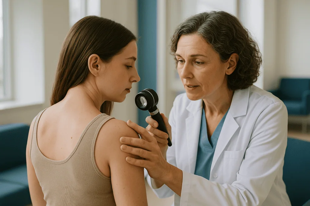
Then the full examination. Good light, the whole surface of the nevus exposed, and a tape measure laid along its longest and widest axes with a ruler or scale sticker placed beside it for the photograph. The clinician runs fingertips across the surface as well as looking at it, because a nodule under the skin of a thick nevus can be felt before it is seen. Any satellite nevi are counted and their positions noted on a body chart. In some cases, the lymph node areas nearby are gently palpated.
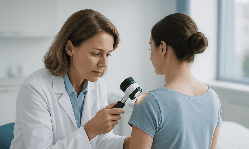
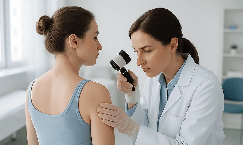
Dermoscopy comes next. The clinician places the dermoscope on several representative zones and on any area that looks or feels different, describing the pattern in each. For a small nevus this may be a single view; for a giant one it is a tour, with each stop labeled.
Photography closes the clinical part: overview shots at a fixed distance, close-ups with the scale in frame, and dermoscopic images saved to the same labeled zones. The final minutes are for the plan, which is where decisions about interval, imaging and any biopsy are made and explained. Nothing about the sequence is dramatic, and that is the point. The value of the check lies in doing exactly the same thing each time.
How does dermoscopy work, and what does it reveal about congenital nevi?
A dermoscope is a handheld magnifier, typically around tenfold, with a built-in light. Ordinary light bounces off the skin’s surface, which is why a mole looks like a flat brown shape to the naked eye. Dermoscopes get past that scatter in one of two ways: polarized light, which filters out surface glare, or a drop of fluid between the lens and skin, which makes the outer layer optically transparent. Either way, pigment structures in the epidermis and the upper dermis become visible.
In a congenital nevus, several patterns are common and considered reassuring in their own right. A globular or cobblestone pattern shows clustered round blobs of pigment, like gravel seen from above, and is frequent in young children and on the trunk. A reticular pattern looks like a brown honeycomb and is seen more on the limbs and with increasing age. Many nevi show a homogeneous, evenly brown background, or a mixture of all three. Features seen more often in congenital than in acquired moles include extra hairs (hypertrichosis), pale rings around hair follicles, tiny white milia-like cysts, pale skin furrow lines running through the pigment, and so-called target structures, where a globule or a network hole contains a central dot.
What dermoscopy does not do is diagnose. It sorts: it tells the clinician whether the lesion’s architecture fits an expected benign pattern, whether a particular zone looks different from the rest, and whether something warrants a biopsy, which is the removal of a sample for a pathologist to examine. For a giant nevus, whose surface is naturally varied, the dermoscope’s main job is to create a detailed map of zones that can be compared with the same zones later.
Photo mapping and digital dermoscopy: how change is actually tracked
Human memory is a poor instrument for moles. A parent sees the nevus every day and adjusts to it; an adult sees their own back almost never. Photo mapping replaces that drift with a fixed baseline.
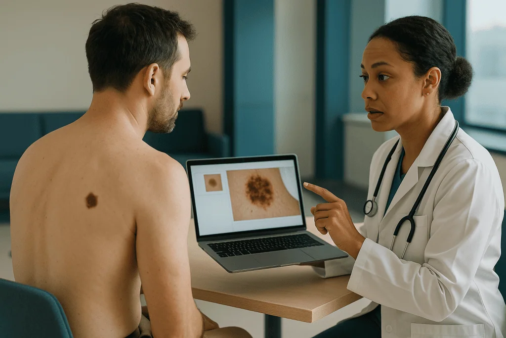
Two tools do most of the work. Total body photography (TBP) is a set of overview images taken in standardized poses, lighting and distances so that the whole skin surface is documented and new or changed lesions can be spotted on comparison. Sequential digital dermoscopy (SDD) stores the magnified dermoscopic image of each chosen zone so that the next image can be placed beside it, or overlaid on it, on a screen. Together they answer the only question that matters at follow-up: is this the same as it was?
Several things make the images useful rather than merely pretty. A scale in every frame. The same camera-to-skin distance. Zones labeled on a body chart so the same spots are re-imaged, not approximations. Attention to any area that was flagged as atypical last time, because a lesion that looked slightly unusual but stable across visits is reassuring in a way a single photograph can never be.
The method has honest limits. A photograph cannot see below the surface, so a deep nodule in a thick nevus may be felt before any image changes. Congenital nevi also change in expected ways as a child grows, so the clinician reads change against what is normal for age, not against a rule that any change is bad. Phone photographs taken at home can supplement the record if they are taken consistently, and many teams will suggest how to do that, but they are a bridge between visits, not a replacement for them.
Who is usually offered structured monitoring, and who is usually asked to wait
Not every congenital nevus needs a formal program. A small CMN, the common kind that one in a hundred newborns carries, is often simply looked at during routine checks, with a lower threshold for a dedicated visit if it changes in a way the family cannot explain. Melanoma within small and medium nevi is uncommon, and when it occurs it tends to arise in the visible skin, which is exactly what inspection and dermoscopy are designed to catch.
Large and giant nevi are different. Here the treating team usually sets up structured monitoring from infancy, often across several specialties: dermatology, pediatrics, sometimes neurology, plastic surgery and psychology. Early imaging of the brain and spine may be discussed, and the skin examinations are more detailed because melanocytes extend deep into the dermis and even beneath it. MedlinePlus Genetics notes that in giant CMN a melanoma may also develop in the brain or spinal cord, which is why surface checks are one part of a wider plan rather than the whole plan.
Who is asked to wait? Infants in whom elective surgery under general anesthesia is being weighed, since the team may prefer to observe before committing a child to staged operations. Families hoping a laser will make a nevus safe, since lightening the surface has limited evidence for changing risk and can make later dermoscopy harder to interpret. Anyone whose nevus is stable and typical on dermoscopy, where watching is a perfectly reasonable plan rather than a failure to act.
Removal is never automatic. Even complete excision of a large nevus does not remove all risk, because of the deep and extracutaneous cells. The reasons teams do operate include a concerning change or nodule, a location that is hard to monitor, and the psychological weight of a visible mark. That balance belongs with the treating team and the family together.
Congenital nevus vs café au lait spot vs ordinary mole: how to tell them apart
Three brown marks on a child can look similar from across a room and mean quite different things up close. The café au lait spot is the one most often confused with a congenital nevus. It is a flat patch the color of milky coffee, with a smooth, even tone and no change in skin texture. It contains more pigment, not more melanocyte clusters, so under the dermoscope it shows a faint, homogeneous tan with none of the globules or network of a nevus. One or two are common and harmless. Several of them, particularly six or more, are a reason for a doctor to consider neurofibromatosis type 1, a genetic condition, which is why the NHS advises getting a child with multiple café au lait spots checked.
| Feature | Congenital melanocytic nevus | Café au lait spot | Common acquired mole |
|---|---|---|---|
| When it appears | At birth or first months | Birth or early childhood | Childhood through about age 40 |
| Color and surface | Light to dark brown, often raised, bumpy or hairy | Uniform light brown, flat, smooth | Tan to dark brown, flat or domed, usually under about 6 mm |
| Dermoscopy | Globular, reticular or mixed; target structures; hair | Faint homogeneous pigment, no network | Regular network or globules, one or two colors |
| Growth | In proportion to the body | In proportion to the body | Appears, enlarges slowly, may fade |
| Associations | Size-related melanoma and neurological risk | Multiple spots: possible NF1 | Many moles raise overall melanoma risk |
The acquired mole, the kind most people have a few dozen of, arrives after birth and usually stays small; Mayo Clinic describes typical moles as under about a quarter of an inch, or 6 mm. A congenital nevus has no such ceiling, and its texture, hair and dermoscopic structures tend to be richer. If the distinction is unclear in a baby, clinicians often simply note the mark and re-examine it a few months later, because the three evolve differently.
Dermoscopy of congenital nevi versus melanoma: the key differences
A nevus is organized; a melanoma is not. That is the shortest version of what a clinician looks for, and dermoscopy turns it into specific observations.
A benign congenital nevus tends to be symmetrical in its architecture even when its outline is irregular, uses one or two patterns across its surface, and shows one or two shades of brown. Features that pull the assessment toward melanoma include an atypical pigment network with thick, irregular lines; streaks or finger-like projections at the edge; a blue-white veil, which is a milky blue haze over part of the lesion; regression structures such as scar-like white areas with grey peppering; dots and globules of uneven size scattered at the periphery; atypical vessels; and three or more colors, especially blue, grey, white or red. Mayo Clinic’s ABCDE guide for the naked eye, asymmetry, border irregularity, color variation, diameter and evolution, maps loosely onto the same ideas.
Here is the honest complication. Congenital nevi, particularly large ones, can show several of these features at baseline without anything being wrong. Their surfaces are heterogeneous by nature, with raised and flat zones, hairy and bald zones, and more than one shade. A single dermoscopic snapshot of a giant nevus can look alarming to an untrained eye and entirely typical to an experienced one. This is why change over time carries more weight than any one image, and why the baseline matters so much.
Two findings tend to override pattern analysis altogether. The first is a new, firm nodule within the nevus, especially one that grows over weeks. The second is a zone that has clearly changed when compared with its own earlier image. Either one usually leads to a biopsy, because dermoscopy narrows the question but pathology answers it.
Acral nevus dermoscopy: palms and soles follow different rules
The skin of the palms and soles is ridged, which is what fingerprints are. Those ridges and the furrows between them change how pigment sits, so a nevus on the sole, whether congenital or acquired, is read with a separate set of patterns.
In a benign acral nevus, pigment runs along the furrows, the narrow grooves between the ridges. Under the dermoscope this appears as fine parallel brown lines separated by paler bands: the parallel furrow pattern. Variants include a lattice-like pattern, where lines also cross the ridges at intervals, and a fibrillar pattern of fine oblique filaments, which is common on weight-bearing parts of the sole where pressure slants the ridges. All three are considered typical of a benign lesion.
The concerning pattern is the mirror image. In the parallel ridge pattern, pigment sits on the broad ridges rather than in the furrows. The ridges can be identified by the tiny white dots along them, which are the openings of sweat ducts. A clinician who sees pigment running through those dotted lines treats the finding seriously, because it is strongly associated with acral melanoma, a form that is unrelated to sun exposure and affects all skin tones.
Congenital acral nevi can be larger than acquired ones and may show more than one benign pattern at once, which is normal for them. What the team watches for is the appearance of a ridge pattern, a diffuse irregular blotch, or growth that outpaces the child’s foot. Nails deserve a glance at the same visit, since a brown streak in a nail can be a pigment band from a nevus in the nail root and is assessed on similar principles. A nevus on the sole is not inherently more dangerous than one elsewhere; it simply requires a reader who knows the local dialect.
Large and giant nevi: when the check looks beyond the skin
For a giant nevus, especially one lying along the back of the body, the neck or the scalp, with many satellite nevi scattered around it, the clinician’s attention extends to the nervous system. Neurocutaneous melanosis is the term for melanocytes also being present in the leptomeninges, the thin membranes covering the brain and spinal cord. MedlinePlus Genetics describes this as a complication of giant CMN that can lead to seizures, a build-up of fluid in the brain, developmental concerns and, less commonly, a melanoma arising in the nervous system itself.
Many children who have these deposits on imaging never develop symptoms. That fact shapes how teams approach the question. Magnetic resonance imaging (MRI), a scan using magnets rather than radiation, may be offered in infancy because small deposits are easier to see before the brain’s white matter fully matures; in other families the team may choose to monitor clinically, watching for the symptoms above. Neither path is wrong by default, and the decision takes into account nevus size, location, satellite count and the family’s preferences.
The skin examination in a giant nevus also deepens. The nevus is thicker, with cells extending into the fat beneath, so the clinician palpates for lumps as carefully as they look for color change. Small bumps called proliferative nodules can arise in early life, often in the first years, and can look worrying while being benign; distinguishing them from melanoma sometimes requires a biopsy, and families should expect that this may happen without it meaning the worst.
Finally, teams frequently include psychological support from the start. A visible mark covering a large part of a child’s body shapes how that child is seen and how they come to see themselves. Addressing that is part of care, not an afterthought.
Congenital nevus dermoscopy monitoring over the years: what normal change looks like
A congenital nevus is not a photograph of a birthmark frozen on the day of delivery. It is living tissue on a growing body, and a monitoring program spends a good deal of its energy learning what this particular nevus does when nothing is wrong.
Growth in proportion to the child is expected. So is a shift in color: many nevi darken in the first years, then lighten gradually through childhood and adolescence, sometimes patchily. Hair often becomes coarser and darker. The surface may thicken into a cobblestone texture, and a nevus that was flat in infancy can become raised. Under the dermoscope, a globular pattern in a toddler commonly matures into a more reticular one by the teenage years, particularly on the limbs.
Some changes are benign but worth noting in the record. A pale halo can appear around the nevus. The skin of a nevus is often dry and prone to eczema-like inflammation, with redness, scale and small fissures that itch; this is a skin-barrier problem rather than a cancer signal, though persistent raw or weeping areas are still examined. In large nevi, the proliferative nodules described earlier can appear and sometimes shrink on their own.
What is not expected is a change that stands out from the nevus’s own history: a firm new lump that keeps enlarging over weeks, a zone that turns a distinctly different color from everything around it, an ulcer or crust that does not heal, or growth in one part that outpaces the rest. These are the findings that move a visit from documentation to biopsy. The purpose of all those earlier photographs is to make such a departure obvious rather than debatable.
What the days and weeks after a check usually look like
After a visit that involved only examination, dermoscopy and photography, the honest answer is that very little happens. Skin that has had a dermoscope pressed against it looks exactly as it did before. Families typically leave with a written plan: the agreed interval until the next visit, any imaging to be arranged, guidance on home photographs and sun care, and a clear list of changes that should prompt an earlier appointment.
If a biopsy was taken, the timeline is a little fuller. A punch biopsy removes a small cylinder of skin; a shave biopsy removes the surface; an excisional biopsy removes the whole lesion. All are done under local anesthetic, the numbing injection stings briefly, and the wound is closed with stitches or left to heal on its own depending on size and site. The team will say when any stitches come out and how to keep the area clean. Pathology results take time, and clinicians usually explain in advance how and when they will be communicated, so that a wait is not mistaken for bad news.
Where removal of a larger nevus has been chosen, the path stretches over months rather than weeks. Staged excision removes the nevus in sections at separate operations, allowing the surrounding skin to stretch between them. Tissue expansion places a balloon-like device under nearby healthy skin and gradually inflates it to grow extra skin for coverage. Each stage carries the ordinary risks of surgery, including infection, bleeding, scarring and anesthesia, and the team will set out alternatives, including continued observation, at every decision point.
For parents, the weeks after a first specialist visit are often heavier emotionally than medically. Asking the team for a point of contact for questions between appointments is reasonable and routine.
Living between checks: sun care, home photos and skin comfort
Monitoring does not pause between appointments, but the between-visit work is simpler than people fear.
Sun protection comes first because ultraviolet exposure is the one modifiable contributor to melanoma risk, and nevus skin is no exception. The CDC’s advice is the ordinary kind done consistently: seek shade in the middle of the day, use clothing and wide-brimmed hats, and apply broad-spectrum sunscreen to exposed skin, remembering that infants under six months are best kept out of direct sun rather than relying on sunscreen. Children with large nevi often prefer rash vests at the pool anyway, which conveniently doubles as protection.
Home photographs help if they are taken the same way each time. Use the same room and light, the same distance, and place a coin or ruler beside the nevus for scale. Keep them in one folder with the date in the file name. The team can suggest a sensible cadence; the goal is a short series that shows whether something changed, not a daily diary.
Skin comfort matters more than most guides admit. Nevus skin tends to be dry, and the extra hair can trap sweat; regular fragrance-free emollient keeps fissures and itch down, and the team can advise on managing flares of eczema over the nevus. Hair removal is cosmetic and does not alter risk, but any method should be discussed first because irritation can muddy the next examination.
Conversations with the child are part of care, too. Children who understand that their mark is a birthmark being looked after, in plain words at their level, tend to handle both curious classmates and clinic visits with more ease. Many teams can connect families with peer support, which is a different kind of monitoring: of how everyone is coping.
What people often get wrong about congenital nevi
Fear and reassurance both breed myths, so it is worth clearing a few.
“It will turn into cancer.” For the common small nevus, melanoma is uncommon, and most people with one live their whole lives without any problem from it. Risk rises with size, which is why giant nevi are followed closely, but even there an increased risk is not a certainty. MedlinePlus Genetics describes the risk in giant CMN as increased, with figures in the literature commonly falling between the low single digits and roughly one in ten over a lifetime; most people with a giant nevus will not develop melanoma.
“Removing it removes the risk.” For small nevi, excision does remove the lesion. For large and giant ones, nevus cells extend deep and may also sit in the nervous system, so surgery can reduce surface risk and address appearance but does not end the need for follow-up.
“Dermoscopy can diagnose melanoma.” It refines suspicion, sometimes dramatically, but a pathologist’s examination of tissue is what diagnoses.
“A mole that grows is dangerous.” A congenital nevus grows with the child. The worrying kind of growth is disproportionate, localized or fast.
“Hair on a mole is a bad sign.” Hair is typical of congenital nevi and, if anything, points toward the ordinary.
“Laser will make it safe.” Laser can lighten surface pigment. Evidence that this changes melanoma risk is limited, and lightened skin is harder to read on dermoscopy, so most teams treat it as a cosmetic option to weigh carefully rather than a safety measure.
“Café au lait spots are the same thing.” They are flat pigment patches with no nevus structures, and their significance lies in their number, not their size.
Questions to ask your care team about congenital melanocytic nevus monitoring
Clinic time is short and the questions that matter tend to arrive in the parking lot. Writing a few down beforehand changes the visit. These are the ones that tend to earn their place.
- How do you classify this nevus by projected adult size, and what does that category mean for the plan?
- What patterns did you see on dermoscopy today, and were any zones flagged as needing closer comparison next time?
- How are the baseline photographs stored, and can we have copies so that any clinician who sees us has the same reference?
- What interval are you suggesting between checks, and what would make you shorten it?
- For a large nevus: is MRI being considered, and if not, what symptoms are you asking us to watch for instead?
- Is removal something you are recommending, considering, or not advising for now, and what are the reasons either way?
- If a biopsy is needed, how will the result reach us and roughly how long does your laboratory usually take?
- What is the best way to contact the team between appointments if we notice something?
- Which specialists are involved, and who coordinates the overall plan?
- Is there psychological or peer support available for our child, or for us?
- How should we photograph the nevus at home, and how often do you actually want us to do it?
- What everyday skin care do you suggest for dryness, itch or eczema over the nevus?
None of these questions has a single right answer, and a good team will say so where the evidence is thin. The aim is not to audit the clinician but to leave the room knowing what is being watched, why, and what would change the plan. That shared understanding is the quiet backbone of monitoring, and it is worth the extra five minutes.
When to call your doctor
Most changes in a congenital nevus are expected and can wait for the scheduled visit. A few should prompt a call for an earlier appointment, and a smaller number need urgent assessment.
Arrange an earlier review if you notice any of the following in or around the nevus:
- A new lump or nodule, particularly one that feels firm and keeps enlarging over days to weeks.
- Bleeding, an open sore, or a crust that does not heal within the time the team has told you to expect for minor skin injury.
- A patch that has become a distinctly different color from the rest of the nevus, especially black, blue, grey, white or red.
- Growth in one part of the nevus that outpaces the child’s overall growth, or an outline that has clearly changed on comparison with your photographs.
- A satellite nevus that looks or behaves differently from the others.
- Pain, tenderness or a new type of itch that does not settle with the emollient routine you have been advised.
For a child with a large or giant nevus, seek urgent medical care the same day for seizures, repeated vomiting with headache, unusual drowsiness or irritability, a rapidly enlarging head in an infant, new weakness, loss of skills the child previously had, or any sudden change in behavior. These can be signs of pressure or disease in the nervous system and are evaluated promptly whether or not an MRI has been done before.
At any age, a mole that is changing in a way you cannot explain is a reason to be seen, and the NHS and Mayo Clinic both advise getting such changes checked rather than waiting to see what happens. Calling and being told it is nothing is a good outcome of monitoring, not a wasted appointment. Every decision about what happens next, from a reassuring look to a biopsy to imaging, sits with the treating team, who will have your baseline images to compare against.
Frequently asked questions
What does dermoscopy reveal about congenital nevi?
Dermoscopy shows the internal architecture of a congenital nevus: whether its pigment is arranged in globules, a network or an even background, and whether features typical of congenital lesions are present, such as hair, pale rings around follicles, small white cysts and target structures. It also lets the clinician document specific zones so that any later change can be compared against the same spot. It does not confirm or rule out melanoma on its own; that requires a biopsy.
What is the difference between café au lait spots and congenital nevi?
A café au lait spot is a flat, evenly light brown patch with normal skin texture and no nevus structures on dermoscopy, caused by extra pigment rather than clustered melanocytes. A congenital nevus is a true mole, often raised, textured or hairy, with globular or network patterns under the dermoscope. Café au lait spots matter mainly by number: the NHS advises a check if a child has several, because multiple spots can be a sign of neurofibromatosis type 1.
What are the key differences between dermoscopic findings of melanoma and nevus?
A benign nevus tends to show a symmetrical architecture, one or two patterns and one or two shades of brown. Melanoma is more likely to show an atypical network, irregular streaks at the edge, a blue-white veil, scar-like regression areas, unevenly scattered dots, atypical vessels and three or more colors. Congenital nevi can look varied at baseline, so clinicians weigh change over time, and especially a new firm nodule, more heavily than a single image.
What are the dermoscopic findings of an acral nevus?
On palms and soles, a benign acral nevus usually shows the parallel furrow pattern, with pigment running in the narrow grooves between skin ridges, or related benign variants called lattice-like and fibrillar patterns. The concerning finding is the parallel ridge pattern, where pigment sits on the broad ridges marked by tiny white sweat-duct dots, because it is strongly associated with acral melanoma. Congenital acral nevi may combine more than one benign pattern, which is expected.
How often should congenital melanocytic nevus monitoring happen?
There is no single interval that fits everyone, and the treating team sets it based on the nevus’s projected size, location, dermoscopic appearance and the person’s age. Small, typical nevi are often simply reviewed at routine health visits, while large and giant nevi usually have a structured schedule from infancy that may include imaging. Any unexplained change between visits is a reason to bring the next check forward rather than wait.
Does a congenital nevus always need to be removed?
No. Many congenital nevi, particularly small and medium ones with typical dermoscopic features, are monitored rather than removed. Teams consider surgery when there is a concerning change or nodule, when the location makes monitoring difficult, or when the visible mark carries a heavy psychological burden. For large and giant nevi, removal does not eliminate all risk because nevus cells extend deep and may also be present around the brain and spinal cord, so monitoring continues afterward.
Why does a congenital nevus grow, and when is growth a problem?
A congenital nevus grows because the child grows; it keeps roughly the same proportion to the body throughout childhood, which is why clinicians classify it by projected adult size. Expected changes also include darkening or lightening, thickening and hair growth. Growth becomes a concern when one part enlarges faster than the rest, when a firm lump appears and keeps growing over weeks, or when the outline changes clearly compared with earlier photographs.
Is dermoscopy safe for a baby, and does it hurt?
Dermoscopy is painless and involves no radiation; the clinician rests a lit magnifier against the skin, sometimes with a drop of gel, and looks or photographs. Babies are usually examined on a parent’s lap, and the main challenge is keeping a wriggling child still long enough for a clear image. Photography is likewise harmless. If a biopsy is ever needed, that is a separate, small procedure under local anesthetic that the team would discuss first.
What is neurocutaneous melanosis and does every large nevus need an MRI?
Neurocutaneous melanosis means melanocytes are also present in the membranes covering the brain and spinal cord. It is more likely with a giant nevus over the back, neck or scalp and with many satellite nevi, and it can cause seizures or fluid build-up, though many children with it never have symptoms. MRI in infancy is one option teams discuss; others monitor clinically. The choice depends on the nevus’s size and position and is made with the family.
Can I use laser or hair removal on a congenital nevus?
Both are cosmetic choices to raise with the team before acting. Laser can lighten surface pigment, but the evidence that it changes melanoma risk is limited, and lighter skin can be harder to interpret on dermoscopy, so it is not a safety measure. Hair removal does not alter risk either, but methods that irritate the skin can muddy the next examination. Neither should be started without the monitoring team’s awareness.
References
- MedlinePlus Genetics: Giant congenital melanocytic nevus
- MedlinePlus Medical Encyclopedia: Giant congenital nevus
- NHS: Birthmarks
- NHS: Moles
This article is for general information only and is not a substitute for professional medical advice. Please consult a qualified doctor about your individual situation.

More from the Blog
How Does Phototherapy Help Atopic Dermatitis? What Sessions Involve and Who May Be Offered Them
Phototherapy for atopic dermatitis exposes affected skin to carefully measured ultraviolet light, usually narrowband UVB, in a clinic cabinet several times a week for…
A Daily Routine That Supports Atopic Dermatitis Treatment: Bathing, Moisturizing and Nail Care
A supportive atopic dermatitis daily routine rests on three anchors: a short, lukewarm bath or shower with a gentle, fragrance-free cleanser; a generous layer…

Is Coconut Oil Good for Your Hair? The One Oil That Penetrates
Coconut oil can genuinely help hair, and the reason is unusual: its main fatty acid, lauric acid, is small enough to penetrate the hair…
Peptides in Skincare: What Signal Peptides Do and What the Studies Show
Peptides in skincare are short chains of amino acids applied topically to signal skin cells, most often to encourage collagen production or soften expression…
How to Get Clear Skin: What Actually Works, According to Dermatology Trials
Dermatology trials show that clear skin comes mainly from treating acne consistently: a gentle cleanser, benzoyl peroxide, an adapalene retinoid, daily sunscreen, and patience…

Why Is Cellulitis Not Improving? Spreading Redness, Fever and When to Contact Your Care Team
Cellulitis usually begins to improve within about 48 hours of starting antibiotics, although redness can spread slightly at first. When it keeps worsening after…