Congenital Nevi
Learn what congenital nevi are, their symptoms and causes, how doctors diagnose them, and the treatment options from monitoring to surgical removal.
Quick answer
Congenital nevi are moles present at birth or appearing in the first months of life, formed by clusters of pigment cells that developed in the womb. Most are small and harmless, needing only monitoring and sun protection. Large or giant nevi carry a higher risk of melanoma and are followed more closely, sometimes with surgery.
What is congenital nevi?
Congenital nevi (the singular is congenital nevus) are moles that are present at birth or that appear during the first months of life. The full medical name is congenital melanocytic nevus. A nevus is the medical word for a mole, and melanocytic means the mole is made of melanocytes, the skin cells that produce the brown pigment called melanin. Because these moles form before or shortly after birth, they are considered a type of birthmark rather than an ordinary mole that develops later in childhood or adulthood.
When people ask what is congenital nevi, the short answer is that they are pigmented birthmarks made of clusters of pigment cells that grouped together while the skin was still developing in the womb. They occur in babies of every ethnic background and in both boys and girls. Most are small, cause no health problems, and are mainly a cosmetic concern. A smaller number are large or very large, and these carry a higher chance of complications, so they are usually followed more closely by doctors.
Doctors often classify congenital nevi by how large they are expected to become once the child is fully grown, because a mole grows in proportion to the body. A commonly used system describes them as small (under about 1.5 centimeters in projected adult size), medium (about 1.5 to under 20 centimeters), large (about 20 to 40 centimeters), and giant (more than about 40 centimeters). Your child’s doctor may use slightly different cut-offs, but the general idea is the same: the larger the nevus, the more attention it usually receives.
Congenital nevi symptoms and signs
Congenital nevi do not usually cause symptoms in the sense of pain or illness. Instead, the main congenital nevi symptoms are visible changes in the skin. Common features include:
- A flat or slightly raised patch of skin that is light brown, dark brown, black, or sometimes bluish or reddish in a newborn.
- A border that may be smooth and even or irregular and uneven.
- A surface that can be smooth, bumpy, pebbly, or wrinkled, especially in larger nevi.
- Coarse, dark hair growing from the nevus, which often becomes more noticeable with age.
- Growth in proportion to the child’s body, so the mole gets bigger as the child grows.
- Darkening or lightening of the color over the years; many nevi become lighter and softer in appearance during childhood.
- Smaller spots of pigment around a large nevus, called satellite nevi.
- Dryness, itching, or a tendency to irritation in some larger nevi, because the skin in these areas may have fewer oil glands and be more fragile.
How a congenital nevus looks and behaves often depends on its size and location. Small and medium nevi usually look like an ordinary mole that happens to have been present since birth. They may be flat at first and become slightly raised or hairy over time. Large and giant nevi can cover a substantial part of the trunk, a limb, the scalp, or the face. They often have a more textured surface, a mix of colors, and many satellite spots. In very large nevi, the skin may be thinner, may sweat less, and may be more prone to cracking or infection.
A congenital nevus on the scalp may look alarming to parents but often lightens considerably in the first years of life. Nevi on the palms, soles, or under a nail may be harder to monitor and are sometimes followed more carefully. In all cases, the most important thing doctors look for is not the appearance at birth but whether the nevus changes in a way that is out of step with the child’s normal growth.
Causes and risk factors
The exact congenital nevi causes are not fully understood, but the current understanding is that they arise from a change in the genetic code of a single pigment cell while the baby is developing in the womb. This change happens after conception, in the developing skin cells themselves, and is called a somatic mutation. Because it occurs in the skin cells rather than in the egg or sperm, it is not inherited from a parent and is not usually passed on to the next generation.
The altered pigment cell multiplies faster than its neighbors, and the resulting cluster of melanocytes forms the nevus. Researchers have found that changes in certain genes involved in cell growth signaling, most often a gene called NRAS and less commonly a gene called BRAF, are present in many congenital nevi. The earlier in development the change occurs, the larger the area of skin that can be affected, which is thought to explain why some nevi are small and others cover a large part of the body.
Important points about risk factors:
- Nothing a parent did or did not do during pregnancy is known to cause congenital nevi. They are not linked to diet, stress, sun exposure during pregnancy, or medications in any established way.
- They are not contagious and cannot spread from one person to another.
- Having a family member with a congenital nevus does not appear to meaningfully raise a baby’s chance of having one, because the genetic change is not inherited.
- Congenital nevi occur in all skin types and ethnic groups.
It is helpful to separate the causes of the nevus itself from the risk factors for complications. Once a congenital nevus exists, the factors that raise the chance of future problems, such as melanoma (a serious form of skin cancer that starts in pigment cells) or nervous system involvement, are mainly its size, the number of satellite nevi, and its location on the body. Large and giant nevi, especially those on the back or trunk with many satellite spots, carry the highest risk. Small nevi carry a low risk, and any melanoma that does develop in them tends to occur in adulthood rather than childhood.
Congenital nevi diagnosis
In most cases, congenital nevi diagnosis is clinical, meaning a doctor can identify the nevus by looking at it and by knowing that it was present at or soon after birth. A pediatrician, family doctor, or dermatologist (a doctor who specializes in skin) will examine the size, color, shape, surface, and location of the nevus and will ask about any changes since birth.
Tools and steps that may be part of the assessment include:
- Skin examination and measurement. The doctor measures the nevus and estimates its projected adult size. Photographs are often taken so that changes can be compared at future visits.
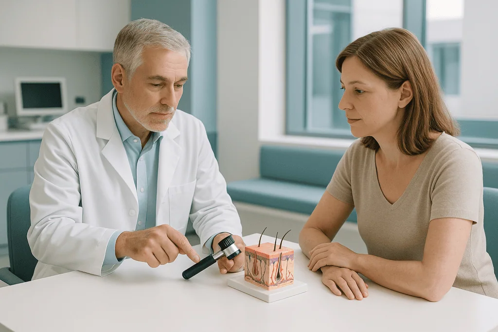
- Dermoscopy. A handheld magnifying device with a light is used to look at the pattern of pigment beneath the skin surface. This is painless and helps distinguish typical features from concerning ones.
- Full skin check. The whole body is examined to count and record satellite nevi and to look for other birthmarks.
- Skin biopsy. If part of a nevus looks unusual, a small sample of tissue may be removed under local anesthetic (numbing medicine) and examined under a microscope by a pathologist. A biopsy is not needed for most congenital nevi and is reserved for areas that raise concern.
- MRI of the brain and spine. In children with a large or giant nevus, or with many satellite nevi, doctors may recommend magnetic resonance imaging (a scan that uses magnets rather than radiation) in early infancy. This looks for pigment cells in the brain or spinal cord, a condition called neurocutaneous melanosis. Not every child with a large nevus needs this scan, and your doctor will explain whether it applies.
- Neurological examination. Developmental milestones, muscle tone, and head size may be monitored more closely in children considered at higher risk.
There is no blood test that diagnoses a congenital nevus. Genetic testing of the nevus tissue is sometimes done in research or specialist settings but is not part of routine care. The main purpose of the diagnostic process is to record a clear baseline, decide how closely the child should be followed, and identify the minority of children who may benefit from imaging or early treatment.
Congenital nevi treatment options
There is no single correct approach, and congenital nevi treatment options depend on the size and location of the nevus, the risk of complications, the child’s age, and the wishes of the family. Many congenital nevi never need any treatment at all. In cases where treatment is considered, the reasons are usually to reduce the long-term risk of melanoma, to improve appearance, or to manage skin problems such as itching and fragility.
- Observation and monitoring. For small and many medium nevi, the standard approach is regular skin checks, often once a year, with photographs to track changes. Parents are taught what changes to watch for at home. This is a genuine treatment plan, not a lack of one, and it avoids surgical scars in children who are unlikely to develop problems.
- Skin care and sun protection. Keeping the nevus moisturized, using gentle cleansers, and protecting the area from sunburn with clothing and sunscreen are recommended for all congenital nevi. Sun protection does not remove the mole but reduces additional skin damage.
- Surgical excision. Surgery to cut out the nevus is the main way to remove it. Small nevi can often be removed in one procedure with the edges of skin stitched together. Larger nevi may require staged excision, in which portions are removed over several operations months apart, allowing the surrounding skin to stretch between sessions.
- Tissue expansion. For large nevi, a balloon-like device called a tissue expander may be placed under nearby healthy skin and gradually filled with saline over weeks. The stretched skin is then used to cover the area after the nevus is removed. This often gives a better color and texture match than a graft.
- Skin grafts and flaps. When there is not enough neighboring skin, surgeons may move skin from another part of the body. Grafts can leave visible differences in color and texture and are usually chosen when other methods are not practical.
- Laser treatment and dermabrasion. These methods remove or lighten the upper layers of the nevus. They may improve appearance but do not remove the deeper pigment cells, so the color can return and the cancer risk is not considered to be eliminated. They are used selectively, often for areas where surgery would be difficult.
- Hair removal. Laser hair reduction may be considered for hairy nevi when the hair causes distress, usually in older children or adults.
Surgery on a child carries the usual risks of anesthesia, bleeding, infection, and scarring, and very large nevi cannot always be completely removed. For this reason, decisions are ideally made by a team that includes a dermatologist, a pediatrician, and a plastic surgeon, with the family fully involved. At Acibadem, congenital nevi that may need surgical removal or reconstruction are usually assessed within the Plastic, Reconstructive & Aesthetic Surgery department, working alongside dermatology and pediatrics.
Timing is also a consideration. Some surgeons prefer to operate in early childhood when skin is more elastic and scars tend to mature well, while others advise waiting until the child can take part in the decision, particularly when the reason for surgery is appearance rather than medical risk. Your doctor may discuss both approaches with you.
Living with congenital nevi and outlook
For the majority of people, the long-term outlook with a congenital nevus is good. Small and medium nevi carry a low lifetime risk of melanoma, and most people live with them without any medical problems. Many nevi lighten and become less noticeable over time, although hair growth may increase around puberty.
People with large or giant nevi face a higher, though still uncertain, risk of melanoma, and this risk is highest in the first years of life and again in adulthood. They also have a small chance of neurocutaneous melanosis, which can in some cases lead to seizures, developmental concerns, or pressure inside the head. Most children with pigment cells found on an MRI never develop symptoms, but those who do need care from a pediatric neurologist. Lifelong skin checks are recommended for anyone with a large nevus, regardless of whether it has been partly or fully removed.
The emotional side of living with a visible birthmark is real and should not be dismissed. Children may face questions or teasing, and parents often feel guilt even though nothing they did caused the nevus. Talking openly with the child in age-appropriate language, connecting with support groups for families affected by large birthmarks, and, where needed, involving a psychologist can help. Decisions about cosmetic treatment are personal, and there is no wrong choice between accepting the nevus and pursuing removal.
Practical day-to-day advice usually includes protecting the skin from the sun, keeping the nevus moisturized to prevent cracking, avoiding harsh scrubbing, and reporting any wound that does not heal or any new lump within the nevus. Photographs taken at home every few months can make it easier to notice gradual changes.
Frequently asked questions
Are congenital nevi dangerous?
Most congenital nevi are not dangerous. Small and medium nevi carry a low risk of turning into melanoma, and this usually happens, if at all, in adulthood. Large and giant nevi carry a higher risk and are therefore monitored more closely. The risk is real but should be kept in perspective, and regular skin checks are the main safeguard.
What do congenital nevi symptoms look like in a newborn?
In a newborn, a congenital nevus usually appears as a flat or slightly raised brown, black, or sometimes bluish patch that was there at birth or appeared in the first weeks. It may have irregular edges and may or may not have hair. Redness or a different shade in the first days is common and often settles as the skin matures.
What causes congenital nevi, and could I have prevented it?
Congenital nevi are caused by a genetic change that occurs in a single pigment cell while the skin is forming in the womb. This change is not inherited and is not linked to anything a parent ate, did, or took during pregnancy. There is no known way to prevent it, and parents should not feel responsible.
How is congenital nevi diagnosis confirmed?
Diagnosis is usually made by a doctor examining the skin and confirming that the mole was present at or soon after birth. A dermatoscope may be used to look at the pigment pattern. A biopsy is only needed if part of the nevus looks unusual, and an MRI of the brain and spine may be suggested for children with large nevi or many satellite spots.
Should a congenital nevus be removed?
Not necessarily. Many congenital nevi are simply observed for life. Removal may be considered when the nevus is large, when it shows worrying changes, when it causes recurring skin problems, or when its appearance causes significant distress. The decision weighs the benefits against surgical risks and scarring, and it is best made together with a dermatologist and a plastic surgeon.
Can congenital nevi treatment options remove the cancer risk completely?
Complete surgical removal of a small nevus substantially reduces the risk from that mole, though no procedure can promise zero risk. In large nevi, pigment cells may extend deeply or into areas that cannot be removed, so surgery lowers but does not eliminate the risk. Laser or dermabrasion treat the surface only and are not considered to remove the cancer risk. Ongoing skin checks remain important after any treatment.
Will a congenital nevus grow or change as my child gets older?
Yes, a congenital nevus normally grows in proportion to the child. It may also become thicker, darker or lighter, more raised, or hairier over the years, and these gradual changes are usually expected. Doctors are concerned about changes that are rapid, uneven, or out of proportion, such as a new lump, bleeding, or a sore that does not heal.
When to see a doctor
All congenital nevi should be shown to a pediatrician or dermatologist at least once so that a baseline record can be made and a follow-up plan agreed. After that, arrange a review sooner than planned if you notice any of the following red-flag signs:
- A new lump, bump, or nodule forming within the nevus, especially one that feels firm or grows quickly.
- Rapid growth of the nevus that is clearly out of proportion to the child’s overall growth.
- A new or changing area of very dark, uneven, or unusual color inside the nevus.
- Bleeding, oozing, crusting, or an open sore on the nevus that does not heal within a few weeks.
- Persistent pain, tenderness, or intense itching in the nevus.
- In a child with a large or giant nevus: seizures, unusual sleepiness, repeated vomiting, a rapidly enlarging head, delays in development, or weakness in the arms or legs, which can point to nervous system involvement and need urgent assessment.
- Signs of skin infection such as spreading redness, warmth, swelling, pus, or fever.
Seek emergency care immediately if a child has a seizure, becomes very drowsy or difficult to wake, or develops sudden weakness. For non-urgent changes, a prompt appointment with the treating dermatologist or pediatrician is appropriate. Early evaluation of any change is the most reliable way to catch the uncommon complications of congenital nevi at a stage when they can be managed.
Medically reviewed by the Acıbadem International Medical Board September 9, 2026
See our medical review board →
Update history
- PublishedSeptember 9, 2026
- Medical review approvedSeptember 9, 2026
- Last content updateSeptember 9, 2026
References2
Treatments for This Condition
Care at Acibadem
Doctors Who Treat This Condition



Aesthetic Plastic & Reconstructive Surgery
Prof. Mehmet Veli Karaaltın, MD
Aesthetic Plastic & Reconstructive Surgery