

What a CT Scan Shows, and Why Doctors Order One
Key Takeaways
- A CT scan reconstructs hundreds of X-ray measurements into cross-sectional slices, showing bone, organs, blood vessels and fresh bleeding in detail that a single X-ray cannot.
- The most common reason for an urgent CT is to rule dangerous conditions such as brain bleeding, lung clots, aortic tears or a perforated organ in or out within minutes.
- Head CT can show a bleed within minutes of a stroke, but a blockage-type stroke may be invisible in its first hours, so a normal scan means no bleed was seen, not that the brain is fine.
- Harvard Health estimates a chest CT at about 7 millisieverts and an abdominal CT at about 8, compared with roughly 3 millisieverts of natural background radiation per year.
- CT shows structure rather than function, so nerve problems, early cartilage damage, seizures, migraine and most blood cancers are not detected by it and need other tests.
- No major guideline body recommends whole-body CT screening for healthy adults because it finds many harmless incidental lesions that lead to further tests without proven benefit.
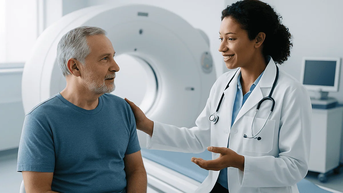
A CT scan shows detailed cross-sectional images of bones, organs, blood vessels and soft tissue by combining many X-ray views into slices. Doctors most often order one to look for bleeding, blood clots, fractures, infection, tumors or the cause of sudden pain, especially in emergencies. It cannot reliably show nerve function, early cartilage damage or some small or flat lesions, so results are always read alongside symptoms and other tests.
The room is cooler than you expected. A technologist tucks a pillow under your knees, a voice from a speaker asks you to hold your breath, and a ring the size of a large tire hums as the table glides through it. Somewhere down the hall, a radiologist is already scrolling through several hundred pictures of your insides before you have even sat up.
That gap between how brief a CT scan feels and how much information it produces is exactly why so many people leave the department with more questions than they arrived with. What did it see? What did it miss? Why did the emergency doctor order it within minutes, while your regular physician spent weeks deciding not to?
The honest answer is that CT is one of medicine’s sharpest tools for some jobs and a poor fit for others. Knowing which is which makes the whole experience far less mysterious, and the conversation with your care team far more useful.
What does a CT scan actually show?
Picture a loaf of bread. A standard X-ray photographs the whole loaf from one side, so the crust, the crumb and anything baked into the middle all pile up on top of each other. A CT scan slices the loaf and lets the radiologist look at each piece in turn, then stacks the slices back into a three-dimensional model that can be rotated on a screen.
The physics is still X-ray. According to the National Institute of Biomedical Imaging and Bioengineering, the CT machine spins a narrow X-ray beam around the body while detectors on the opposite side record how much radiation passes through at every angle. A computer then reconstructs those measurements into images in which dense material such as bone appears white, air appears black and organs sit in shades of gray between them.
That gray scale is where the diagnostic value lives. Fresh blood, a fluid-filled cyst, a solid tumor, fatty tissue and normal liver all absorb X-rays slightly differently, so a trained eye can tell them apart. Modern scanners resolve structures of a few millimeters and can capture the entire chest in a single held breath.
What a CT scan shows, then, is anatomy in high definition: the shape, size, density and position of structures, and whether something is where it should not be. It is a map of the territory. It is not, on its own, a report of how well that territory is working, which is a distinction that matters for the rest of this article.
How is a CT scan different from an X-ray or MRI?
Patients often assume the three main imaging tests are interchangeable, or that MRI is simply the premium version of CT. Neither is true. Each answers a different question, and a good clinician chooses based on what needs to be seen, how quickly and in whom.
| Feature | X-ray | CT scan | MRI |
|---|---|---|---|
| How it works | Single X-ray projection | Rotating X-rays reconstructed into slices | Magnetic field and radio waves, no ionizing radiation |
| Typical scan time | Seconds | Minutes; the imaging itself often under a minute | Commonly 30 to 60 minutes or longer |
| Best for | Bone fractures, chest screening for pneumonia, foreign objects | Bleeding, trauma, lung and abdominal organs, blood vessels, kidney stones | Brain and spinal cord detail, ligaments, cartilage, joints, some tumors |
| Limits | Overlapping shadows, poor soft-tissue detail | Radiation dose; less contrast between similar soft tissues | Slow, noisy, sensitive to motion, not suitable with some implants |
Speed is the quiet hero here. The Mayo Clinic notes that a CT examination is usually completed in well under half an hour, and the actual scanning portion is often a matter of seconds. For a person who cannot lie still, a child who is frightened or someone whose blood pressure is dropping, that speed can matter more than the finer soft-tissue detail an MRI would offer.
MRI earns its reputation in the brain, spinal cord and joints, where subtle differences in water content matter. CT wins in the chest, in trauma and whenever calcium or fresh blood is the question. Choosing between them is a clinical judgment, not a ranking.
What is the most common reason for a CT scan?
Ask an emergency physician and the answer arrives quickly: to find out, fast, whether something dangerous is happening inside a person who cannot be examined from the outside. Head injury, sudden severe headache, chest pain that could be a torn vessel or a lung clot, abdominal pain that might be appendicitis or a perforated organ. In each of these, the difference between waiting and acting is measured in minutes, and CT delivers answers on that timescale.
Outside the emergency department the pattern shifts. The NHS lists the everyday reasons a CT is requested: diagnosing conditions such as damage to bones, injuries to internal organs, problems with blood flow, stroke and cancer; guiding further tests or treatments, including where to place a biopsy needle or how to plan radiotherapy; and monitoring conditions over time, such as checking whether a tumor has shrunk during treatment.
MedlinePlus adds a fourth category that people rarely think about: procedure planning. Surgeons use CT to measure the exact dimensions of a blood vessel before repairing it, to model a fractured pelvis before fixing it, or to check the position of a kidney stone before deciding whether it can be broken up from the outside.
So the single most common reason is not a single disease. It is uncertainty that carries risk. When a clinician needs to rule something serious in or out and the clock is running, CT is often the first tool reached for. When time is on the patient’s side and the question is about soft tissue, a slower test may be the better choice.
What does a CT scan show in the brain?
A head CT is the workhorse of neurological emergencies, and it earns that role because of one thing it does exceptionally well: it shows fresh blood. Bleeding inside or around the brain appears bright white against the gray of brain tissue within minutes of the event, which is why a person arriving with a sudden, thunderclap headache or after a fall is scanned before almost anything else.
The Mayo Clinic describes head CT as the usual first test in suspected stroke. The logic is precise. Most strokes are caused by a blocked vessel, but a minority are caused by bleeding, and the treatments for those two situations are opposites. CT can tell them apart quickly and safely, clearing the way for the right decision. In the earliest hours, a blockage-type stroke may be invisible on plain CT, which is why a normal scan does not always mean a normal brain; it means no bleed was seen.
Beyond blood, a head CT shows skull fractures, the fluid-filled ventricles deep in the brain and whether they have enlarged, larger tumors, abscesses, calcium deposits and the sinuses. With contrast added, or with a specialized CT angiogram, the arteries and veins of the head and neck can be traced for narrowing, aneurysm or clot.
What it shows poorly is the fine architecture of the brain itself. Early multiple sclerosis plaques, small tumors at the base of the skull, subtle changes of dementia and most causes of seizures or chronic headache are better assessed with MRI, if imaging is needed at all. A head CT answers the question "is something acutely wrong?" more than "why has this been happening for months?"
What does a CT scan of the chest show?
Lungs are mostly air, and air is nearly transparent to X-rays. That accident of anatomy makes the chest one of CT’s finest performances. Against the black background of aerated lung, a small nodule, a patch of pneumonia, scarring, fluid or a collapsed segment stands out in a way that no other test matches.
Cleveland Clinic lists the structures a chest CT evaluates: the lungs, the heart and its vessels, the esophagus, the ribs and spine, and the lymph nodes clustered around the airways. A radiologist reading a chest CT is looking for infection, inflammation, tumors, enlarged lymph nodes, fluid collections and the pattern of any scarring, which can hint at the underlying cause.
Add contrast dye timed to the arteries, and the same scan becomes a CT pulmonary angiogram, the standard test for a blood clot that has traveled to the lungs. Timed to the aorta instead, it can reveal a tear in the wall of the body’s largest artery, one of the most time-critical diagnoses in medicine.
A specialized low-dose chest CT is also used to screen for lung cancer in adults with a significant smoking history. The CDC describes this as a recommended annual test for people who meet age and smoking-history criteria, which is a decision to make with a clinician rather than on a whim, since the scan can also detect harmless nodules that lead to further testing.
The heart is a special case. Coronary CT angiography can show plaque and narrowing in the heart’s arteries, and a simpler calcium-score CT counts calcified plaque without contrast. Both are useful in specific people, and neither is a substitute for the blood pressure, cholesterol and lifestyle conversation that prevents most heart disease.
What does an abdominal and pelvic CT show?
Abdominal pain is medicine’s great impersonator. Appendicitis, a kidney stone, a twisted bowel, a burst cyst, diverticulitis and a leaking aneurysm can all begin with a similar ache, and a hand on the belly can only tell so much. This is why CT of the abdomen and pelvis has become one of the most frequently requested scans in adult emergency care.
In a single pass, the scan shows the liver, gallbladder, pancreas, spleen, kidneys, adrenal glands, bladder, bowel, uterus or prostate and the major vessels running through the middle of it all. MedlinePlus notes that it is used to find the cause of pain, detect tumors, infections, abscesses or blockages, and evaluate injuries after trauma.
Kidney stones deserve a special mention because CT sees them so well. Nearly all stones, regardless of what they are made of, are dense enough to appear bright even without contrast, and the scan shows exactly where a stone sits and whether it is blocking the flow of urine, information that guides whether a person can wait it out or needs intervention.
Contrast changes what can be seen. Given by mouth, it outlines the bowel so a leak or blockage becomes obvious. Given intravenously, it lights up the blood supply of the liver, kidneys and any mass, helping distinguish a benign cyst from a solid growth that needs further attention.
There are blind spots. The lining of the bowel, where polyps and early colon cancers begin, is far better examined by colonoscopy. Small gallstones can hide from CT and show clearly on ultrasound. Endometriosis, most causes of chronic pelvic pain and many gynecologic conditions are better assessed with ultrasound or MRI.
What cancers show up on a CT scan?
Most solid tumors of any meaningful size can be seen on CT, which is why it is the backbone of cancer staging worldwide. A mass in the lung, liver, kidney, pancreas, adrenal gland or lymph nodes appears as a lump of tissue with a shape, density and blood supply that differ from the organ around it. With contrast, many cancers enhance, meaning they soak up dye differently from normal tissue, and that pattern helps the radiologist narrow down what the lump might be.
The scan is also how clinicians answer the question that follows every cancer diagnosis: has it spread? A CT of the chest, abdomen and pelvis in one sitting can survey lymph nodes, liver, lungs and bones for secondary deposits, and repeat scans over months are how treatment response is tracked. The NHS specifically lists monitoring cancer treatment among CT’s core uses.
Honesty about limits is essential here. CT does not diagnose cancer; it identifies a suspicious area. Only a biopsy examined under a microscope can confirm what a mass is made of, and many findings that look alarming on a scan turn out to be benign cysts, scars or harmless nodules. The reverse is also true. Very small tumors, flat lesions along the lining of the bowel or bladder, early cancers of the cervix or skin, and blood cancers such as leukemia are often invisible to CT or better found by other means.
Breast cancer is a good example of mismatch. CT can show a large breast mass incidentally, but mammography and ultrasound are far better at finding the small, early changes that matter for screening. The right test depends on the cancer being looked for, not on which machine is most impressive.
What cannot be detected on a CT scan?
This is the question people most want answered and are least often told. A normal CT is reassuring, but it is not a certificate of health, and understanding what the scan does not see prevents both false comfort and unnecessary worry.
Function is the first gap. CT shows structure. A heart can look anatomically perfect on CT and still beat irregularly; a brain can appear normal and still have epilepsy, migraine, depression or early dementia. Nerves are too fine to see in detail, so numbness, tingling and nerve pain are rarely explained by CT alone. Blood tests, electrical studies and clinical examination fill that space.
Soft-tissue subtlety is the second. Cartilage, ligaments, tendons, the discs between vertebrae and the spinal cord itself are better shown by MRI. Early damage in a knee, a torn rotator cuff or a slipped disc pressing on a nerve may show nothing on CT and be obvious on MRI. Small tumors in the brain, pituitary or pelvis fall into the same category.
Timing is the third. A blockage-type stroke can be invisible on CT in its first hours. A very recent bone stress fracture may not yet show. Some infections begin before they change the tissue enough to be seen.
Finally, CT cannot see what it was not pointed at. A scan of the head does not evaluate the neck; a scan of the abdomen without contrast may miss vascular problems that a contrast study would catch. When a scan comes back normal but symptoms persist, the right response is not to assume nothing is wrong. It is to revisit the question with the clinician, who may choose a different test.
What does contrast dye do in a CT scan, and is it safe?
Contrast is what turns a black-and-white map into one with the rivers drawn in. The iodine-based liquid injected into a vein absorbs X-rays strongly, so for a minute or two after injection, blood vessels and any tissue with a rich blood supply appear bright. That transient brightness lets radiologists see a clot in a lung artery, a tear in the aorta, an abscess wall, or the enhancement pattern that separates a solid tumor from a fluid-filled cyst.
Expect a few odd sensations. The NHS describes a warm flush spreading through the body and a metallic taste in the mouth, both of which pass within a minute or so. Some people feel briefly as if they have wet themselves; they have not. These are not allergic reactions, simply the dye moving through.
True reactions are uncommon. Most are mild, such as itching or a rash, and settle without treatment. Severe allergic reactions are rare, which is why the department asks about previous contrast reactions, asthma and allergies before the scan, and why the NHS notes you may be asked to wait in the department for up to an hour afterward so any reaction can be dealt with promptly.
The kidneys clear the dye, so kidney function is often checked with a blood test beforehand in people who are older, have diabetes or known kidney disease. Some medications, including certain diabetes tablets, may be paused around the scan; how and whether to do so is a decision for the prescribing clinician, not something to guess at.
Drinking normally afterward helps the kidneys flush the contrast, and most people are back to their usual routine the same day. Breastfeeding can generally continue; discuss any concerns with the radiology team before the scan.
How much radiation is in a CT scan, and should you worry?
CT uses more radiation than a plain X-ray, and pretending otherwise would be a disservice. Harvard Health puts the figures side by side: a chest X-ray delivers roughly 0.1 millisieverts, a CT of the chest about 7, and a CT of the abdomen around 8. For scale, the same source notes that natural background radiation from soil, air and space adds up to about 3 millisieverts per year for the average person. One abdominal CT, then, is in the region of two to three years of background exposure.
What does that mean for risk? Harvard Health cites an estimated additional lifetime cancer risk of roughly 1 in 2,000 from a 10-millisievert dose, layered onto a baseline lifetime risk that is already many times higher for everyone. The estimate is derived from population studies and carries uncertainty at low doses; it is a modeled risk, not a measured certainty.
Two facts put this in perspective. First, the diseases CT is used to find or exclude, such as bleeding in the brain, a clot in the lung or a burst appendix, carry immediate risks that dwarf a small theoretical future one. Second, radiology departments now tailor dose to body size and clinical question, and low-dose protocols exist for tasks such as kidney-stone imaging and lung screening.
The sensible position is neither fear nor indifference. Children and younger adults are more radiosensitive and have more years ahead in which any effect could emerge, so clinicians are rightly more cautious with them. Pregnancy should always be disclosed before a scan. For everyone, the question worth asking is simple: will the result change what we do? If the answer is yes, the scan is almost always worth it. If no, it is fair to ask about alternatives.
What happens during a CT scan, and how long does it take?
The machine looks intimidating and behaves gently. It is a wide, open ring, not a tunnel, which is one reason people who struggle with claustrophobia in MRI often find CT far easier.
Preparation depends on the scan. For many, there is none at all. If contrast is planned, the NHS advises you may be asked not to eat for a few hours beforehand and to drink a preparation liquid for some bowel studies. Metal fasteners, jewelry and hearing aids are removed because they scatter X-rays and blur the picture; a gown is usually provided.
On the table, a technologist positions you, often on your back with arms above your head for chest and abdominal studies. The table slides through the ring while the X-ray tube spins inside the housing. You will hear whirring and feel nothing. A recorded voice may ask you to hold your breath for a few seconds so the lungs and organs stay still.
The Mayo Clinic notes that the whole appointment typically takes less than 30 minutes, with the actual imaging lasting only seconds to a few minutes. The NHS gives a similar range of around 10 to 20 minutes for the scan itself. Contrast studies run slightly longer because of the injection and the short observation period afterward.
Results do not come from the technologist, who is not permitted to interpret images. A radiologist reviews the pictures and sends a report to the clinician who requested the scan. In emergencies that happens within the hour; for routine scans the NHS advises results are usually available within a few weeks, though many departments are faster. Ask at the time how and when you will hear.
Is there anything you shouldn't do after a CT scan?
For a scan without contrast, the honest answer is nothing. You can drive, eat, work, exercise and hold a baby immediately. The radiation does not linger in the body; once the tube stops, exposure is over. There is no residue to wash off and no need to keep distance from other people, a myth borrowed from nuclear medicine tests that use injected radioactive tracers, which CT does not.
After a contrast injection, a few small adjustments are reasonable. Drinking fluids as you normally would, or a little more, supports the kidneys as they clear the dye over the following day. The NHS suggests waiting in the department for up to an hour so any delayed reaction can be managed. If you were asked to pause a medication around the scan, restart it only as your prescribing clinician directed, since the timing depends on your kidney function and the reason it was paused.
Watch the injection site. Mild bruising is normal; spreading redness, swelling or increasing pain over the next day or two is not and should be reported.
Driving is generally fine unless you were given a sedative, which is uncommon for adults but sometimes used for children or very anxious patients. In that case, arrange a lift.
Breastfeeding parents are often told to pump and discard milk after contrast. Mainstream guidance no longer requires this in most situations because the amount of contrast reaching milk, and then absorbed by the infant, is tiny; raise it with the radiology team if you want individual advice. Beyond these points, the most useful thing to do after a CT is to make sure you know how the result will reach you and who will explain it.
Should you get a full-body CT scan just to check?
Advertisements for whole-body scans promise peace of mind. The evidence, and the position of every major health organization, points the other way for people without symptoms or a specific risk.
The problem is not that CT misses things; it is that it finds too many. Scan enough healthy adults from neck to pelvis and the majority will have something: a small lung nodule, a liver cyst, a kidney lesion, an adrenal nodule. The overwhelming majority of these are harmless, yet once seen they cannot be unseen. Each triggers follow-up scans, sometimes biopsies, sometimes surgery, along with months of anxiety, for a finding that would never have caused harm.
Harvard Health frames the radiation trade-off plainly: a whole-body scan delivers a substantial dose to a person with no medical reason to receive it, and repeated annual screening compounds that exposure without proven benefit in reducing deaths. No major guideline body recommends whole-body CT screening for the general population.
Targeted screening is a different matter, and the distinction is worth holding onto. Low-dose lung CT for people with a heavy smoking history, calcium scoring in selected people at intermediate cardiovascular risk, and CT colonography as one option for bowel cancer screening are all evidence-based because they look for a specific disease in a group with a defined risk, where the balance of benefit and harm has been studied.
The opinion this magazine will stand behind is simple. A scan should answer a question. When there is no question, a normal result is not reassurance; it is a snapshot with an expiry date, and an abnormal one is often a problem manufactured rather than found. Time and money are better spent on the boring, proven things: blood pressure, cholesterol, age-appropriate screening, movement and sleep.
When to see a doctor: symptoms that may warrant a CT scan
Most people do not decide to have a CT scan; a clinician decides for them after listening and examining. Recognizing the symptoms that tend to lead there helps you seek care at the right speed.
Call emergency services or go to an emergency department immediately for any of the following red flags: a sudden, severe headache unlike any before, especially with vomiting, stiff neck or confusion; sudden weakness, numbness or drooping on one side of the face or body, slurred speech or loss of vision, which are stroke warning signs that the American Heart Association summarizes as face drooping, arm weakness and speech difficulty; a head injury followed by vomiting, drowsiness, worsening headache or a seizure; sudden tearing chest or back pain; sudden severe shortness of breath with chest pain, particularly after surgery, a long journey or with a swollen leg; severe abdominal pain with a rigid belly, fever or fainting; or major trauma such as a fall from height or a road collision.
See a doctor promptly, within days, for persistent unexplained abdominal pain, a cough lasting more than a few weeks, coughing up blood, unexplained weight loss, blood in the urine, a new lump, or pain that wakes you from sleep. These do not automatically mean cancer or anything serious, and many will be investigated with blood tests or ultrasound first, but they deserve a proper assessment rather than watchful waiting.
Bring a list of your medications and any kidney problems or allergies to the appointment, since these affect whether contrast can be used. Tell the team if you are or might be pregnant. If a scan is proposed, it is entirely reasonable to ask what question it is meant to answer and what will happen if it is normal. The best imaging decisions are shared ones.
Frequently asked questions
What cannot be detected on a CT scan?
CT cannot show how organs function, so conditions such as epilepsy, migraine, irregular heart rhythms and most nerve problems are invisible to it. It also sees cartilage, ligaments, spinal discs and the spinal cord poorly compared with MRI, can miss very small or flat tumors, and may not show a blockage-type stroke in its first hours. A normal CT excludes what was looked for, not everything.
What is the most common reason for a CT scan?
Ruling out something dangerous quickly is the most common reason, particularly in emergency care: head injury, suspected stroke, chest pain that might be a clot or aortic tear, and severe abdominal pain. Outside emergencies, CT is widely used to find the cause of symptoms, stage and monitor cancer, plan surgery or radiotherapy, and guide biopsy needles to the right spot.
What cancers show up on a CT scan?
Most solid tumors large enough to distort tissue can be seen, including lung, liver, kidney, pancreatic, adrenal, ovarian and many bowel and lymph node cancers, along with spread to the liver, lungs and bones. CT does not diagnose cancer; a suspicious area still needs a biopsy. Small, flat or early cancers of the bowel lining, cervix, skin and breast, and blood cancers such as leukemia, are usually found by other tests.
Is there anything you shouldn't do after a CT scan?
Nothing after a scan without contrast; you can drive, eat and be around others straight away because no radiation stays in the body. After contrast, drink fluids as usual to help the kidneys clear the dye, stay in the department for the short observation period if asked, restart any paused medication only as your clinician directed, and report spreading redness or swelling at the injection site.
How long does a CT scan take?
The imaging itself usually lasts seconds to a few minutes. The Mayo Clinic notes the whole appointment typically takes less than 30 minutes, and the NHS gives a range of about 10 to 20 minutes for the scan. Contrast studies run a little longer because of the injection and a short wait afterward. Results go to the requesting clinician after a radiologist reads the images.
What does a CT scan show in the brain?
A head CT shows bleeding inside or around the brain, skull fractures, enlarged fluid spaces, larger tumors, abscesses and the sinuses, and with contrast or angiography it can trace the arteries for narrowing or aneurysm. It is the first test in suspected stroke because it separates bleeding from blockage. It is less useful for subtle, chronic problems such as early dementia or multiple sclerosis, where MRI is preferred.
Is a CT scan safe, and how much radiation does it use?
CT is considered safe when there is a clear medical reason, but it does use more radiation than an X-ray. Harvard Health puts a chest CT at about 7 millisieverts and an abdominal CT at about 8, against roughly 3 millisieverts of yearly natural background exposure, with an estimated added lifetime cancer risk around 1 in 2,000 per 10 millisieverts. Clinicians weigh that small theoretical risk against the immediate value of the answer.
What does contrast dye do in a CT scan?
Iodine-based contrast absorbs X-rays strongly, so for a short window after injection blood vessels and well-supplied tissues appear bright, revealing clots, vessel tears, abscesses and the enhancement pattern that distinguishes solid tumors from cysts. A warm flush and metallic taste are normal and brief. Serious allergic reactions are rare, and kidney function is often checked beforehand because the kidneys clear the dye.
Is a CT scan better than an MRI?
Neither is better overall; they answer different questions. CT is faster, more open and superior for bone, fresh bleeding, lungs, kidney stones and trauma, which makes it the emergency tool of choice. MRI uses no ionizing radiation and shows the brain, spinal cord, cartilage, ligaments and some tumors in finer detail but takes far longer and is unsuitable with certain implants. Clinicians pick based on what needs to be seen.
Can a CT scan be done if I am pregnant?
It can when the benefit clearly outweighs the risk, such as suspected major trauma or a life-threatening clot, but the team must know beforehand. Scans that do not include the abdomen or pelvis expose the fetus to very little radiation, and shielding and dose adjustments are used when possible. In many situations ultrasound or MRI can answer the question instead, so always disclose a known or possible pregnancy.
References
- CT Scans: MedlinePlus, National Library of Medicine
- CT scan: NHS
- CT (Computed Tomography) Scan: Cleveland Clinic
- Computed Tomography (CT): National Institute of Biomedical Imaging and Bioengineering, NIH
This article is for general information only and is not a substitute for professional medical advice. Please consult a qualified doctor about your individual situation.

More from the Blog

Can an Ultrasound Detect Cancer? What It Can Show, Its Limits and the Next Steps
An ultrasound can detect abnormal lumps, masses and changes in organs that may be cancer, and it is especially good at telling a fluid-filled…
What a PET-CT Scan Shows That a CT Alone Cannot: Metabolic Activity in Cancer Staging
A PET-CT scan shows how actively tissues are using energy, mapped onto a detailed anatomical picture. The PET part tracks a mildly radioactive sugar…
When Does a Brain Lesion Need a Stereotactic Biopsy Instead of Open Surgery?
A brain lesion is usually sampled with a stereotactic needle biopsy when the tissue diagnosis will change treatment but removing the lesion is not…

Stereotactic Brain Biopsy Step by Step: Planning Scans, Coordinates and the Needle Path
A stereotactic brain biopsy uses a detailed MRI or CT scan, a three-dimensional coordinate system and a thin needle to remove a tiny sample…
What Happens During an EMG Test? Nerve Conduction Study, Needle Electrodes and Timing
During an EMG test, a clinician first performs a nerve conduction study, placing sticky surface electrodes on the skin and delivering brief electrical pulses…
How an Echocardiogram Is Done, How Long It Takes, and Echo vs ECG
A standard echocardiogram is an ultrasound scan of the heart. You undress from the waist up, wear a gown, lie on your left side,…