

What a Hysteroscopy Can Show: Polyps, Fibroids, Adhesions and How Findings Guide the Plan
Key Takeaways
- Hysteroscopy inspects only the cavity and lining of the uterus, so it can show polyps, submucosal fibroids, adhesions, a septum, retained tissue and a displaced coil, but never the ovaries, endometriosis or fibroids buried in the muscle wall.
- A diagnostic hysteroscopy typically lasts about 5 to 10 minutes within an appointment of up to 30 minutes, according to the NHS, and many people have it without a general anesthetic.
- Polyps look soft, pink and stalked while fibroids look firm, pale and rounded, yet only the laboratory report on removed tissue can say what either is made of.
- Adhesions are poorly seen on standard ultrasound, which is why hysteroscopy is the reference test when very light periods, cyclical pain or repeated miscarriage raise suspicion of scarring.
- The NHS reports that feeling faint affects about 1 in 200 people having the test without anesthetic, and that most people return to normal activities the next day.
- A normal hysteroscopy is a useful result because it redirects the search for bleeding or fertility problems toward hormonal, ovarian, clotting or muscle-wall causes.
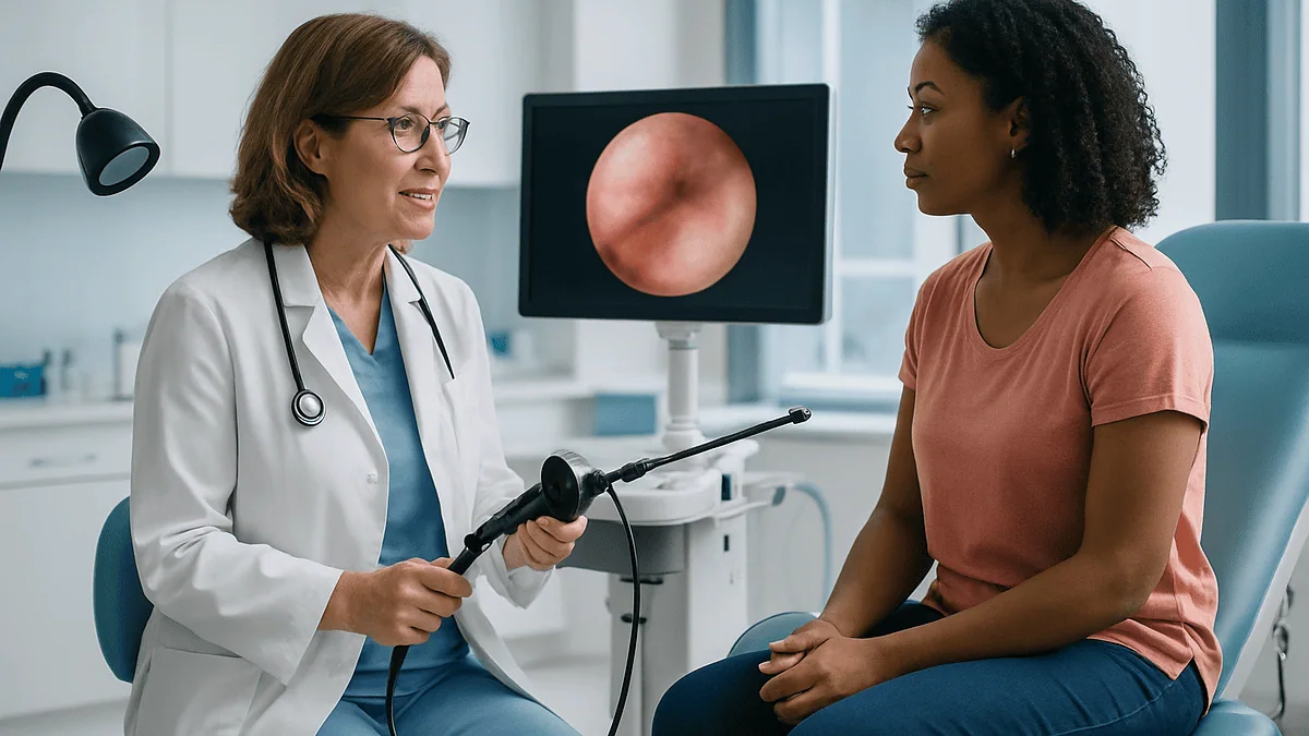
A hysteroscopy lets a clinician look directly inside the uterus with a thin camera, so it can show endometrial polyps, fibroids that bulge into the cavity, scar tissue (adhesions), a dividing wall called a septum, retained tissue, a misplaced coil and areas of lining that need a biopsy. What it cannot see is the ovaries, the outer uterine wall or endometriosis. Findings help the treating team choose between monitoring, removal or further tests.
The letter arrives with a referral code and a two-line explanation: a camera test to look inside the womb. For many people the first reaction is not fear but a very practical question. What is the doctor expecting to find, and what happens if they find it?
That question deserves a straight answer, because what hysteroscopy can show is narrower and more specific than most people assume. It is a look at one room in the house: the lining and the cavity of the uterus. Inside that room it is remarkably good at spotting the small growths and scars that drive heavy or unpredictable bleeding and some fertility problems. Outside that room, it sees nothing at all.
This explainer walks through what the camera can and cannot reveal, how a polyp looks different from a fibroid, why a stretch of scar tissue matters, and how each finding tends to shape the plan your care team proposes.
What hysteroscopy can show: a short, honest list
A hysteroscopy is a procedure in which a slim telescope with a light and camera, called a hysteroscope, is passed through the vagina and cervix into the uterus. The cavity is gently opened with fluid or gas so the lining can be seen on a screen. According to the NHS, the test is used to investigate heavy periods, bleeding between periods, bleeding after menopause, pelvic pain, repeated miscarriage and difficulty getting pregnant.
Here is what the camera is designed to reveal:
- Endometrial polyps: soft overgrowths of the uterine lining, often on a stalk.
- Fibroids that protrude into the cavity: firm, muscular growths pressing the lining inward.
- Adhesions: bands of scar tissue that stick the walls of the cavity together.
- A uterine septum or other difference in the shape of the cavity.
- Retained pregnancy tissue after miscarriage, birth or termination.
- A contraceptive coil (IUD) that has shifted or whose threads cannot be found.
- Lining that looks unusually thick, irregular or patchy and needs a sample taken.
Notice what is missing from that list. Ovarian cysts, endometriosis, fibroids buried deep in the muscle wall and anything in the pelvis outside the uterus are invisible to this instrument. That is not a weakness of the technique; it is simply the boundary of the room it looks into. Clinicians pair hysteroscopy with ultrasound, and sometimes MRI, precisely because each tool answers a different question.
The value of a direct view is that it turns a shadow on an ultrasound into something a clinician can describe by size, position and texture, and often remove in the same sitting. Those three details, more than the label alone, are what shape the plan.
How does a hysteroscopy actually work?
Picture a drinking straw with a camera lens at the tip. Modern diagnostic hysteroscopes are only a few millimeters across, which is why the NHS notes that many people have the test in an outpatient room without a general anesthetic, sometimes with a local anesthetic or simple pain relief beforehand.
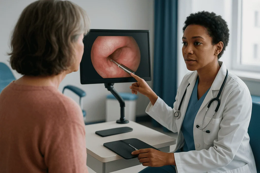
You lie on an examination couch, usually with your legs supported. A speculum, the same instrument used for cervical screening, may be placed to see the cervix, although with the thinnest scopes it can often be skipped. The clinician then guides the hysteroscope through the cervical canal. A warmed sterile salt solution, called saline, flows through the scope to open the cavity slightly, in the same way air inflates a collapsed balloon so you can see its inner surface.
What you feel most is often the fluid and the stretch, which many people describe as period-type cramping. The NHS reports that a diagnostic look typically takes about 5 to 10 minutes, with the whole appointment lasting up to 30 minutes if a small procedure is added.
On the screen, the clinician works systematically: the canal, the top of the cavity, the openings to each fallopian tube, then every wall. If something needs sampling, a tiny instrument passes through a channel in the scope. A biopsy, a small piece of tissue sent to a laboratory, takes seconds. Small polyps can often be removed through the same channel, an approach clinicians call “see and treat”.
When larger fibroids or extensive scar tissue are expected, the test is usually planned as a short operation under general or regional anesthetic, using a slightly wider operating hysteroscope. The principle is identical; only the setting and the tools change.
What can a hysteroscopy diagnose? Polyps up close
Endometrial polyps are the single most common finding when hysteroscopy is done for abnormal bleeding. A polyp is a localized overgrowth of the endometrium, the lining that thickens and sheds each cycle. Cleveland Clinic describes them as usually noncancerous growths that become more common in the 40s and 50s, though they can appear at any age.
On ultrasound a polyp may be nothing more than a thickened stripe of lining. Through the hysteroscope it becomes unmistakable: a smooth, pink or reddish projection, often on a slender stalk, that sways gently in the saline flow. Its surface shows a fine network of vessels, and it has the same soft, lining-like texture as the wall it grows from. That softness is the first clue separating it from a fibroid.
Why do polyps matter? They can bleed irregularly, extend periods, cause spotting between cycles or after menopause, and occasionally interfere with implantation of an embryo. Cleveland Clinic notes that a small proportion contain precancerous or cancerous cells, which is why clinicians generally recommend that polyps found after menopause, or those causing symptoms, be removed and examined under a microscope rather than simply watched.
Hysteroscopy adds detail no scan can: how many polyps there are, where each one sits, and whether the stalk is narrow enough to remove in the outpatient room. A single pea-sized polyp on a thin stalk is a very different plan from several broad-based polyps spread across the cavity.
The camera cannot tell a benign polyp from a worrying one by appearance alone. Only the laboratory can. So the honest answer to “what can a hysteroscopy diagnose” is that it identifies and removes the polyp; the diagnosis of what that polyp is made of comes a week or two later, in the pathology report.
Hysteroscopy for fibroids: which ones show and which stay hidden
Fibroids are firm, noncancerous growths made of muscle and fibrous tissue that develop in or on the uterus. Mayo Clinic notes that many people will have fibroids at some point in their lives, often without symptoms. The NHS estimates that around 1 in 3 women develop them, most commonly between the ages of 30 and 50.
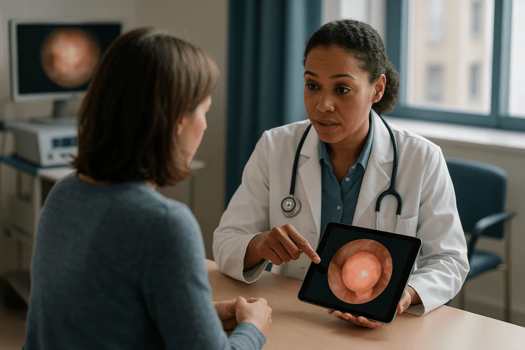
Here is the crucial point for anyone offered hysteroscopy for fibroids: the camera sees only the fibroids that touch or distort the cavity. Clinicians describe fibroids by location. Submucosal fibroids sit just beneath the lining and bulge inward; these are the ones a hysteroscope sees clearly. Intramural fibroids grow within the muscle wall, and subserosal fibroids sit on the outer surface. The latter two may be enormous on an ultrasound and completely invisible on hysteroscopy.
Through the scope, a submucosal fibroid looks quite different from a polyp. It is pale, often white or yellowish, firm, and rounded like a marble pushing against a sheet. Larger vessels may cross its surface. The clinician will assess how much of it protrudes into the cavity. A fibroid that is mostly inside the cavity can often be shaved away hysteroscopically; one that is mostly buried in the wall may need a different approach, or more than one session, or no surgery at all if it causes no trouble.
This is why the plan after a fibroid finding so often depends on a measurement rather than a label. A small fibroid that is almost entirely in the cavity may be a short day-case removal. A larger one that is mostly intramural might be better managed with medicines that shrink fibroids or control bleeding, or with procedures that do not go through the cervix at all. Those choices rest on the full picture from scan and scope together, and on your own priorities, which the treating team will weigh with you.
Adhesions and scar tissue: what the camera reveals about Asherman's syndrome
Intrauterine adhesions are bands or sheets of scar tissue that form inside the cavity and stick its walls together. When they are extensive and cause symptoms, clinicians use the term Asherman’s syndrome. Scarring most often follows surgery on the uterine lining, especially procedures after miscarriage or birth, and sometimes infection.
Unlike polyps and fibroids, adhesions rarely show well on a standard ultrasound. A thin lining or an unusual outline may raise suspicion, but hysteroscopy is widely regarded as the reference test, because scar tissue is best appreciated by looking at it directly. Through the scope, adhesions appear as pale, bloodless strands, like cobwebs across a doorway, or as dense white walls that narrow the cavity into a small pocket. The clinician notes whether they are filmy or thick, how much of the cavity is involved, and whether the tubal openings are visible.
Those details matter because the symptoms of adhesions are easy to misread. Periods may become very light or stop altogether, which can be mistaken for a hormonal problem. Some people experience cyclical pain when menstrual blood is trapped behind scar tissue. Others only discover adhesions during investigations for repeated miscarriage or difficulty conceiving, when a scarred cavity offers little healthy lining for an embryo to settle into.
Treatment, where it is needed, involves carefully dividing the adhesions under direct vision, usually with small scissors or an electrosurgical instrument passed through an operating hysteroscope. The procedure is delicate: too little and the cavity stays restricted; too much and the healthy lining can be damaged. Some teams follow surgery with measures intended to reduce re-scarring, and a second look to confirm the result. Whether any of this is appropriate depends on symptoms and goals, and that judgment belongs with the treating team.
Shape differences, retained tissue and lost coils
Beyond growths and scars, hysteroscopy answers several other questions that frustrate scans.
The first is the shape of the cavity itself. During development, the uterus forms from two tubes that fuse. If the central wall between them does not fully dissolve, a septum remains: a wedge of tissue dividing the cavity partly or completely. A septum is the most common congenital difference in uterine shape and has been associated with miscarriage. Through the hysteroscope it appears as a pale ridge running down from the top of the cavity, and because the view is two-dimensional, clinicians usually combine it with three-dimensional ultrasound or MRI to confirm whether the outer contour of the uterus is normal, which distinguishes a septum from other variants.
The second is retained tissue. After a miscarriage, delivery or termination, small fragments of placenta or pregnancy tissue can remain attached to the wall. They may cause prolonged bleeding or infection, and on ultrasound they can be hard to tell from blood clot. Under direct vision, retained tissue has a distinctive shaggy, often yellowish appearance, and it can frequently be removed precisely while leaving the surrounding lining intact.
The third is the wandering coil. When the threads of an intrauterine device cannot be felt or seen at the cervix, the device may have rotated, embedded in the wall or, rarely, passed through it. An ultrasound usually locates it. If the coil is still inside the cavity, hysteroscopy lets the clinician see it and retrieve it with a small grasper, which is far gentler than searching blindly.
Finally, the camera inspects the cervical canal on the way in, where polyps and narrowing can also hide. It is a short journey, but a thorough one.
What hysteroscopy cannot show, and why ultrasound still comes first
An honest explainer has to spend as much time on limits as on strengths, because disappointment after a “normal” hysteroscopy usually comes from expecting it to see things it never could.
Hysteroscopy does not see the ovaries. Ovarian cysts, polycystic ovaries and ovarian masses are assessed by ultrasound and blood tests. It does not see endometriosis, a condition in which tissue resembling the lining grows outside the uterus; that diagnosis typically requires laparoscopy, a keyhole camera placed through the abdomen. It does not see adenomyosis, where lining tissue grows into the muscle wall, except as indirect hints. It cannot assess whether the fallopian tubes are open, although it can see their entrances; tubal patency is tested with dye studies. Fibroids within or on the outer wall, as discussed, may be entirely out of view.
This is why most pathways begin with a transvaginal ultrasound, a scan performed with a probe placed in the vagina to give a close view of the pelvis. Ultrasound maps the whole uterus and both ovaries in minutes. Hysteroscopy is then reserved for the specific job of inspecting, sampling or treating the cavity when the scan or the symptoms point there. The NHS lists both as routine investigations for abnormal bleeding, used in sequence rather than in competition.
There is also a limit on interpretation. A hysteroscopy can show that lining looks thickened or irregular, but the camera cannot grade cells. Endometrial hyperplasia, an overgrowth of lining cells that can sometimes progress toward cancer, and endometrial cancer itself are laboratory diagnoses made on biopsy tissue. The scope’s contribution is to make sure the sample comes from the right place, rather than from a random sweep that might miss a small area.
Knowing these boundaries helps you ask the right follow-up question when a result comes back: not “why didn’t the camera see it?” but “which test looks at that?”
Who is usually offered hysteroscopy, and who is asked to wait
Guidelines in the United Kingdom and United States converge on a similar list of reasons to look inside the cavity. According to the NHS and Johns Hopkins Medicine, hysteroscopy is commonly offered for:
- Heavy, prolonged or irregular periods, particularly when ultrasound suggests a polyp or submucosal fibroid.
- Any bleeding after menopause, because the cavity must be assessed and the lining sampled.
- Bleeding between periods or after intercourse once cervical causes have been checked.
- Repeated miscarriage or difficulty conceiving, to exclude adhesions, a septum or polyps.
- A suspected lost coil or retained tissue.
- Abnormal thickening of the lining seen on a scan, with or without symptoms, depending on age and risk factors.
Equally important is who is usually asked to wait or offered something else. Hysteroscopy is generally postponed during a known or possible pregnancy, so most units ask for a pregnancy test on the day. It is deferred during an active pelvic infection or heavy bleeding that would obscure the view, and often rescheduled if you are mid-period for the same reason. People with a very narrow or scarred cervix may be offered medicine to soften it beforehand, or a procedure under anesthetic rather than an outpatient attempt.
Cervical cancer that has already been diagnosed is a reason not to pass instruments through the cervix. Significant heart or lung conditions do not rule the test out, but they shape the choice of anesthetic and setting.
Timing within the cycle matters too. For people who still menstruate, clinicians often prefer the first half of the cycle, after bleeding stops, when the lining is thin and small polyps are easiest to see. None of these rules are absolute. They are the starting points from which your care team personalizes the decision.
How findings guide the plan: a summary table
The most useful way to think about a hysteroscopy report is as a set of decision points rather than a verdict. Each finding opens a small menu of reasonable next steps, and your symptoms, age, plans for pregnancy and preferences decide which is chosen. The table below summarizes the usual directions. It is a map, not a prescription; the route is always set with the treating team.
| Finding on hysteroscopy | What it typically looks like | How it commonly guides the plan |
|---|---|---|
| Endometrial polyp | Soft, pink, often on a stalk | Removal, frequently in the same session, with tissue sent for analysis |
| Submucosal fibroid | Firm, pale, rounded, bulging inward | Hysteroscopic removal if mostly in the cavity; other options if mostly in the wall |
| Adhesions | Pale bands or sheets narrowing the cavity | Division under direct vision if symptomatic or affecting fertility; monitoring otherwise |
| Septum | Ridge dividing the cavity from the top | Confirmation with 3D ultrasound or MRI; surgery considered in selected cases |
| Thick or irregular lining | Patchy, uneven surface | Targeted biopsy; further plan depends on laboratory result |
| Retained tissue or displaced coil | Shaggy tissue or visible device | Removal under direct vision |
| Normal cavity | Smooth walls, both tubal openings seen | Attention turns to hormonal, ovarian or other causes |
Two patterns are worth drawing out. First, a normal result is genuinely useful. It rules the cavity out as the cause and redirects effort toward hormones, ovulation, clotting or the muscle wall, each of which has its own treatments. Second, the laboratory result often matters more than the camera’s impression. A polyp that looks entirely innocent may still return a report that changes the plan, and the team will usually wait for pathology before finalizing anything.
Hysteroscopy recovery time: the following days and weeks
Recovery after a diagnostic hysteroscopy is usually brief, which surprises people who expected something closer to an operation. The NHS advises that most people feel able to return to normal activities the following day, and some the same day if no general anesthetic was used. Cleveland Clinic similarly describes a quick return to routine for simple outpatient procedures.
In the first hours, cramping similar to period pain is common, and simple pain relief recommended by your clinician is usually enough. Light bleeding or spotting for a few days is expected, particularly if a biopsy or polyp removal was done; the NHS notes this can continue for several days. A watery discharge, sometimes tinged pink, reflects the saline used to open the cavity and settles quickly.
If you had a general anesthetic, the NHS advises arranging for someone to take you home and not driving, operating machinery or making important decisions for the next 24 hours, because the drugs take time to clear.
Most units ask you to avoid intercourse, tampons and swimming for about a week, or until bleeding has stopped, to reduce the chance of infection while the cervix settles. Showers are fine. Exercise can usually resume as soon as you feel comfortable, with heavier training delayed a little longer after operative procedures.
Where a larger fibroid or extensive adhesions were treated, expect a longer tail: a week or so of cramps and bleeding, and a follow-up appointment to review the result and discuss any further steps. If a biopsy was taken, results typically arrive within a few weeks, and the team will explain how they will be shared.
These are typical ranges from mainstream guidance, not promises. Your own recovery will depend on what was done and on how your body responds, and your discharge instructions take priority over any general article.
Risks of hysteroscopy, and the alternatives your team may discuss
Hysteroscopy is considered a low-risk procedure, but no procedure is risk-free, and understanding the small risks makes the consent conversation meaningful rather than ceremonial.
The NHS lists the main complications as accidental damage to the uterus or cervix, which is uncommon and may need antibiotics or, rarely, a further operation to repair; excessive bleeding, more likely after operative procedures; and infection, which typically presents with a smelly discharge, fever or persistent pain in the days afterward. The NHS also notes that feeling faint affects about 1 in 200 people who have the test without anesthetic, a reflex response to instruments passing through the cervix rather than a sign of harm. Fluid overload, where the body absorbs too much of the saline, is a recognized risk of longer operative procedures and is monitored closely by the surgical team.
Pain is a risk worth discussing in advance. Experience varies widely: some people feel little more than mild cramping, while others find the outpatient procedure very uncomfortable. Good units offer local anesthetic, a choice of pain relief, and the option to stop at any point or to rebook under general anesthetic. Asking about these options beforehand is entirely reasonable.
Alternatives depend on the question being asked. For abnormal bleeding, a blind endometrial biopsy taken in clinic without a camera can sample the lining, though it may miss small focal lesions. Saline infusion sonography, an ultrasound performed while fluid fills the cavity, outlines polyps and fibroids without a scope, but cannot remove or sample them. MRI gives detailed images of fibroids and the muscle wall. Hormonal treatments and a hormone-releasing coil can manage bleeding without any investigation of the cavity in appropriate cases.
None of these is automatically better or worse. The right choice depends on what needs to be seen, sampled or treated, which is a conversation for you and your clinician.
What people often get wrong about what hysteroscopy can show
Myths cluster around this test, and clearing them away takes some of the anxiety out of the appointment.
“If the hysteroscopy is normal, nothing is wrong.” A normal cavity rules out polyps, submucosal fibroids and adhesions as the cause of symptoms. It says nothing about hormones, ovulation, clotting, the muscle wall, the ovaries or endometriosis. Normal is informative, not dismissive.
“A polyp means cancer.” Cleveland Clinic is clear that most uterine polyps are benign. A small proportion contain abnormal cells, which is why polyps are removed and analyzed, especially after menopause, but the finding itself is not a diagnosis of anything sinister.
“The camera can tell what a growth is.” Experienced clinicians recognize typical appearances, but the definitive answer comes from the laboratory. Treat the pathology report, not the screen, as the final word.
“Hysteroscopy finds all fibroids.” Only those touching the cavity. A person can have several large fibroids and a completely normal hysteroscopy if all of them sit in the wall or on the outside.
“It diagnoses endometriosis.” It does not. Endometriosis lives outside the uterus and is assessed by examination, imaging and, when needed, laparoscopy.
“Scar tissue is rare and only follows major surgery.” Adhesions most often follow relatively routine procedures on the lining, particularly after pregnancy, and can be the hidden reason behind very light periods or difficulty conceiving.
“It is a major operation.” For most diagnostic purposes it is an outpatient test lasting minutes, with a return to normal activity the next day according to the NHS. Operative procedures take longer and may need an anesthetic, but even these are usually day cases.
Each of these corrections points the same way: hysteroscopy is a precise tool with a defined job, and it works best when expectations match that job.
Questions to ask your care team before and after the procedure
A good consultation is a two-way exchange, and a short list of questions turns a referral letter into a shared plan. Consider taking these with you.
Before the procedure:
- What are you expecting to find, based on my symptoms and scan, and what would change the plan if you find it?
- Will this be a diagnostic look only, or will you remove polyps or take a biopsy in the same session if needed?
- What pain relief or anesthetic options are available, and can I stop or rebook under general anesthetic if it is too uncomfortable?
- Should I take anything beforehand, and is there a best point in my cycle for the test?
- Do I need someone to accompany me home?
After the procedure:
- Can you describe exactly what you saw, including size and position of anything found?
- Was a sample sent to the laboratory, when will the result be available, and how will I receive it?
- Does this finding explain my symptoms, or might something else be contributing?
- What are my options now, including monitoring, and what happens if I choose to do nothing for the moment?
- How might this affect plans for pregnancy, if that is relevant to me?
- What symptoms in the coming days should prompt me to call, and whom do I call?
Write the answers down or bring someone who can. People routinely leave appointments remembering the emotional tone and forgetting the specifics, and the specifics are what you will need when the pathology result arrives or when a follow-up decision is due. If anything in the explanation feels rushed or unclear, asking for a written summary or a further conversation is a reasonable request, not an imposition.
When to call your doctor after a hysteroscopy
Mild cramping and light bleeding are part of normal recovery. Certain symptoms, however, are red flags that need prompt attention, because they can signal infection, injury to the uterus or significant bleeding. The NHS and Cleveland Clinic advise contacting your care team or seeking urgent care if you notice any of the following in the days after the procedure:
- Heavy bleeding, such as soaking through a pad every hour or passing large clots, or bleeding that is getting heavier rather than lighter.
- Severe or worsening pelvic or abdominal pain that is not relieved by the pain relief you were advised to take.
- A fever, chills or feeling generally unwell and shivery.
- Vaginal discharge that is foul-smelling, green or yellow.
- Difficulty passing urine, or pain when doing so that is new since the procedure.
- Feeling faint, breathless, or having chest pain or a racing heartbeat.
- Any symptom that frightens you or does not fit what you were told to expect.
If you had a general anesthetic, also report persistent vomiting, confusion or drowsiness beyond the first day.
In the longer term, call if bleeding that the procedure was meant to investigate continues or returns, if periods stop or become unusually light after a procedure on the lining, which can be an early sign of new adhesions, or if you have not heard about a biopsy result within the time frame you were given.
Severe pain, heavy bleeding with dizziness, or a high fever should be treated as an emergency rather than a matter for the next routine clinic. Your discharge paperwork should include a direct number; keep it to hand for the first week. Acting early on a red flag is almost always simpler than waiting, and no clinician minds a call that turns out to be unnecessary.
Frequently asked questions
What can a hysteroscopy diagnose?
A hysteroscopy can identify endometrial polyps, fibroids that bulge into the uterine cavity, scar tissue (adhesions), a uterine septum, retained pregnancy tissue and a displaced contraceptive coil, and it allows a targeted biopsy of lining that looks thick or irregular. It cannot diagnose conditions outside the cavity such as ovarian cysts or endometriosis, and the final diagnosis of any growth depends on laboratory analysis of the tissue removed.
What is the difference between uterine polyps vs fibroids on hysteroscopy?
Polyps are soft overgrowths of the lining, usually pink, often on a stalk and easily moved by the saline flow. Fibroids are firm muscular growths that appear pale and rounded, pushing the lining inward from the wall. Polyps can frequently be removed through a diagnostic scope, while fibroids often need an operating hysteroscope, and how much of a fibroid sits in the cavity shapes whether hysteroscopic removal is suitable.
Can a hysteroscopy detect cancer?
It can show areas of lining that look abnormal and guide a biopsy from exactly that spot, which is how endometrial cancer and precancerous changes are diagnosed. The camera itself cannot confirm or exclude cancer by appearance. Bleeding after menopause is a common reason for hysteroscopy precisely because sampling the lining under direct vision is more reliable than a blind sample.
Does a hysteroscopy show endometriosis?
No. Endometriosis involves tissue growing outside the uterus, on the ovaries, pelvic lining or bowel, where a hysteroscope cannot reach. It is assessed by examination, ultrasound or MRI and, when needed, laparoscopy. A normal hysteroscopy does not rule endometriosis in or out, so continued pelvic pain after a normal result should be raised again with your clinician.
Can a hysteroscopy see scar tissue in the uterus?
Yes, and it is regarded as the reference test for intrauterine adhesions, which often hide from standard ultrasound. Scar tissue appears as pale bands or sheets that narrow the cavity. Hysteroscopy shows how thick and widespread the adhesions are and whether the openings to the fallopian tubes are visible, details that guide whether dividing the scar tissue is likely to help.
What is the typical hysteroscopy recovery time?
According to the NHS, most people feel able to return to normal activities the day after a hysteroscopy, and some the same day if no general anesthetic was used. Cramping and light bleeding may last a few days, and intercourse and tampons are usually avoided for about a week. Recovery after removal of a large fibroid or extensive adhesions tends to take longer, and your own discharge advice takes priority.
Does a hysteroscopy hurt?
Experiences vary widely. Many people describe period-like cramping during the few minutes the scope is inside, while others find it considerably more uncomfortable. Pain relief taken beforehand, local anesthetic and the option to stop or rebook under general anesthetic are all commonly offered, so it is worth discussing what matters to you before the appointment rather than during it.
Will the doctor remove a polyp during the hysteroscopy?
Often yes, if the appointment was planned as a see-and-treat session and the polyp is small enough to remove through the instrument channel. Larger polyps, multiple polyps or fibroids may need a separate operative procedure. Ask in advance whether removal is planned for the same visit, since this affects how long the appointment takes and what to expect afterward.
Can a hysteroscopy find the cause of infertility or miscarriage?
It can identify cavity problems associated with these difficulties, including adhesions, a septum, polyps and submucosal fibroids, and correcting some of these may be recommended. It does not assess the ovaries, egg supply, tubal patency or hormonal factors, so it forms one part of a wider fertility work-up rather than a complete answer on its own.
What happens if the hysteroscopy is normal?
A normal result means the cavity has smooth walls and no polyps, fibroids, adhesions or shape differences, which rules these out as the cause of your symptoms. Attention then turns to other explanations such as hormonal imbalance, problems with ovulation, bleeding disorders or conditions in the muscle wall, each of which has its own tests and treatments to discuss with your care team.
References
This article is for general information only and is not a substitute for professional medical advice. Please consult a qualified doctor about your individual situation.

More from the Blog

Excision vs Ablation for Endometriosis: How the Two Surgical Techniques Differ
Excision removes endometriosis tissue by cutting it out at its full depth so a pathologist can examine it, while ablation destroys the surface of…

Preparing for a PCOS Evaluation: Why Tracking Cycles, Weight Changes and Symptoms Helps
Preparing for a PCOS evaluation means bringing a dated record of your menstrual cycles (first day of each period, cycle length, skipped months), a…
PCOS Treatment Planning: How Cycle Regulation, Skin Care and Fertility Goals Are Prioritized
A PCOS treatment plan is built around the goal that matters most to the person right now: regulating cycles, calming skin and hair changes,…

After an Ovarian Cyst Ruptures: What to Expect
After an ovarian cyst ruptures, most people feel a sudden sharp pelvic pain on one side that eases over several days as the released…

Who Is a Candidate for an IUD? Heavy Bleeding, Pelvic Infection History and What Doctors Check
Most people who want long-acting, reversible contraception can get an IUD, including teenagers, people who have never been pregnant, and those with heavy periods,…

Diagnostic vs Operative Hysteroscopy: When Looking Becomes Treating in the Same Session
A diagnostic hysteroscopy uses a thin lighted telescope passed through the cervix to look inside the uterus and find the cause of symptoms such…